. 2002 Jul 23;99(15):9801-6.
doi: 10.1073/pnas.132182099. Epub 2002 Jul 1.

The crystal structure of a tetrameric hemoglobin in a partial hemichrome state

Affiliations

The crystal structure of a tetrameric hemoglobin in a partial hemichrome state

Antonio Riccio et al. Proc Natl Acad Sci U S A. .

Abstract

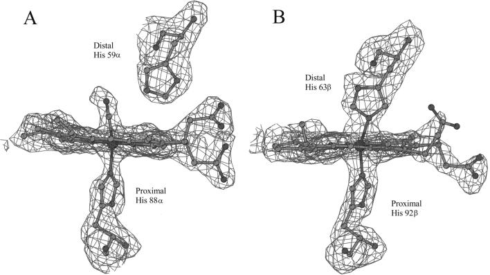
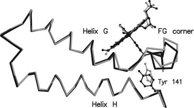
Tetrameric hemoglobins are the most widely used systems in studying protein cooperativity. Allosteric effects in hemoglobins arise from the switch between a relaxed (R) state and a tense (T) state occurring upon oxygen release. Here we report the 2.0-A crystal structure of the main hemoglobin component of the Antarctic fish Trematomus newnesi, in a partial hemichrome form. The two alpha-subunit iron atoms are bound to a CO molecule, whereas in the beta subunits the distal histidine residue is the sixth ligand of the heme iron. This structure, a tetrameric hemoglobin in the hemichrome state, demonstrates that the iron coordination by the distal histidine, usually associated with denaturing states, may be tolerated in a native-like hemoglobin structure. In addition, several features of the tertiary and quaternary organization of this structure are intermediate between the R and T states and agree well with the R --> T transition state properties obtained by spectroscopic and kinetic techniques. The analysis of this structure provides a detailed pathway of heme-heme communication and it indicates that the plasticity of the beta heme pocket plays a role in the R --> T transition of tetrameric hemoglobins.


Figures

Figure 1
Figure 1
Electron density maps (contoured at 2.5 σ) corresponding to the α (A) and β (B) heme regions.
Figure 2
Figure 2
Stereo drawing showing the scissors-like motion of the β chain EF corner occurring upon hemichrome formation. The EF regions of HbTn[α(CO)β(hemi)]2 (cyan) and HbTnCO (red) are shown after superimposition of the F helix. For clarity the propionate groups of heme have been omitted.
Figure 3
Figure 3
Distribution of the Cα–Cα distances between the proximal and the distal histidine residues in 276 α and β chains belonging to 80 tetrameric Hbs reported in the PDB (38). The value corresponding to the β chain of HbTn[α(CO)β(hemi)]2 is colored in black.
Figure 4
Figure 4
Stereo drawing showing the relative orientation of His-97β side chain and helix C of the α chain at the α1β2 of liganded and unliganded Hbs. HbTnCO, HbTn[α(CO)β(hemi)]2, HbTb-deoxy and the R2 carbonmonoxy form of human Hb are colored in red, cyan, green, and yellow, respectively. The residues of helix C of the α chains have been used in the superimposition. For clarity only helix C of HbTn[α(CO)β(hemi)]2 is shown.
Figure 5
Figure 5
Structural modifications of the α chain produced by hemichrome formation in the β chain. HbTn[α(CO)β(hemi)]2 and HbTnCO are colored in gray and black, respectively.
Figure 6
Figure 6
Cα trace of HbTn[α(CO)β(hemi)]2 (cyan), HbTbCO (purple), and HbTb-deoxy (green). The residues of the α1β1 dimers have been superimposed. Residues belonging to the α1β1 and α2β2 dimers are mostly located in the lower and in the upper part of the figure, respectively. The labeled residues Asp-5, together with Lys-95 and Pro-47, belong to β2 and α2 chains, respectively. The axis required to superimpose the α2β2 dimers of HbTbCO and HbTb-deoxy is normal to the figure and its position is marked by a black circle. The gray circle represents the intersection of the axis required to superimpose the α2β2 dimers of HbTn[α(CO)β(hemi)]2 and HbTb-deoxy with a plane parallel to the figure and passing through the center of mass of the tetramer. The two axes form an angle of 15°.

Similar articles

Cited by

References

    1. Perutz M F. Nature (London) 1970;228:726–739. - PubMed
    1. Perutz M F, Wilkinson A J, Paoli M, Dodson G G. Annu Rev Biophys Biomol Struct. 1998;27:1–34. - PubMed
    1. Rifkind J M, Abugo O, Levy A, Heim J. Methods Enzymol. 1994;231:449–480. - PubMed
    1. Levy A, Rifkind J M. Biochemistry. 1985;24:6050–6054. - PubMed
    1. Levy A, Kuppusamy P, Rifkind J M. Biochemistry. 1990;29:9311–9316. - PubMed

Publication types

Associated data

LinkOut - more resources