. 2002 Jul 9;99(14):9421-6.
doi: 10.1073/pnas.132285699. Epub 2002 Jul 1.

Direct visualization of distinct T cell epitopes derived from a melanoma tumor-associated antigen by using human recombinant antibodies with MHC- restricted T cell receptor-like specificity

Affiliations

Direct visualization of distinct T cell epitopes derived from a melanoma tumor-associated antigen by using human recombinant antibodies with MHC- restricted T cell receptor-like specificity

Galit Denkberg et al. Proc Natl Acad Sci U S A. .

Abstract

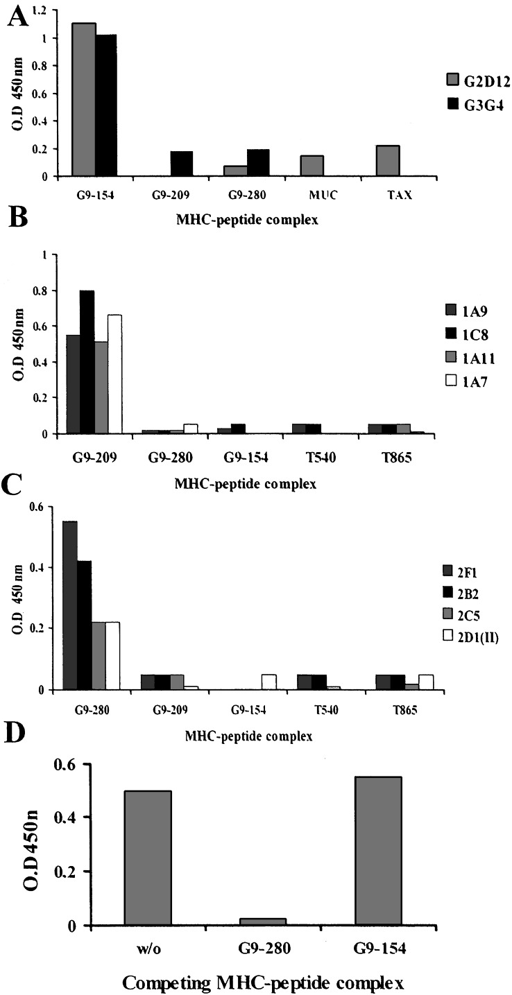
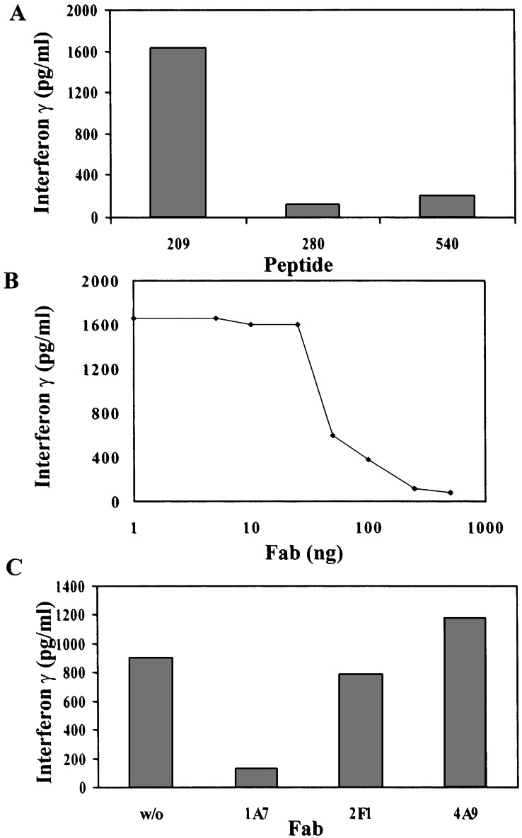
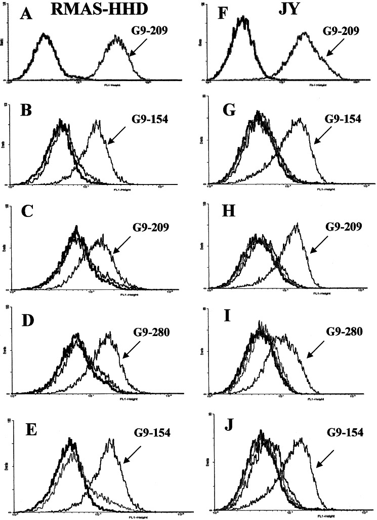
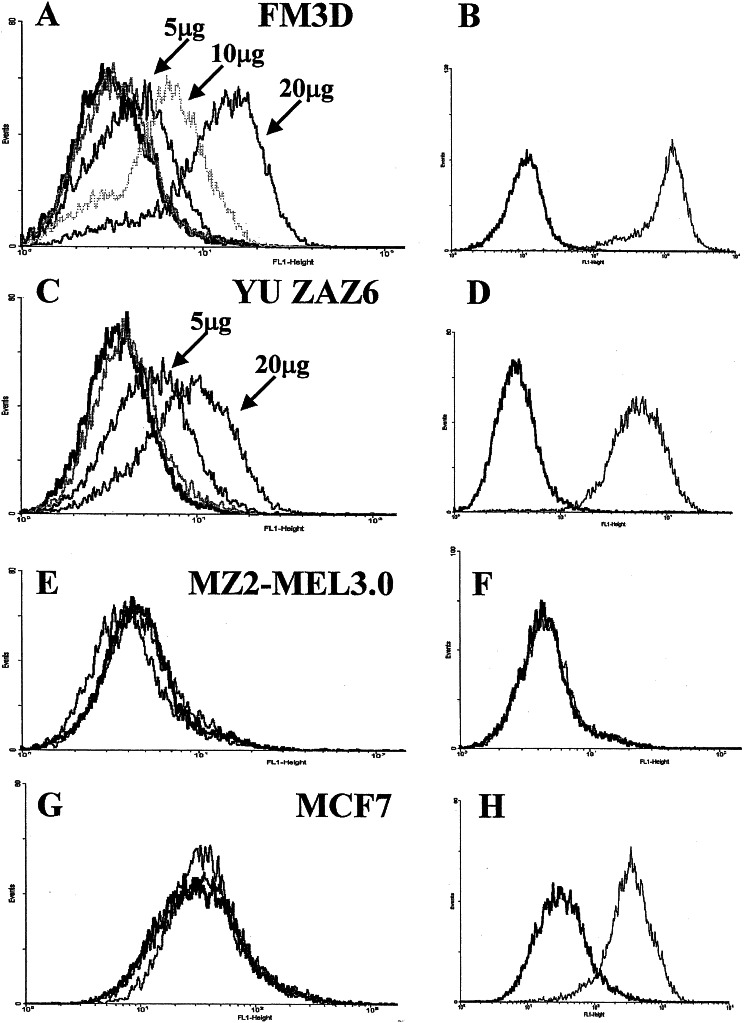
Specificity in the cellular immune system is controlled and regulated by the T cell antigen receptor (TCR), which specifically recognizes peptide/major histocompatibility complex (MHC) molecules. In recent years many cancer-associated MHC-restricted peptides have been isolated and because of their highly restricted fine specificity, they are desirable targets for novel approaches in immunotherapy. Antibodies that would recognize tumor-associated MHC-peptide complexes with the same specificity as the TCR would be valuable reagents for studying antigen presentation by tumor cells, for visualizing MHC-peptide complexes on cells, and eventually for monitoring the expression of specific complexes during immunotherapy. To generate molecules with such a unique fine specificity, we selected a large nonimmune repertoire of phage Fab antibodies on recombinant HLA-A2 complexed with three common antigenic T cell, HLA-A2-restricted epitopes derived from the melanoma differentiation antigen gp100. We were able to isolate a surprisingly large panel of human recombinant Fab antibodies that exhibit a characteristic TCR-like binding specificity to each of the three gp100-derived epitopes, yet unlike TCRs, they did so with an affinity in the nanomolar range. These TCR-like antibodies recognize the native MHC-peptide complex expressed on the surface of antigen-presenting cells. Moreover, they can detect the specific MHC-peptide complexes on the surface of melanoma tumor cells. These results demonstrate the ability to isolate high-affinity human recombinant antibodies with the antigen-specific, MHC-restricted specificity of T cells, and this ability was demonstrated for three different epitopes of the same melanoma-derived antigen.


Figures

Figure 1
Figure 1
Binding in ELISA of soluble purified Fabs to recombinant scMHC-HLA-A2-peptide complexes. Binding of soluble purified Fab clones specific for the gp100-derived epitopes G9-154 (A), G9-209 (B), and G9-280 (C) to immobilized scMHC-HLA-A2-peptide complexes is indicated. Shown are the specificities of several Fab clones to the gp100-derived epitopes for which they were selected, but not to the indicated control MHC–peptide complexes containing other gp100 and telomerase-derived HLA-A2-restricted epitopes. (D) Competition of MHC–peptide complexes for binding of Fab 2F1 to immobolized G9-280–HLA-A2 complexes; W/O, without competitior.
Figure 2
Figure 2
Specific inhibition of peptide-specific, MHC-restricted T cell activation with TCR-like Fab. T2 cells were pulsed with peptide as indicated and incubated with the G9-209-specific HLA-A-restricted CTL clone R6C12 in the presence of various concentrations of Fab 1A7 or control Fabs as indicated. T cell stimulation was measured by the release of IFN-γ to the culture medium. IFN-γ was determined by a double sandwich ELISA. (A) Stimulation of R6C12 CTLs with the G9-209 peptide but not control peptides. (B) Inhibition of T cell response with Fab 1A7 (C) Inhibition of T cell response is specific to Fab 1A7 but not to control Fabs 2F1 or 4A9.
Figure 3
Figure 3
Binding of Fab antibodies to APCs. RMAS-HHD or JY cells were loaded with the indicated HLA-A2-restricted peptides. Peptide-loaded cells were then incubated with the specific soluble purified Fab antibodies as shown. Detection of binding was with FITC-labeled anti-human Fab. (A) RMAS-HHD cells loaded with the G9-209 peptide and control unloaded cells were stained with the anti-HLA-A2 antibody BB7.2 to demonstrate the stabilization/expression of HLA-A2 complexes on the surface of peptide-loaded but not on peptide-unloaded cells. (B) RMAS-HHD cells were loaded with the G9-154 (marked) and control G9-280 peptides or control unloaded cells. Cells were stained with the G9-154-specific Fab G2D12. (C) RMAS-HHD cells were loaded with peptides G9-209-specific (marked) and G9-154 (control). Loaded and unloaded cells were stained with the G9-209-specific Fab 1A7. (D) RMAS-HHD cells were loaded with G9-280-specific (marked) and G9-209 (contol) peptides. Loaded and unloaded cells were stained with the G9-280-specific Fab 2F1. (E) RMAS-HHD cells were loaded with peptide G9-154 and incubated with Fabs G2D12 (marked), 1A7, and 2F1 specific for G9-154, G9-209, and G9-280, respectively. (F) JY cells were loaded with peptides G9-209 and stained with anti-HLA-A2 BB7.2 antibody. Controls are cells incubated with secondary anti-mouse-FITC antibody. (G) JY cells were loaded with peptides G9-154 (marked), G9-209, G9-280, or unloaded and then reacted with the G9-154/HLA-A-specific Fab G2D12. (H) JY cells were loaded with peptides G9-209 (marked), G9-154, G9-280, or unloaded and then reacted with the G9-209/HLA-A-specific Fab 1A7. (I) JY cells were loaded with peptides G9-280 (marked), G9-154, G9-209, or unloaded and then reacted with the G9-280/HLA-A-specific Fab 2F1. (J) JY cells were loaded with the peptide G9-154 and incubated with Fabs G2D12 (marked), 1A7, and 2F1 specific for G9-154, G9-209, and G9-280 in complex with HLA-A2, respectively. Control unloaded cells are represented by black traces.
Figure 4
Figure 4
Detection of MHC–peptide complexes on the surface of tumor cells. Melanoma FM3D (A) and YU ZAZ6 (C), which express HLA-A2 (B and D), as determined by reactivity with monoclonal antibody BB7.2, were stained with 5, 10, or 20 μg of Fab G2D12 specific for the melanoma gp100-derived G9-154 epitope, or with a Fab TCR-like antibody specific for the viral epitope TAX. Detection of binding was with FITC-labeled anti-human Fab. The melanoma HLA-A2- MZ2-MEL3.0 cells were not stained with G2D12 (E) or BB7.2 (F) (indication for HLA-A2). MCF7 HLA-A2+ breast carcinoma cells were stained with BB7.2 (H) but not with Fab G2D12 or the TAX-specific Fab (G). Black traces represent cells incubated with the secondary FITC-labeled antibody.

References

    1. Boon T, van der Bruggen P. J Exp Med. 1996;183:725–729. - PMC - PubMed
    1. Rosenberg S A. Nature (London) 2001;411:380–384. - PubMed
    1. Renkvist N, Castelli C, Robbins P F, Parmiani G. Cancer Immunol Immunother. 2001;50:3–15. - PMC - PubMed
    1. Anichini A, Maccalli C, Mortarini R, Salvi S, Mazzocchi A, Squarcina P, Herlyn M, Parmiani G. J Exp Med. 1993;177:989–998. - PMC - PubMed
    1. Coulie P G, Brichard V, Van Pel A, Wolfel T, Schneider J, Traversari C, Mattei S, De Plaen E, Lurquin C, Szikora J P. J Exp Med. 1994;180:35–42. - PMC - PubMed

Publication types

MeSH terms