. 2002 Jul 9;99(14):9196-201.
doi: 10.1073/pnas.142459399. Epub 2002 Jul 1.

The protofilament structure of insulin amyloid fibrils

Affiliations

The protofilament structure of insulin amyloid fibrils

José L Jiménez et al. Proc Natl Acad Sci U S A. .

Abstract

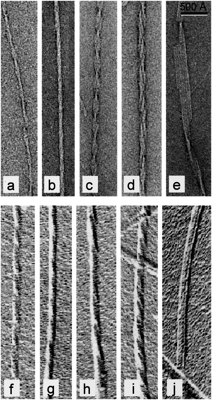
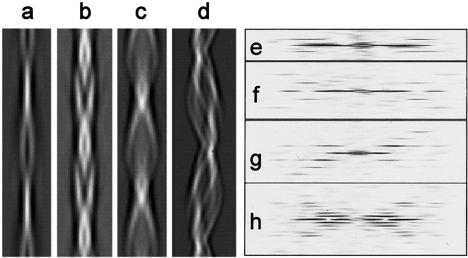
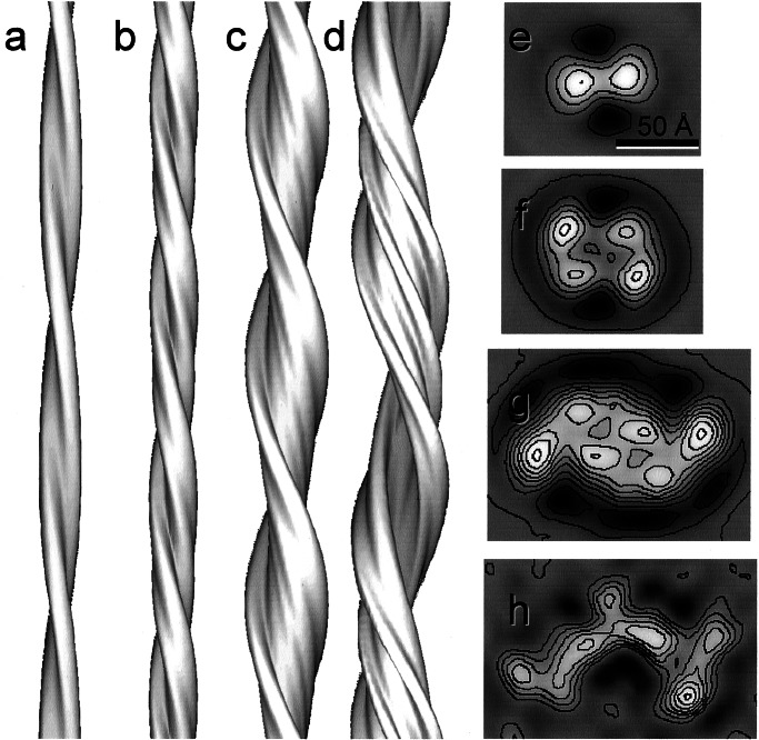
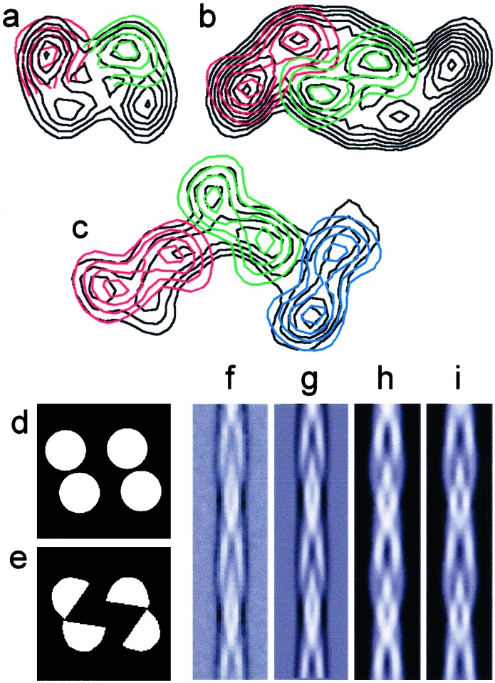
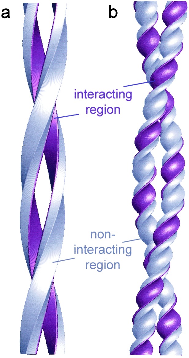
Under solution conditions where the native state is destabilized, the largely helical polypeptide hormone insulin readily aggregates to form amyloid fibrils with a characteristic cross-beta structure. However, there is a lack of information relating the 4.8 A beta-strand repeat to the higher order assembly of amyloid fibrils. We have used cryo-electron microscopy (EM), combining single particle analysis and helical reconstruction, to characterize these fibrils and to study the three-dimensional (3D) arrangement of their component protofilaments. Low-resolution 3D structures of fibrils containing 2, 4, and 6 protofilaments reveal a characteristic, compact shape of the insulin protofilament. Considerations of protofilament packing indicate that the cross-beta ribbon is composed of relatively flat beta-sheets rather than being the highly twisted, beta-coil structure previously suggested by analysis of globular protein folds. Comparison of the various fibril structures suggests that very small, local changes in beta-sheet twist are important in establishing the long-range coiling of the protofilaments into fibrils of diverse morphology.


Figures

Figure 1
Figure 1
Gallery of negatively stained (ae) and shadowed (fj) insulin fibrils, showing the diversity of fibril structures. The protofilament substructure is visible in e. The shadowing shows that all of the fibrils are left handed (gj). (Bar = 500 Å.)
Figure 2
Figure 2
Reprojected images and calculated diffraction patterns of the helical repeat from cryo-EM images of four different insulin amyloid fibril types. (a and e) Filtered image and diffraction pattern of the smallest diameter fibril. (b and f) For the compact fibril. (c and g) A larger fibril. (d and h) A twisted ribbon. The images are reprojected from the 3D maps in Fig. 3. Distances between crossovers (layer line spacings) are: a, 525 Å; b, 355 Å; c, 426 Å; d, 940 Å. Note that ac are double helices, so that their layer line spacings correspond to one half of a complete turn.
Figure 3
Figure 3
Surface representation of 3D maps and contoured density cross sections of the four insulin fibril structures shown in Fig. 2. (a and e) Structure of the fibril with a pair of protofilaments twisting around each other. (b and f) The four-protofilament compact fibril. (c and g) The six-protofilament fibril. (d and h) The twisted ribbon. The protofilaments are well resolved in the first three structures, but are less clear in the twisted ribbon. (eh, bar = 50 Å.)
Figure 4
Figure 4
Superposition of cross sections from the different fibril morphologies suggests the presence of a common protofilament structure. (a) The protofilaments fit well when compared individually between the two- and four-protofilament fibrils. (b) The pair of protofilaments fits well in size with pairs of protofilaments in the six-protofilament fibril. (c) The ribbon could hold six-protofilaments although their boundaries are not clear. (d and e) Two possible, simple models for the cross section of the compact, four-protofilament fibril. (f) Average of the repeats extracted from cryo-EM images of the compact fibrils and aligned as single particles. (g) Reprojection of the 3D map of the compact fibril. (h and i) Reprojections of the 3D models whose cross sections are shown in d and e, respectively.
Figure 5
Figure 5
(a) Insulin structure showing the three native disulfide bonds. A chain, green; B chain, blue; disulfide bonds, gold. (b) Topology diagram of insulin color coded as in a. (c) Possible topology for the amyloid protofilament. Orientations of the termini and disulfide bonds within the curved structure are arbitrary. The C terminus of chain B (dashed) is not required for amyloid fibril formation (see ref. 43). (d) β-strand model of a protofilament. Each chain is shown in two segments, a straight and a curved β-strand (PDB accession no. 1umu, residues 93–100). Each insulin molecule would occupy two layers, connected by the interchain disulfide bonds. (e) A possible β-strand model docked into the EM density of the compact fibril (transparent gray surface). The four protofilaments are colored separately.
Figure 6
Figure 6
Models for protofilament packing. (a) A twisted pair of rectangular protofilaments in which an interactive surface is colored purple. The protofilament twist accompanies the filament twist. (b) A supercoiled pair of protofilaments in which the regions involved in packing interactions rotate around each protofilament. In the correlated twist model a, interacting regions would be fixed relative to the cross-β structure, and other regions could accommodate large loops and/or folded domains that would not interfere with protofilament packing. Similar models can be constructed with more than two protofilaments, in which the cross section rotates as a rigid unit in the helical structure. Keeping the cross section fixed means that all packing contacts can be preserved in the helical fibril. This is the case for the four-protofilament model in Fig. 5e.

References

    1. Pepys M B. In: The Oxford Textbook of Medicine. 3rd Ed. Weatherall D J, Ledingham J G G, Warell D A, editors. Vol. 2. Oxford: Oxford Univ. Press; 1996. pp. 1512–1524.
    1. Sunde M, Serpell L C, Bartlam M, Fraser P E, Pepys M B, Blake C F. J Mol Biol. 1997;273:729–739. - PubMed
    1. Dobson C M. Trends Biochem Sci. 1999;24:329–332. - PubMed
    1. Cohen A S, Shirahama T, Skinner M. In: Electron Microscopy of Proteins. Harris J R, editor. Vol. 3. London: Academic; 1982. pp. 165–205.
    1. Serpell L C, Sunde M, Benson M D, Tennent G A, Pepys M B, Fraser P E. J Mol Biol. 2000;300:1033–1039. - PubMed

Publication types