. 2002 Jul 9;99(14):9404-9.
doi: 10.1073/pnas.152147499. Epub 2002 Jul 1.

Crystal structure of human cytomegalovirus IL-10 bound to soluble human IL-10R1

Affiliations

Crystal structure of human cytomegalovirus IL-10 bound to soluble human IL-10R1

Brandi C Jones et al. Proc Natl Acad Sci U S A. .

Abstract

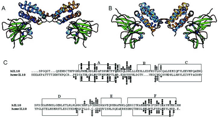
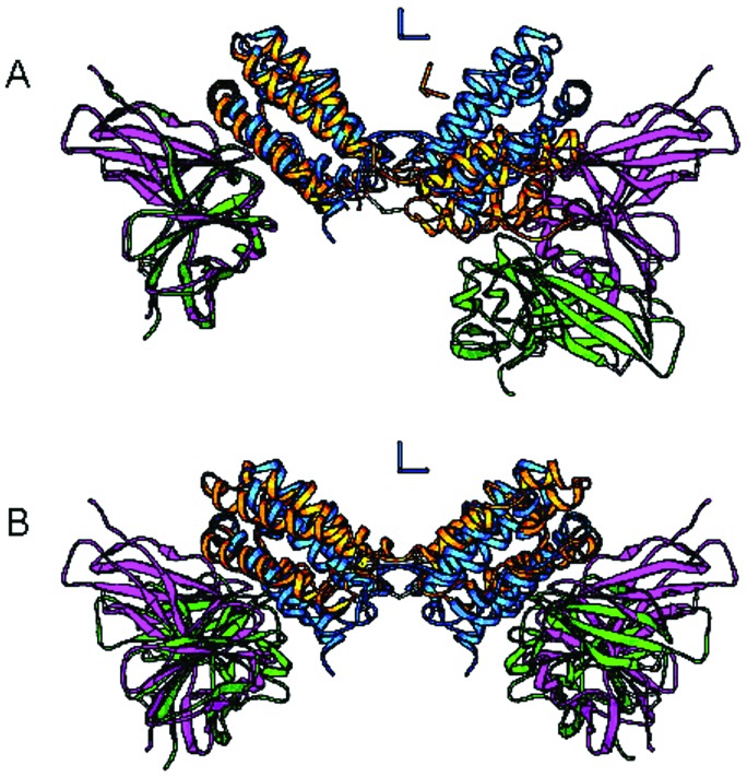
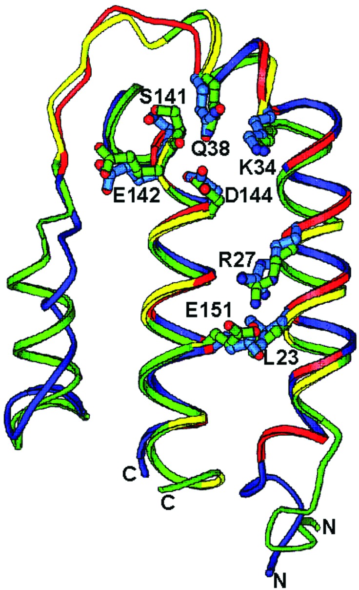
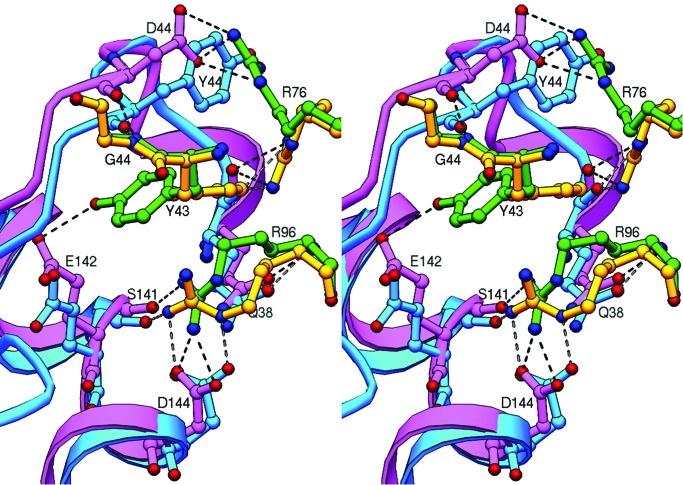
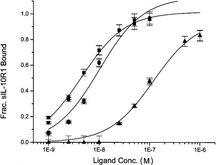
Human IL-10 (hIL-10) modulates critical immune and inflammatory responses by way of interactions with its high- (IL-10R1) and low-affinity (IL-10R2) cell surface receptors. Human cytomegalovirus exploits the IL-10 signaling pathway by expressing a functional viral IL-10 homolog (cmvIL-10), which shares only 27% sequence identity with hIL-10 yet signals through IL-10R1 and IL-10R2. To define the molecular basis of this virus-host interaction, we determined the 2.7-A crystal structure of cmvIL-10 bound to the extracellular fragment of IL-10R1 (sIL-10R1). The structure reveals cmvIL-10 forms a disulfide-linked homodimer that binds two sIL-10R1 molecules. Although cmvIL-10 and hIL-10 share similar intertwined topologies and sIL-10R1 binding sites, their respective interdomain angles differ by approximately 40 degrees. This difference results in a striking re-organization of the IL-10R1s in the putative cell surface complex. Solution binding studies show cmvIL-10 and hIL-10 share essentially identical affinities for sIL-10R1 whereas the Epstein-Barr virus IL-10 homolog (ebvIL-10), whose structure is highly similar to hIL-10, exhibits a approximately 20-fold reduction in sIL-10R1 affinity. Our results suggest cmvIL-10 and ebvIL-10 have evolved different molecular mechanisms to engage the IL-10 receptors that ultimately enhance the respective ability of their virus to escape immune detection.


Figures

Figure 1
Figure 1
Structure and sequence of the cmvIL-10/sIL-10R1 complex. (A) Ribbon diagram of the 1:2 cmvIL-10/sIL-10R1 complex viewed perpendicular to the twofold axis of cmvIL-10. (B) Ribbon diagram of 1:2 hIL-10/sIL-10R1 complex in the same orientation as A. (C) Structure-based sequence alignment of hIL-10 and cmvIL-10. α-Helices are defined by brackets and labeled. cmvIL-10 or hIL-10 residues that bury surface into sIL-10R1 or into their respective dimer interfaces are marked with circles and crosses, respectively. The amount of buried surface area is denoted by different numbers of circles or crosses with 1 > 5 Å2, 2 > 10 Å2 < 35 Å2, 3 > 35 Å2 < 60 Å2, 4 > 60 Å2 < 85 Å2, and 5 > 85 Å2.
Figure 2
Figure 2
Superposition of cmvIL-10 and hIL-10 1:2 receptor complexes. (A) Superposition of sIL-10R1 bound to cmvIL-10 (green) and hIL-10 (magenta). cmvIL-10 and hIL-10 are colored yellow and blue, respectively. Axes, in the same color as their respective cytokines, are shown representing the location of the twofold axes of cmvIL-10 and hIL-10 dimers. (B) Superposition of cmvIL-10 and hIL-10 1:2 complexes by way of the twofold axes of the cmvIL-10 and hIL-10 dimers. Color scheme is as in A.
Figure 3
Figure 3
The sIL-10R1 binding epitope. cmvIL-10 and hIL-10 scaffold residues (10–62 and 137–157 for cmvIL-10, 11–62 and 137–160 for hIL-10) are shown in green and blue, respectively. cmvIL-10 residues that bury surface area into sIL-10R1 are shown in red whereas hIL-10 residues are yellow. Side chains are shown for conserved side chain residues that bury surface area into each interface.
Figure 4
Figure 4
Stereoview of conformational changes in cmvIL-10 and hIL-10 Site Ia interfaces. cmvIL-10 and hIL-10 residues are shown in cyan and magenta, respectively. sIL-10R1 residues Tyr-43, Arg-76, and Arg-96 are colored yellow in the cmvIL-10/sIL-10R1 complex and green in the hIL-10/sIL-10R1 complex. The second conformation of sIL-10R1 Arg-76 in the hIL-10/sIL-10R1 is not shown for clarity but adopts the same conformation as sIL-10R1 Arg-76 bound to cmvIL-10 (yellow).
Figure 5
Figure 5
sIL-10R1 binding properties of the IL-10 homologs. Fraction of bound sIL-10R1 is plotted against increasing concentrations of cmvIL-10 (■), hIL-10 (●), and ebvIL-10 (▴) (log10 scale). Lines are nonlinear fits of the data to a 1:1 binding model.

References

    1. Moore K W, de Waal Malefyt R, Coffman R L, O'Garra A. Annu Rev Immunol. 2001;19:683–765. - PubMed
    1. Liu Y, Wei S H-Y, Ho A S-Y, de Waal Malefyt R, Moore K W. J Immunol. 1994;152:1821–1929. - PubMed
    1. Kotenko S V, Krause C D, Izotova L S, Pollack B P, Wu W, Pestka S. EMBO J. 1997;16:5894–5903. - PMC - PubMed
    1. Walter M R, Nagabhushan T L. Biochemistry. 1995;34:12118–12125. - PubMed
    1. Zdanov A, Schalk-Hihi C, Gustchina A, Tsang M, Weatherbee J, Wlodawer A. Structure (London) 1995;3:591–601. - PubMed

Publication types

MeSH terms

Associated data