. 2002 Jul 2:2:8.
doi: 10.1186/1471-213x-2-8.

A transcriptional response to Wnt protein in human embryonic carcinoma cells

Affiliations

A transcriptional response to Wnt protein in human embryonic carcinoma cells

Jennifer Willert et al. BMC Dev Biol. .

Abstract

Background: Wnt signaling is implicated in many developmental decisions, including stem cell control, as well as in cancer. There are relatively few target genes known of the Wnt pathway.

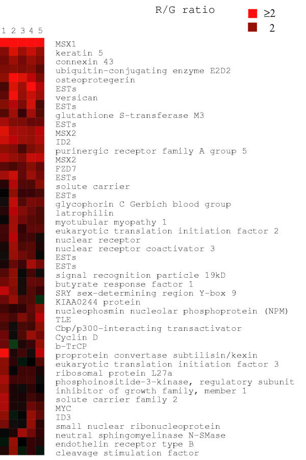
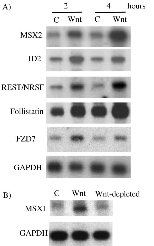
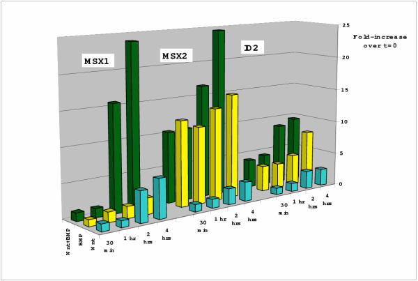
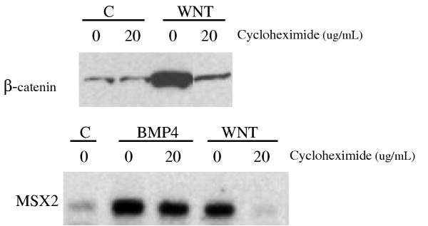
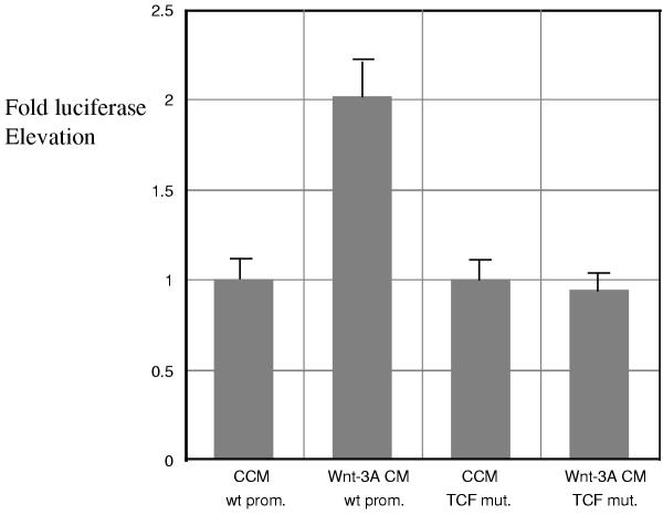
Results: We have identified target genes of Wnt signaling using microarray technology and human embryonic carcinoma cells stimulated with active Wnt protein. The ~50 genes upregulated early after Wnt addition include the previously known Wnt targets Cyclin D1, MYC, ID2 and betaTRCP. The newly identified targets, which include MSX1, MSX2, Nucleophosmin, Follistatin, TLE/Groucho, Ubc4/5E2, CBP/P300, Frizzled and REST/NRSF, have important implications for understanding the roles of Wnts in development and cancer. The protein synthesis inhibitor cycloheximide blocks induction by Wnt, consistent with a requirement for newly synthesized beta-catenin protein prior to target gene activation. The promoters of nearly all the target genes we identified have putative TCF binding sites, and we show that the TCF binding site is required for induction of Follistatin. Several of the target genes have a cooperative response to a combination of Wnt and BMP.

Conclusions: Wnt signaling activates genes that promote stem cell fate and inhibit cellular differentiation and regulates a remarkable number of genes involved in its own signaling system.


Figures

Figure 1
Figure 1
Microarray cluster analysis demonstrating increased expression of Wnt-3A responsive genes, including MSX1, MSX2, ID2, Versican, NPM, Frizzled-7, TLE/Groucho, Cyclin D1, and MYC. NCCIT cells were exposed to control conditioned medium (CCM) and Wnt-3A CM [8] for 4 hours. Shown here are 5 separate experiments. Note also that MSX 2 is included on this particular array in duplicate which provides an internal check on the precision of the measurements. Additional array experiments led to the identification of Follistatin and REST/NRSF as elevated genes (which were confirmed in Figure 2A). Brightest red corresponds to ratio of = 2:1 and dimmer red corresponds to ratio of 2:1. The full data set will be available at the Wnt homepage and the Stanford Microarray database
Figure 2
Figure 2
A) Confirmation of microarray data using Northern analysis. Northern blot analysis using MSX2, ID2, REST/NRSF, FZD7 (Frizzled7) and Follistatin cDNA probes. NCCIT cells were treated with CCM (-) or Wnt-3A CM (+) for the designated number of hours. 1 ug mRNA was loaded into each lane. GAPDH is shown as a sample loading control. B) MSX1 induction by Wnt-3A CM is dependent on the presence of soluble Wnt-3A protein. C = CCM. Wnt = Wnt-3A CM. Wnt-depleted = Wnt-3A CM. Cells were incubated with the various media for 4 hours.
Figure 3
Figure 3
Time course of Wnt induced gene expression and cooperation with BMP. NCCIT cells were exposed to Wnt-3A CM, BMP-4 (10 ng/ml final concentration) or the combination of Wnt-3A and BMP-4 for the specified number of hours. Shown here are MSX1, MSX2 and ID2 demonstrating that Wnt-3A induced effects are not seen until 2 hours (which corresponds to β-catenin accumulation). BMP-4 mediated induction of MSX1, MSX2 and ID2 occurs rapidly, as early as 30 minutes and stays relatively constant. Cooperative effects of Wnt-3A and BMP-4 are not evident until approximately 2 hours. The height of the bars presents the ratio induced/non-induced, quantified by phosphoimager analysis of Northern blots and normalized to GAPDH expression measured in the same experiment. Each bar represents average values obtained from 3–5 experiments for each mRNA tested.
Figure 4
Figure 4
A) Western blot demonstrating cycloheximide effects of Wnt-3A mediated accumulation of β-catenin. NCCIT cells were stimulated for 4 hours with CCM (C) or Wnt-3A CM (Wnt) in the presence (20 microgram/ml) or absence (0) of cycloheximide. Note that β-catenin does not accumulate in response to Wnt-3A CM when cycloheximide is present. B) Northern analysis demonstrating that MSX2 induction by Wnt-3A is abolished in the presence of cycloheximide. Cells were stimulated for 4 hours with CCM (C), BMP-4 or Wnt-3A CM (Wnt) in the presence (20 microgram/ml media) or absence of cycloheximide. As expected, BMP-4 mediated induction of MSX2 is not affected by cycloheximide which contrasts with the inhibition of Wnt-3A mediated induction of MSX2 in the presence of cycloheximide. GAPDH was used to verify equal loading. The inhibitory effect was also seen for MSX1, ID2, Follistatin, Versican and Cyclin D1 (not shown).
Figure 5
Figure 5
Response of a reporter containing the Follistatin promoter to Wnt-3A protein in NCCIT cells [37]. The Follistatin promoter [36] contains one putative TCF binding site (CTTTGAT). This promoter linked to luciferase was transfected in NCCIT cells, which were then exposed to Wnt-3A CM and CCM for 8 hours. This assay showed a significant increase in activity when cells were stimulated directly with Wnt-3A CM or co-transfected with activated β-catenin (not shown). This effect was abrogated by co-transfection of dominant-negative TCF-4 or axin (not shown). When the TCF site within the Follistatin promoter was mutated into CATCGAT, the Wnt-3A response was abolished.

References

    1. Cadigan K, Nusse R. Wnt signaling: a common theme in animal development. Genes & Dev. 1997;11:3286–3305. - PubMed
    1. Taipale J, Beachy PA. The Hedgehog and Wnt signalling pathways in cancer. Nature. 2001;411:349–54. doi: 10.1038/35077219. - DOI - PubMed
    1. Bhanot P, Brink M, Harryman Samos C, Hsieh JC, Wang YS, Macke JP, Andrew D, Nathans J, Nusse R. A new member of the frizzled family from Drosophila functions as a Wingless receptor. Nature. 1996;382:225–230. - PubMed
    1. Wehrli M, Dougan ST, Caldwell K, O'Keefe L, Schwartz S, Vaizel-Ohayon D, Schejter E, Tomlinson A, DiNardo S. arrow encodes an LDL-receptor-related protein essential for Wingless signalling. Nature. 2000;407:527–30. doi: 10.1002/(SICI)1096-9861(19990517)407:4<527::AID-CNE5>3.0.CO;2-H. - DOI - PubMed
    1. Moon RT, Brown JD, Yang-Snyder JA, Miller JR. Structurally related receptors and antagonists compete for secreted Wnt ligands. Cell. 1997;88:725–8. - PubMed

Publication types

MeSH terms