. 2002 Jul 15;11(15):1785-95.
doi: 10.1093/hmg/11.15.1785.

SMN, the spinal muscular atrophy protein, forms a pre-import snRNP complex with snurportin1 and importin beta

Affiliations

SMN, the spinal muscular atrophy protein, forms a pre-import snRNP complex with snurportin1 and importin beta

Usha Narayanan et al. Hum Mol Genet. .

Abstract

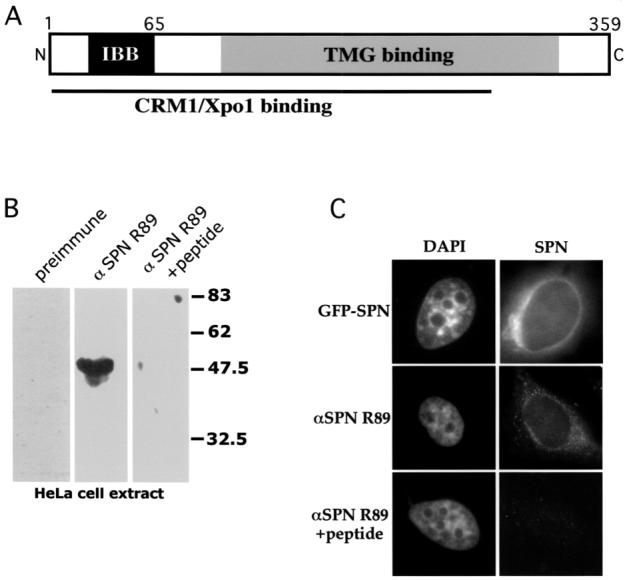
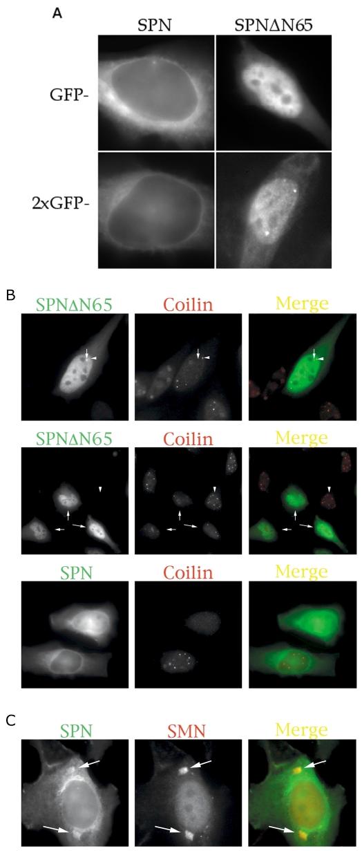
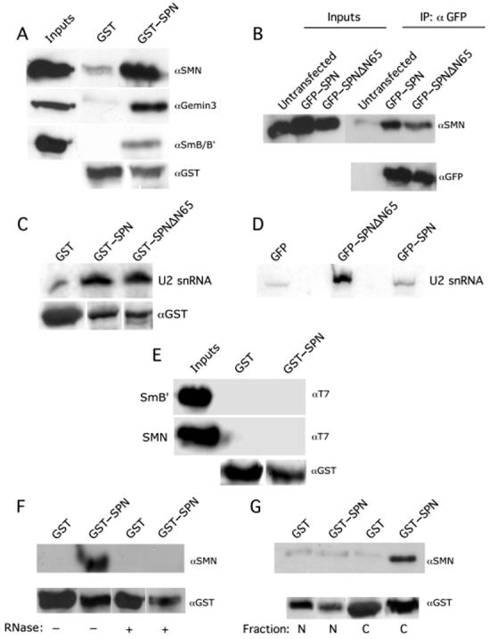
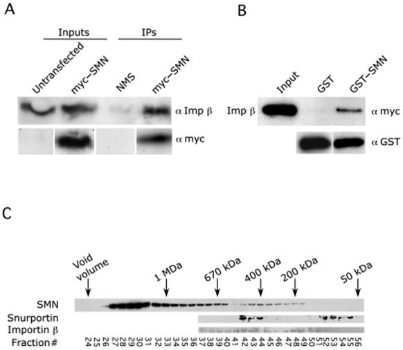
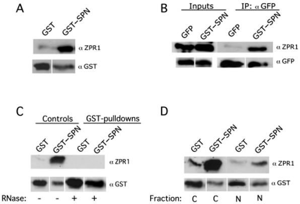
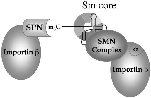
The survival of motor neuron (SMN) protein is mutated in patients with spinal muscular atrophy (SMA). SMN is part of a multiprotein complex required for biogenesis of the Sm class of small nuclear ribonucleoproteins (snRNPs). Following assembly of the Sm core domain, snRNPs are transported to the nucleus via importin beta. Sm snRNPs contain a nuclear localization signal (NLS) consisting of a 2,2,7-trimethylguanosine (TMG) cap and the Sm core. Snurportin1 (SPN) is the adaptor protein that recognizes both the TMG cap and importin beta. Here, we report that a mutant SPN construct lacking the importin beta binding domain (IBB), but containing an intact TMG cap-binding domain, localizes primarily to the nucleus, whereas full-length SPN localizes to the cytoplasm. The nuclear localization of the mutant SPN was not a result of passive diffusion through the nuclear pores. Importantly, we found that SPN interacts with SMN, Gemin3, Sm snRNPs and importin beta. In the presence of ribonucleases, the interactions with SMN and Sm proteins were abolished, indicating that snRNAs mediate this interplay. Cell fractionation studies showed that SPN binds preferentially to cytoplasmic SMN complexes. Notably, we found that SMN directly interacts with importin beta in a GST-pulldown assay, suggesting that the SMN complex might represent the Sm core NLS receptor predicted by previous studies. Therefore, we conclude that, following Sm protein assembly, the SMN complex persists until the final stages of cytoplasmic snRNP maturation and may provide somatic cell RNPs with an alternative NLS.


Figures

Figure 1
Figure 1
Schematic of snurportin1 (SPN) and characterization of antibody R89. (A) A cartoon of SPN, indicating the importin β (IBB), the CRM1/Xpo1 and trimethylguanosine (TMG) cap binding domains (24). (B) Western blot analysis of HeLa extract with SPN antibody R89. The left lane was probed with preimmune serum, the middle lane with R89 and the right lane with R89 in the presence of a 10-fold excess of the cognate peptide. The antibody specifically detected a band of approximately 45 kDa that was competed by the peptide. (C) The subcellular localization of SPN was studied by transient transfection of HeLa cells with GFP-SPN (top panels). In the middle panels, cells were immunostained with anti-SPN R89. Competition with a 20-fold excess of peptide (lower panels) resulted in background levels of fluorescence. Both the exogenous and endogenous SPN localized to the cytoplasm with enrichment at the nuclear periphery.
Figure 2
Figure 2
A) SPN is actively imported into the nucleus. Transient transfections of HeLa cells with 1 × or 2 × GFP tags. SPN (left panels) and SPNΔN65 (right) are shown. Surprisingly, SPNΔN65, which lacks an IBB domain, localizes primarily to the nucleus. (B) Comparison of mutant and wild-type proteins and their effects on Cajal bodies (CBs). Transient overexpression of GFP-SPN or GFP-SPNΔN65 causes a reduction in the number of CBs (see Table 1). The larger foci that were occasionally observed upon transfection with GFP-SPNΔN65 (top panels, arrow) did not strictly correspond to CBs (arrowhead). The low-magnification image in the second set of panels shows that cells expressing GFP-SPNΔN65 displayed fewer CBs (arrows) than did the untransfected cells (e.g. the cell marked by the arrowhead). Cells expressing lower levels of GFP-SPN (third panel, lower cell) typically displayed higher numbers of CBs than did those cells expressing higher levels of the construct (upper cell). (C) Cytoplasmic accumulations of SMN (arrows) could sometimes be detected in the GFP-SPN channel (arrows) and were not visible in the DAPI channel (not shown).
Figure 3
Figure 3
SPN interacts indirectly with SMN in the cytoplasm. (A) SPN interacts with the cytoplasmic SMN complex in vitro. Pulldown assays with GST-SPN or GST alone were performed using HeLa cytoplasmic extracts. The pulldowns were analyzed by western blotting with monoclonal antibodies against SMN (7B10), Gemin3 (11G9) and SmB/B′(Y12). (B) SPN interacts with SMN in vivo. HeLa cells were transiently transfected with GFP-SPN or GFP-SPNΔN65; untransfected cells were used as a negative control. Immunoprecipitations (IPs) were performed from total HeLa cell lysates with polyclonal antibodies against GFP. The IPs were then analyzed by western blotting with anti-SMN. (C) SPN and SPNΔN65 interact with U2 snRNA with similar affinities in vitro. GST-pulldown assays were performed using GST-SPN, GST-SPNΔN65 and GST (negative control) from total Hela cell lysate. RNA was isolated from the pulldowns; a U2 snRNA specific radiolabeled probe was used for analysis by northern blotting. The lower panel shows the loading controls; note that the GST-only lane is slightly overloaded relative to the experimental lanes. (D) SPNΔN65 and SPN bind U2 snRNA in vivo. HeLa cells were transiently transfected with GFP-SPN, GFP-SPNΔN65 or GFP (negative control). IPs were performed from total HeLa cell lysates with monoclonal antibodies against GFP. RNA was isolated from the IP, and analyzed by northern blotting. (E) SPN and SMN do not interact directly. Pulldown assays were performed using GST-SPN and GST (negative control), along with His-T7-SmB′or His-T7-SMN. The pulldowns were analyzed by western blotting with anti-T7. (F) RNA mediates the interaction of SPN with SMN. Pulldown assays were performed using GST-SPN or GST (negative control) from HeLa cell lysates in the absence or presence of ribonucleases. The pulldowns were analyzed by western blotting with anti-SMN. (G) SPN interacts with cytoplasmic SMN. Pulldown assays were performed using GST-SPN and GST (negative control) from HeLa cells (nuclear or cytoplasmic fractions). The pulldowns were analyzed by western blotting with anti-SMN. Anti-GST and anti-GFP were used as loading controls where indicated. Inputs show 12% of the total lysate used in the pulldowns.
Figure 4
Figure 4
SMN interacts directly with importin β. (A) HeLa cells were transfected with myc-SMN; untransfected cells were used as a negative control. Immunoprecipitations (IPs) were performed from total HeLa cell lysates with monoclonal antibodies against myc. The IPs were then analyzed by western blotting with anti-Imp β. (B) SMN and Imp β interact directly using purified recombinant proteins. Pulldown assays were performed using GST-SMN or GST alone and His-myc-importin β. The pulldowns were analyzed by western blotting with anti-myc and anti-GST (loading control). Input shows approximately 50% of the total lysate used in the pulldowns. (C) Gel filtration column of HeLa cytoplasm. Fractions were analyzed by western blotting with the indicated antibodies. SPN, SMN and importin β cofractionate in the ∼400 kDa range, consistent with the existence of a complex containing all three proteins.
Figure 5
Figure 5
SPN forms a cytoplasmic complex with ZPR1. (A) SPN interacts with ZPR1 in vitro. Pulldown assays with GST-SPN and GST were performed using HeLa lysate. The pulldowns were analyzed by western blotting with monoclonal antibodies against ZPR1 (LG9). (B) SPN interacts with ZPR1 in vivo. HeLa cells were transiently transfected with GFP-SPN or GFP alone. Immunoprecipitations (IPs) were performed from total HeLa cell lysates with polyclonal antibodies against GFP. The IPs were then analyzed by western blotting with LG9 and GFP (loading control). (C) RNA mediates the SPN-ZPR1 interaction. Pulldown assays were performed using GST-SPN; GST (negative control) from HeLa cell lysates in the presence or absence of ribonucleases. (D) SPN interacts with cytoplasmic ZPR1. Pulldown assays were performed using GST-SPN and GST (negative control) along with nuclear (N) or cytoplasmic (C) HeLa cell extracts.
Figure 6
Figure 6
Model of the U snRNP import complex, adapted from (11). SPN binds to the TMG cap (m3G) of Sm class snRNPs and interacts with importin β via its IBB domain. The hypothetical snRNP core-binding factor (see text) is predicted to be the SMN complex. Since SMN lacks an IBB domain, direct binding to importin β (Fig. 4B) is therefore unlikely to proceed via the SPN/importin α binding pocket (α). Two importin β molecules are shown in the model, as predicted by (11). Alternatively, binding of the Sm core by SMN might stabilize the binding of a single importin β. Additional experiments will be required to distinguish between these two possibilities.

References

    1. Görlich D, Mattaj IW. Nucleocytoplasmic transport. Science. 1996;271:1513–1518. - PubMed
    1. Allen TD, Cronshaw JM, Bagley S, Kiseleva E, Goldberg MW. The nuclear pore complex: mediator of translocation between nucleus and cytoplasm. J. Cell Sci. 2000;113:1651–1659. - PubMed
    1. Ribbeck K, Görlich D. Kinetic analysis of translocation through nuclear pore complexes. EMBO J. 2001;20:1320–1330. - PMC - PubMed
    1. Smith AE, Slepchenko BM, Schaff JC, Loew LM, Macara IG. Systems analysis of Ran transport. Science. 2002;295:488–491. - PubMed
    1. Ryan KJ, Wente SR. The nuclear pore complex: a protein machine bridging the nucleus and cytoplasm. Curr. Opin. Cell Biol. 2000;12:361–371. - PubMed

Publication types

MeSH terms