Review
. 2002 Jul 1;542(Pt 1):17-29.
doi: 10.1113/jphysiol.2002.023218.

Transport of volatile solutes through AQP1

Affiliations
Review

Transport of volatile solutes through AQP1

Gordon J Cooper et al. J Physiol. .

Abstract

For almost a century it was generally assumed that the lipid phases of all biological membranes are freely permeable to gases. However, recent observations challenge this dogma. The apical membranes of epithelial cells exposed to hostile environments, such as gastric glands, have no demonstrable permeability to the gases CO2 and NH3. Additionally, the water channel protein aquaporin 1 (AQP1), expressed at high levels in erythrocytes, can increase membrane CO2 permeability when expressed in Xenopus oocytes. Similarly, nodulin-26, which is closely related to AQP1, can act as a conduit for NH3. A key question is whether aquaporins, which are abundant in virtually every tissue that transports O2 and CO2 at high levels, ever play a physiologically significant role in the transport of small volatile molecules. Preliminary data are consistent with the hypothesis that AQP1 enhances the reabsorption of HCO3- by the renal proximal tubule by increasing the CO2 permeability of the apical membrane. Other preliminary data on Xenopus oocytes heterologously expressing the electrogenic Na+-HCO3- cotransporter (NBC), AQP1 and carbonic anhydrases are consistent with the hypothesis that the macroscopic cotransport of Na+ plus two HCO3- occurs as NBC transports Na+ plus CO3(2-) and AQP1 transports CO2 and H2O. Although data - obtained on AQP1 reconstituted into liposomes or on materials from AQP1 knockout mice - appear inconsistent with the model that AQP1 mediates substantial CO2 transport in certain preparations, the existence of unstirred layers or perfusion-limited conditions may have masked the contribution of AQP1 to CO2 permeability.


Figures

Figure 1
Figure 1. Model of aquaporin 1
A, schematic representation of the structure of AQP1. The molecule has intracellular amino and carboxy termini, six membrane-spanning segments, and five loops. Loops B and E fold back into the membrane, contain the tandem repeat of the amino-acid sequence NPA. B, the hourglass model predicts that loops B and E overlap in the membrane to allow the formation of a channel pore. (Data from Nielsen et al. 2002.)
Figure 2
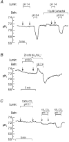
Figure 2. Unique permeability properties of the apical membranes of the gastric gland
A, low H+ permeability. The experiments in all three panels were performed at 37 °C on isolated perfused gastric glands from the rabbit, and the records are from a single parietal cell. Intracellular pH (pHi) was monitored using the fluorescent pH-sensitive dye BCECF in conjunction with a digital imaging system. Reducing the pH of the luminal fluid from 7.4 to 1.4 in a CO2/HCO3-free system produced no detectable pHi change, whereas reducing bath (i.e. basolateral) pH by only 1 pH unit caused a large and rapid fall in pHi. B, low NH3/NH4+ permeability. Exposing the lumen to a Hepes-buffered solution containing 20 mm NH4+/NH3 at pH 8.0 ([NH3]o ∼ 1.46 mm) produced no detectable pHi increase, whereas exposing the bath to 20 mm NH4+/NH3 at pH 7.4 ([NH3]o ∼ 0.4 mm) produced a rapid pHi increase followed by the usual series of changes. C, low CO2/HCO3 permeability. Exposing the lumen to a solution equilibrated with 100 % CO2 and containing 22 mm HCO3 (pH 6.1, [CO2]o ∼ 24 mm) produced no discernable pHi decrease, whereas exposing the bath to 1 % CO2/4.4 mm HCO3 (pH 7.4, [CO2]o ∼ 0.24 mm) or 5 % CO2/22 mm HCO3 (pH 7.4, [CO2]o ∼ 1.2 mm) caused the expected pHi decreases. The gland was bathed in 200 μM DIDS to eliminate the pHi recovery from the intracellular acid load that would otherwise have occurred. (In A, data from Waisbren et al. 1994a. In B, data from Waisbren et al. 1994b.)
Figure 3
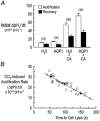
Figure 3. Expression of aquaporin 1 increase the permeability of Xenopus oocytes to CO2
A, experiments on oocytes with vitelline membrane intact. Switching the extracellular buffer from Hepes to 1.5 % CO2/10 mm HCO3 (constant pHo = 7.5) causes a sustained fall of intracellular pH, the initial rate of which is indicated by the filled bars. The initial rate of pHi recovery (i.e. alkalinization) caused by removing the CO2/HCO3 solution is summarized by the open bars. Injection of carbonic anhydrase (CA) caused a significant increase in the initial CO2-induced rate in both water-injected oocytes and those expressing AQP1. Moreover, in CA-injected oocytes, expression of AQP1 significantly increased the CO2-induced acidification rate. B, experiments on oocytes with vitelline membrane removed, and in absence of injected CA. Each symbol represents data from a separate experiment (i.e. oocyte). There was a linear relationship between the initial rate of CO2-induced acidification and lysis time (an index of the AQP1 expression level), with acidification rate increasing as lysis time decreased. (In A, data from Nakhoul et al. 1998. In B, data from Cooper & Boron, 1998a.)
Figure 4
Figure 4. Effect of pCMBS on the CO2-induced acidification
A, H2O-injected control oocyte. Two records represent paired data from an experiment on a single oocyte exposed to 1.5 % CO2/10 mm HCO3 before and after a 15-min exposure to 1 mm pCMBS. B, AQP1-expressing oocyte. C, C189S-expressing oocyte. D, change in CO2-induced acidification rate produced by pCMBS. Each bar represents the mean of paired differences, before and after treating the oocyte with pCMBS. (Reproduced from Cooper & Boron, 1998a with permission of the American Physiological Society.)
Figure 5
Figure 5. Aquaporin 1 increases the CO2 permeability of liposomes made from E. coli membrane lipids
A, effect of incorporating AQP1 into liposomes. In vesicles prepared from E. coli membranes, adding AQP1 produced a significant increase in the rate at which intravesicular pH decreased upon exposure to a CO2-containing solution. B, effect of blocking AQP1 with HgCl2. The AQP1-dependent increase in CO2 permeability was reduced by addition of HgCl2, and reversed by β-mercaptoethanol. (Reproduced from Prasad et al. 1998 with permission of the Journal of Biological Chemistry.)

Comment in

References

    1. Benga G, Pop VI, Popescu O, Ionescu M, Mihele V. Water exchange through erythrocyte membranes: nuclear magnetic resonance studies on the effects of inhibitors and of chemical modifications of human membranes. Journal of Membrane Biology. 1983;76:129–137. - PubMed
    1. Boron WF, Cooper GJ. Effect of DIDS on the CO2 permeability of the water channel AQP1. FASEB Journal. 1998;12:A374.
    1. Burckhardt BC, Frömter E. Pathways of NH3/NH4+ permeation across Xenopus laevis oocyte cell membrane. Pflügers Archiv. 1992;420:83–86. - PubMed
    1. Burg M, Grantham J, Abramow M, Orloff J. Preparation and study of fragments of single rabbit nephrons. American Journal of Physiology. 1966;210:1293–1298. - PubMed
    1. Cooper GJ, Boron WF. Effect of PCMBS on CO2 permeability of Xenopus oocytes expressing aquaporin 1 or its C189S mutant. American Journal of Physiology. 1998a;275:C1481–1486. - PubMed

Publication types

LinkOut - more resources