. 2002 Jul 3;94(13):1010-9.
doi: 10.1093/jnci/94.13.1010.

Effects of interferon beta on transcobalamin II-receptor expression and antitumor activity of nitrosylcobalamin

Affiliations

Effects of interferon beta on transcobalamin II-receptor expression and antitumor activity of nitrosylcobalamin

Joseph A Bauer et al. J Natl Cancer Inst. .

Abstract

Background: The ubiquitous plasma membrane transcobalamin II receptor (TC II-R) mediates uptake of cobalamin (Cbl; vitamin B12), an essential micronutrient. Tumors often require more Cbl than normal tissue, and increased Cbl uptake may result from increased TC II-R expression. To examine whether Cbl could therefore be used as a carrier molecule to target a chemotherapy drug, we tested an analogue of Cbl with nitric oxide as a ligand, nitrosylcobalamin (NO-Cbl). Because interferon beta (IFN-beta) has antitumor effects and increases expression of some membrane receptors, we examined whether it may enhance the effects of NO-Cbl.

Methods: Antiproliferative effects of NO-Cbl were assessed in 24 normal and cancer cell lines. Xenograft tumors of human ovarian cancer NIH-OVCAR-3 cells were established in athymic nude mice, and tumor growth was monitored after treatment with NO-Cbl and IFN-beta, both individually and concomitantly. TC II-R expression and apoptosis was monitored in vitro and in vivo. RNA protection assays and mitochondrial membrane potential assays were used to distinguish the extrinsic and intrinsic apoptotic pathways, respectively.

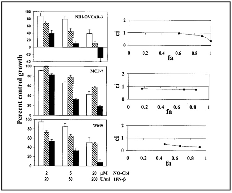
Results: Cancer cell lines were more sensitive to NO-Cbl (with ID(50)s [the dose that inhibits growth by 50%] as low as 2 microM) than normal cell lines (with ID(50)s of 85-135 microM). Single-agent NO-Cbl and IFN-beta treatment of NIH-OVCAR-3 xenografts induced tumor regression, whereas combination treatment induced tumor eradication. IFN-beta treatment increased TC II-R expression in vitro and uptake of [(57)Co]cobalamin in vivo. Compared with NIH-OVCAR-3 cells treated with NO-Cbl, cells treated with NO-Cbl and IFN-beta were more apoptotic and expressed higher mRNA levels of various apoptosis-associated genes. No changes in mitochondrial membrane potential were observed in cells treated with NO-Cbl.

Conclusion: NO-Cbl inhibited tumor growth in vivo by activating the extrinsic apoptotic pathway. The increased expression of TC II-R induced by IFN-beta resulted in enhanced antitumor effects with NO-Cbl both in vitro and in vivo.


Figures

Fig. 1.
Fig. 1.
Effects of nitrosylcobalamin (NO-Cbl), interferon β (IFN-β), or the combination of both agents on the proliferation of the ovarian cancer cell line NIH-OVCAR-3, the breast cancer cell line MCF-7, and the melanoma cell line WM9. (Left panels) Cells were treated with NO-Cbl (open bars), IFN-β (hatched bars), or the combination of the two (solid bars) for 5 days, and growth was measured by the colorimetric sulforhodamine B assay (33,34). Data points represent the mean of eight replicates. Data are expressed as mean percentage of control with 95% confidence intervals. (Right panels) Synergy between NO-Cbl and IFN-β was determined by median effect analysis (36). The y-axis shows the combination index (ci). A ci greater than 1 indicates antagonism between NO-Cbl and IFN-β, a ci equal to 1 indicates additivity, and a ci less than 1 indicates synergy. The x-axis shows the fraction affected (fa). An fa of 0 corresponds to 100% of control growth, whereas an fa of 1 corresponds to 0% of control growth.
Fig. 2.
Fig. 2.
Effect of nitrosylcobalamin (NO-Cbl), interferon β (IFN-β), or the combination of both agents on ovarian cancer NIH-OVCAR-3 xenografts. A) NCR male athymic nude (nu/nu) mice (n = 8 per group) were injected subcutaneously with 2 × 10⁶ NIH-OVCAR-3 cells. Daily drug treatments of control (phosphate-buffered saline), NO-Cbl (100 mg/kg, intraperitoneally), human IFN-β (105 U/day, subcutaneously), or the combination began on day 7 after injection of the cells. Tumor volume was measured three times per week. Points represent the mean tumor volume (in cubic mm) ± 95% confidence intervals. B) NIH-OVCAR-3 tumors were isolated from an additional series of mice after 10 days. Tumors were processed, sectioned, and stained to visualize apoptotic cells. Apoptotic cells were detected by terminal deoxynucleotidyltransferase-mediated dUTP-biotin nick end labeling. Representative sections from control (untreated), NO-Cbl treated, IFN-β treated, and NO-Cbl and IFN-β treated (NO-Cbl + IFN-β) are shown. Light areas identify cells undergoing apoptosis.
Fig. 3.
Fig. 3.
Biochemical analysis of transcobalamin II receptor (TC II-R) expression. A) Established xenograft tumors of ovarian cancer NIH-OVCAR-3 cells (upper panels) and melanoma WM9 cells (lower panels) were treated with and without 105 U interferon-β (IFN-β) subcutaneously (s.c.) for 3 days. Sections from tumors, isolated after 3 days, were analyzed for the presence of TC II-R by immunohistochemistry with a rabbit polyclonal anti-TC II-R antibody. The signal intensity (brown color) was quantified and TC II-R levels were normalized to those in untreated WM9 cells (signal intensity [Int] = 1). The relative intensities of the immunostaining are shown for each panel. B) Expression of TC II-R in NIH-OVCAR-3 cells treated with IFN-β in vitro for 4 hours and for 16 hours was determined by western blot analysis with a rabbit polyclonal anti-TC II-R antibody. TC II-R was detected as a monomer of 62 kd and a corresponding dimer of 124 kd. Lysates of rat liver and kidney are shown as positive controls. Only the 62-kd band was detected in the rat samples because of the presence of the reducing agent dithiothreitol (DTT) in the lysis buffer. The blot was stripped and reprobed with an anti-actin antibody to show that equivalent amounts of lysates were loaded in each lane.
Fig. 4.
Fig. 4.
Effect of interferon β (IFN-β) on the uptake of [57Co]cobalamin in NIH-OVCAR-3 xenografts. NIH-OVCAR-3 cells were injected in the flanks of nude mice and grown for 14 days before the administration of [57Co]cobalamin (day 0). IFN-β (105 U) was administered subcutaneously from days −3 through 5. The biodistribution of the [57Co]cobalamin was examined on days 4 and 6 by placing the anesthetized mice on a phosphor imaging screen and collecting images with the use of the OptiQuant image analysis software. Areas of low [57Co]cobalamin signal intensity are identified by the blue color, and areas of high signal intensity are identified by the red color.
Fig. 5.
Fig. 5.
Effect of nitrosylcobalamin (NO-Cbl) on the induction of apoptosis-related mRNA. A) RNA from NIH-OVCAR-3 cells that were left untreated (U), treated with NO-Cbl (N), interferon β (IFN-β) (β), or treated with the combination (+) for either 4 hours or 8 hours was isolated and used in a ribonuclease protection assay to assess mRNA induction of multiple genes associated with apoptosis. Specific induced mRNAs for death-associated genes are indicated by arrows. DR3 = death receptor 3; TRAIL = tumor necrosis factor-related apoptosis-inducing ligand; TRADD = tumor necrosis factor receptor associated death domain; TNFRp55 = tumor necrosis factor receptor p55 chain; L32 = 50S ribosomal house keeping gene; GAPDH = glyceraldehyde-3-phosphate dehydrogenase. B) Caspase-8 activity was measured in NIH-OVCAR-3 cells treated with NO-Cbl (10 µM) for various times (0–4 h). Equal amounts of total cell protein were assayed for caspase-8 activity. Enzyme activity is expressed in total fluorescence units per each time point. Points represent mean caspase-8 activity ± 95% confidence intervals of triplicate samples.
Fig. 6.
Fig. 6.
Effect of nitrosylcobalamin (NO-Cbl) on mitochondrial membrane potentials. NIH-OVCAR-3 cells were incubated with the mitochondrial membrane binding dye JC-1 for 30 minutes in serum-free medium. The cells were then placed in complete medium and treated with either sodium nitroprusside (SNP; 200 µM) as a positive control or NO-Cbl (200 µM) for 4 hours. The cells were imaged with a laser scanning confocal microscope. Functional mitochondria that have an intact transmembrane potential (ΨΔm) stain red, whereas mitochondria that have lost transmembrane potential stain green. Compared with NO-Cbl treatment, SNP treatment caused a dramatic loss of ΨΔm, indicated by a loss of red signal.
Fig. 7.
Fig. 7.
Intracellular release of nitric oxide (NO) from NO-Cbl. NIH-OVCAR-3 cells were incubated with inducible NO synthase (iNOS) inhibitor NG-monomethyl-l-arginine monoacetate (L-NMMA) (500 µM) for 1 hour to inhibit endogenous iNOS activity. The cells then were incubated with 5 µM DAF-FM diacetate (4-amino-5-methylamino-2′,7′-difluorofluorescein) for 30 minutes. DAF-FM is a dye that diffuses into cells and is trapped there after esterases remove its terminal acetate residues. DAF-FM is strongly fluorescent when it binds NO. The cells were washed, placed in complete medium, and treated with NO-Cbl (100 µM) and L-NMMA (500 µM). Time lapse images were obtained within a custom incubator by confocal fluorescent microscopy. Images were collected every 6 minutes for 5 hours. Color reflects the intensity of the NO signal, which ranges from moderate intensity (violet) to high intensity (green-yellow).

References

    1. Rickes EL, Brink NG, Koniuszy FR, Wood TR, Folkers K. Crystalline vitamin B12. Science. 1948;107:396–397. - PubMed
    1. Linnell JC, Matthews DM. Cobalamin metabolism and its clinical aspects. Clin Sci (Colch) 1984;66:113–121. - PubMed
    1. Seetharam B, Bose S, Li N. Cellular import of cobalamin (vitamin B-12) J Nutr. 1999;129:1761–1764. - PubMed
    1. Weir DG, Scott JM. Vitamin B12 cobalamin. In: Shils ME, Olson JA, Shike M, Ross AC, editors. Modern nutrition in health and disease. Philadelphia (PA): Lippincott Williams, and Wilkins; 1999. pp. 659–728.
    1. Seetharam B, Li N. Transcobalamin II and its cell surface receptor. Vitam Horm. 2000;59:337–366. - PubMed

Publication types

MeSH terms