. 2002 Jul 23;99(15):9807-12.
doi: 10.1073/pnas.152321499. Epub 2002 Jul 2.

Early kinetic intermediate in the folding of acyl-CoA binding protein detected by fluorescence labeling and ultrarapid mixing

Affiliations

Early kinetic intermediate in the folding of acyl-CoA binding protein detected by fluorescence labeling and ultrarapid mixing

Kaare Teilum et al. Proc Natl Acad Sci U S A. .

Abstract

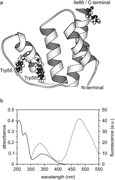
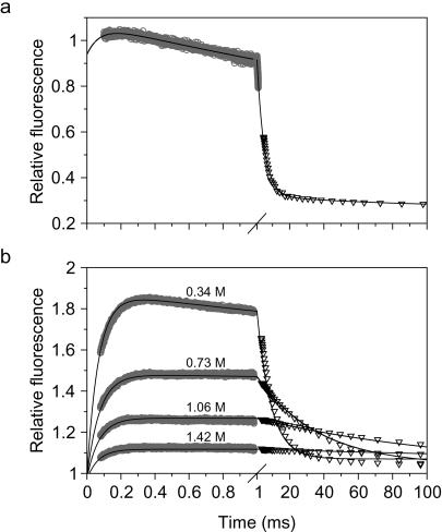
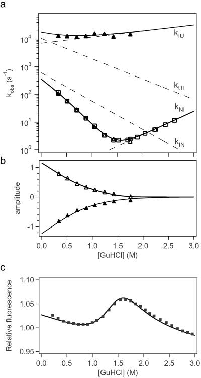
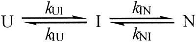
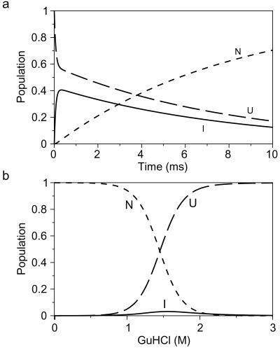
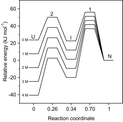
Early conformational events during folding of acyl-CoA binding protein (ACBP), an 86-residue alpha-helical protein, were explored by using a continuous-flow mixing apparatus with a dead time of 70 micros to measure changes in intrinsic tryptophan fluorescence and tryptophan-dansyl fluorescence energy transfer. Although the folding of ACBP was initially described as a concerted two-state process, the tryptophan fluorescence measurements revealed a previously unresolved phase with a time constant tau = 80 micros, indicating formation of an intermediate with only slightly enhanced fluorescence of Trp-55 and Trp-58 relative to the unfolded state. To amplify this phase, a dansyl fluorophore was introduced at the C terminus by labeling an I86C mutant of ACBP with 5-IAEDANS [5-((((2-iodoacetyl)amino)ethyl)amino)naphthalene-1-sulfonic acid]. Continuous-flow refolding of guanidine HCl-denatured ACBP showed a major increase in tryptophan-dansyl fluorescence energy transfer, indicating formation of a partially collapsed ensemble of states on the 100-micros time scale. A subsequent decrease in dansyl fluorescence is attributed to intramolecular quenching of donor fluorescence on formation of the native state. The kinetic data are fully accounted for by three-state mechanisms with either on- or off-pathway intermediates. The intermediate accumulates to a maximum population of 40%, and its stability depends only weakly on denaturant concentration, which is consistent with a marginally stable ensemble of partially collapsed states with approximately 1/3 of the solvent-accessible surface buried. The findings indicate that ultrafast mixing methods combined with sensitive conformational probes can reveal transient accumulation of intermediate states in proteins with apparent two-state folding mechanisms.


Figures

Figure 1
Figure 1
(a) Ribbon diagram (MOLSCRIPT; ref. 38) of ACBP, based on the NMR structure coordinates of Andersen and Poulsen (39). The two tryptophan residues and the mutated C-terminal isoleucine are shown in ball and stick. (b) Absorbance spectrum (solid line) and fluorescence emission spectrum with excitation at 280 nm (dashed line) of 15 μM ACBP,I86C labeled with IAEDANS (in 20 mM Na-acetate, pH 5.3).
Figure 2
Figure 2
Refolding kinetics of unmodified ACBP (a) and dansyl-labeled ACBP,I86C (b) in 20 mM Na-acetate (pH 5.3) containing 0.34 M GuHCl at 26°C. In a and b, data from continuous-flow (○) and stopped-flow (▿) experiments were matched and combined. (a) The tryptophans were excited at 280 nm and the fluorescence was measured with a 324-nm long-pass filter. The solid line represents a fit to a sum of three exponentials. (b) ACBP,I86C labeled with IAEDANS at the C-terminal Cys residue. The tryptophans were excited at 280 nm, and the fluorescence from the dansyl group was measured with a 418-nm long-pass filter. Refolding at four different GuHCl concentrations are shown. The solid lines represent simulated kinetic traces using kinetic parameters in Table 1, based on a three-state folding mechanism with an on-pathway intermediate (Scheme S1).
Figure 3
Figure 3
Fit of experimental folding data for ACBP,I86C-AEDANS. Rate constants (a) and kinetic amplitudes (b) for the fast (▴) and rate-determining (▵) folding phases obtained by exponential fitting of matching continuous-flow and stopped-flow measurements of tryptophan-dansyl energy transfer (Fig. 2). Rate constants for the slower phase were also obtained in an independent experiment where total fluorescence above 324 nm was measured (□). Data were fitted to an on-pathway three-state model (Scheme S1). The solid lines indicate the predicted observable rate constants and amplitudes. The corresponding elementary rate constants are shown as dashed lines. (c) Fluorescence above 418 nm (■) measured in an independent equilibrium experiment. The solid line represents the equilibrium fluorescence change predicted from the kinetic parameters in Table 1. The fluorescence of the three states was slightly different in this simulation compared with the simulation of the kinetic data. U (f0 = 1.05; slope = −0.023), I (f0 = 3.04; slope = −0.06), and N states (f0 = 1.028; slope = −0.035).
Scheme 1
Scheme 1
Figure 4
Figure 4
Populations of unfolded (U), intermediate (I), and native (N) states predicted from the kinetic parameters in Table 1. (a) Time-dependent variation in populations during the first 10 ms of refolding at 0.34 M GuHCl. (b) Populations at equilibrium as function of [GuHCl] predicted by the kinetic parameters.
Figure 5
Figure 5
Free energy levels of intermediate and transition states encountered during ACBP folding relative to the native state. The energy levels were calculated from the fitted kinetic parameters (Table 1) with a preexponential factor of 4.8 × 10⁸ s−1. The reaction coordinate represents the fractional burial of solvent-accessible surface area calculated from the fitted m-values.

References

    1. Park S-H, Shastry M C R, Roder H. Nat Struct Biol. 1999;6:943–947. - PubMed
    1. Dalby P A, Oliveberg M, Fersht A R. J Mol Biol. 1998;276:625–646. - PubMed
    1. Raschke T M, Marqusee S. Nat Struct Biol. 1997;4:298–304. - PubMed
    1. Capaldi A P, Shastry M C, Kleanthous C, Roder H, Radford S E. Nat Struct Biol. 2001;8:68–72. - PubMed
    1. Jackson S E, Fersht A R. Biochemistry. 1991;30:10428–10435. - PubMed

Publication types

LinkOut - more resources