Review
. 2002 Jul;15(3):342-54.
doi: 10.1128/CMR.15.3.342-354.2002.

Cultivation of pathogenic and opportunistic free-living amebas

Affiliations
Review

Cultivation of pathogenic and opportunistic free-living amebas

Frederick L Schuster. Clin Microbiol Rev. 2002 Jul.

Abstract

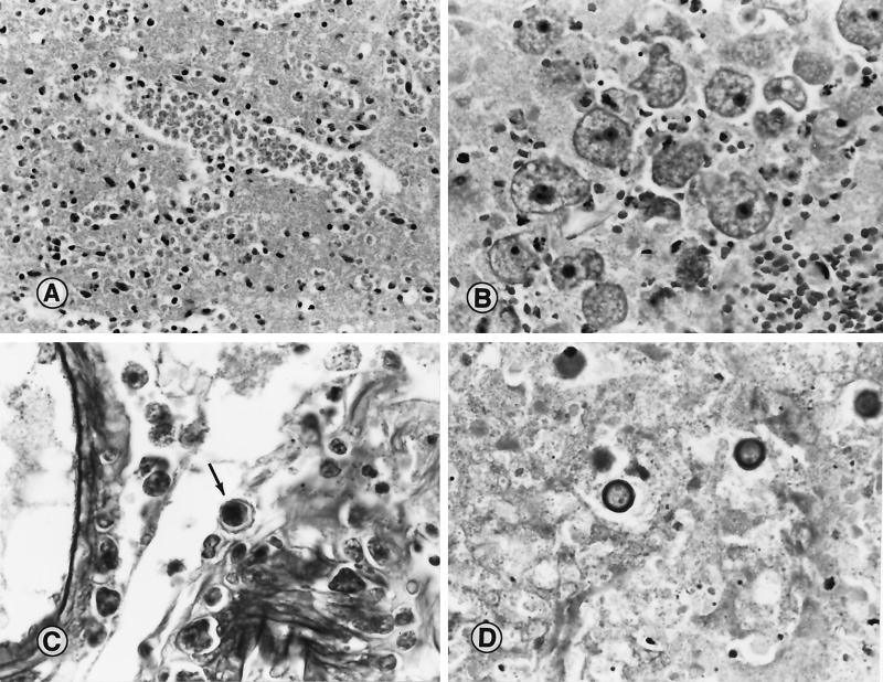
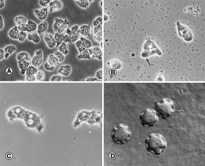
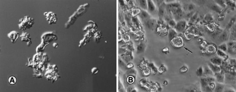
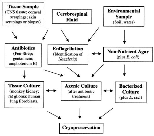
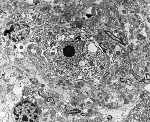
Free-living amebas are widely distributed in soil and water, particularly members of the genera Acanthamoeba and NAEGLERIA: Since the early 1960s, they have been recognized as opportunistic human pathogens, capable of causing infections of the central nervous system (CNS) in both immunocompetent and immunocompromised hosts. Naegleria is the causal agent of a fulminant CNS condition, primary amebic meningoencephalitis; Acanthamoeba is responsible for a more chronic and insidious infection of the CNS termed granulomatous amebic encephalitis, as well as amebic keratitis. Balamuthia sp. has been recognized in the past decade as another ameba implicated in CNS infections. Cultivation of these organisms in vitro provides the basis for a better understanding of the biology of these amebas, as well as an important means of isolating and identifying them from clinical samples. Naegleria and Acanthamoeba can be cultured axenically in cell-free media or on tissue culture cells as feeder layers and in cultures with bacteria as a food source. Balamuthia, which has yet to be isolated from the environment, will not grow on bacteria. Instead, it requires tissue culture cells as feeder layers or an enriched cell-free medium. The recent identification of another ameba, Sappinia diploidea, suggests that other free-living forms may also be involved as causal agents of human infections.


Figures

FIG. 1.
FIG. 1.
(A) Section of cerebral cortex from a patient with PAM due to N. fowleri. A large cluster of trophic amebas is seen in the brain tissue. Magnification, ×210; original magnification, ×230. (B) Section of brain tissue from a patient with Balamuthia GAE. Typical trophic amebas are seen within the necrotic CNS tissue. Magnification, ×210; original magnification, ×230. (C) Tissue section showing cysts of A. castellanii within the wall of a blood vessel and the perivascular space. Note the double wrinkled wall seen around one of the cysts (arrow). Magnification, ×370; original magnification, ×400. (D) Cysts of B. mandrillaris in necrotic brain tissue from a patient with amebic encephalitis. Magnification, ×520; original magnification, ×560. (Micrographs copyright A. J. Martinez.)
FIG. 2.
FIG. 2.
(A) N. fowleri, from a patient with PAM, growing in axenic medium in tissue culture flasks. Amebas contain numerous fluid-filled vacuoles and appear larger than their counterparts growing on bacteria. The cultures were photographed in situ on an inverted microscope. Magnification, ×960; original magnification, ×1,000. (Micrograph copyright T. H. Dunnebacke.) (B) N. fowleri amebas in a wet-mount microscope slide. The prominent clear, ectoplasmic pseudopod is seen (arrow). Adjacent particles are bacteria. Magnification, ×960; original magnification, ×1,000. (C) Trophic A. castellanii ameba from culture. Amebas show the characteristic projecting pseudopods (acanthopodia) over the surface. Magnification, ×960; original magnification, ×1,000. (D) Interference-contrast image of cysts of Acanthamoeba sp. isolated from a human brain biopsy specimen. The cysts are on the surface of an agar plate. Magnification, ×1440; original magnification, ×1,500. (Micrographs B to D copyright G. S. Visvesvara.)
FIG. 3.
FIG. 3.
(A) Interference-contrast micrograph of trophic Balamuthia amebas in a wet-mount preparation. The amebas are pleomorphic and are likely to show varied morphology depending on whether they are in a cell-free medium or feeding on tissue culture cells. Magnification, ×380; original magnification, ×400. (B) Light micrograph of monkey kidney cell monolayer in a tissue culture flask. The open area seen in the micrograph is where the cells have been fed upon by Balamuthia amebas (arrow), about 24 h after the amebas were inoculated into the flask. The photograph was taken in situ on the stage of an inverted microscope. Magnification, ×190; original magnification, ×200.
FIG. 4.
FIG. 4.
The chart presents a general scheme for isolation of free-living pathogenic amebas from clinical samples or from environmental sources. The goal is to establish the amebas either in an axenic culture (cell-free growth medium), with a tissue culture feeder layer, or in a xenic culture with a suitable bacterial strain as a food source. Although two different tissue culture types are noted (monkey kidney or rat glioma cells), other cell types (e.g., human lung fibroblasts) are suitable as feeder layers. When possible, cryopreservation of ameba strains is recommended as an alternative to regular subculturing and as a backup in the event of loss of a culture.
FIG. 5.
FIG. 5.
Transmission electron micrograph of N. fowleri in brain tissue of a mouse that had been experimentally inoculated with a suspension of the amebas. The characteristic nuclear morphology of the ameba can be seen as a large nucleus with a centrally located nucleolus (vesicular nucleus). Original magnification, ×7,500. (Micrograph copyright A. J. Martinez.)

References

    1. Adam, K. M. G. 1959. The growth of Acanthamoeba sp. in a chemically defined medium. J. Gen. Microbiol. 21:519-529. - PubMed
    1. Adam, K. M. G., and D. A. Blewett. 1967. Carbohydrate utilization by the soil amoeba Hartmannella castellanii. J. Protozool. 14:227-282. - PubMed
    1. Anzil, A. P., C. Rao, M. A. Wrzolek, G. S. Visvesvara, J. H. Sher, and P. B. Kozlowsky. 1991. Amebic meningoencephalitis in a patient with AIDS caused by a newly recognized opportunistic pathogen. Arch. Pathol. Lab. Med. 115:21-25. - PubMed
    1. Balamuth, W. 1964. Nutritional studies on axenic cultures of Naegleria gruberi. J. Protozool. 11(Suppl.):19-20.
    1. Band, R. N. 1961. Biotin, a growth requirement for four soil amoebae. Nature 192:674. - PubMed

LinkOut - more resources