Review
. 2002 Jul;15(3):401-13.
doi: 10.1128/CMR.15.3.401-413.2002.

In vitro cultivation of microsporidia of clinical importance

Affiliations
Review

In vitro cultivation of microsporidia of clinical importance

Govinda S Visvesvara. Clin Microbiol Rev. 2002 Jul.

Abstract

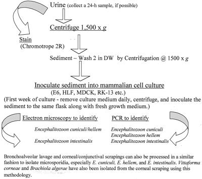
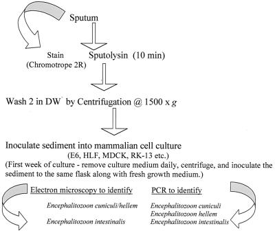
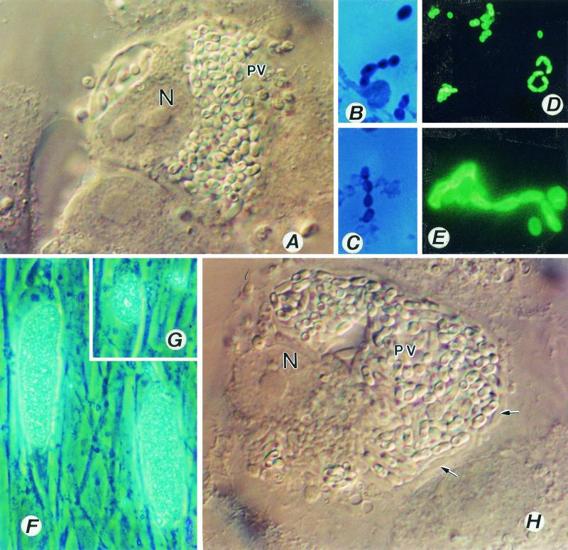
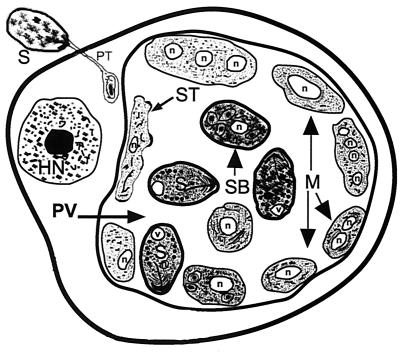
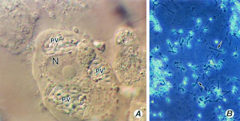
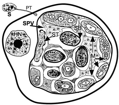
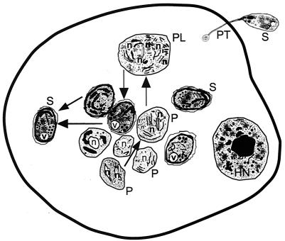
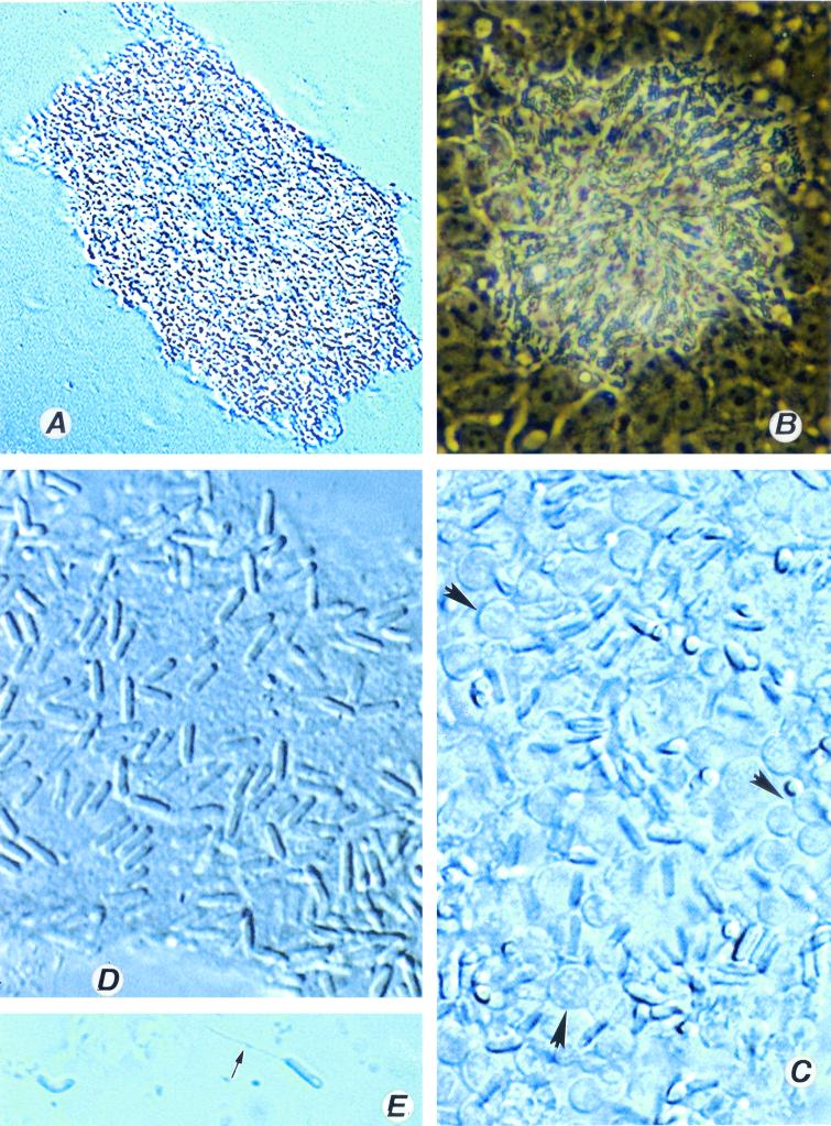
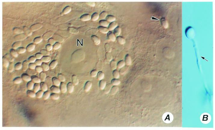
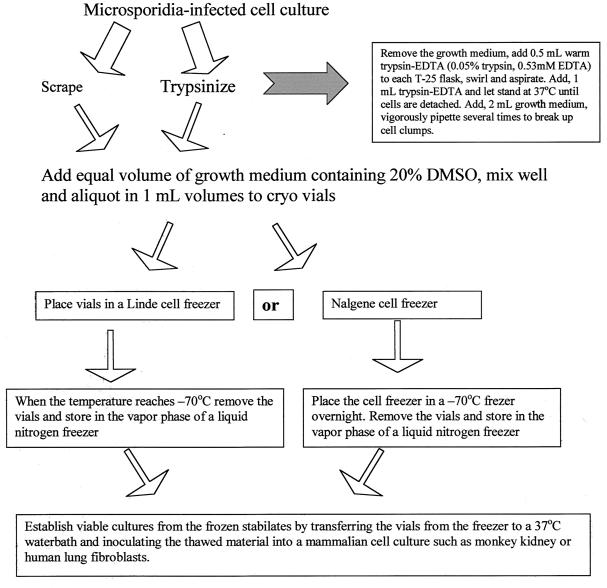
Although attempts to develop methods for the in vitro cultivation of microsporidia began as early as 1937, the interest in the culture of these organisms was confined mostly to microsporidia that infect insects. The successful cultivation in 1969 of Encephalitozoon cuniculi, a microsporidium of mammalian origin, and the subsequent identification of these organisms as agents of human disease heightened interest in the cultivation of microsporidia. I describe the methodology as well as the cell lines, the culture media, and culture conditions used in the in vitro culture of microsporidia such as Brachiola (Nosema) algerae, Encephalitozoon cuniculi, E. hellem, E. intestinalis, Enterocytozoon bieneusi, Trachipleistophora hominis, and Vittaforma corneae that cause human disease.


Figures

FIG. 1.
FIG. 1.
Flow chart to illustrate the isolation and continuous cultivation of microsporidia, especially Encephalitozoon species, from bronchoalveolar lavage samples, corneal/conjunctival scrapings, and urine samples. DW, distilled water.
FIG. 2.
FIG. 2.
Flow chart to illustrate the isolation and continuous cultivation of microsporidia, especially Encephalitozoon species, from sputum. DW, distilled water.
FIG. 3.
FIG. 3.
(A to E) Encephalitozoon cuniculi. (A) A monkey kidney (E6) cell distended with spores of E. cuniculi growing inside a parasitophorous vacuole (PV). Magnification, ×1,200. N, host cell nucleus. (B and C) Gram-chromotrope-stained preparations exhibiting chains of spores with four spores. Magnification, ×1,200. (D and E) Immunofluorescence patterns of spores. A centrifuged pellet obtained from an infected flask reacted first with a 1:1,000 dilution of a rabbit anti-E cuniculi serum and subsequently with a 1:100 dilution of a fluorescein isothiocyanate-conjugated goat anti-rabbit immunoglobulin G. Note spore chains of two, three, and five spores (D). Magnification, ×250. Note chains of two and four spores (E). Magnification, ×1,200. Such chains of spores are also seen in cultures of E. hellem and E. intestinalis, illustrating polysporous sporogony, which is commonly seen in cultures infected with Encephalitozoon species. (F to H) Cell cultures infected with Encephalitozoon hellem. Note two HLF cells distended with spores and developing stages of the parasite, giving the appearance of corn on the cob (F). Magnification, ×600. (G) Early stages depicting the process of invasion and the formation of corn on the cob architecture, Magnification, ×600. (H) An E6 cell distended with spores and developing stages (small arrows) of E. hellem. Magnification, ×1,200.
FIG. 4.
FIG. 4.
Diagrammatic representation of the characteristic features of a host cell after infection with E. cuniculi and/or E. hellem as seen with the transmission electron microscope. A spore discharges its polar tubule, and the infectious sporoplasm travels down the length of the tubule and infects the host cell. The microsporidian undergoes further development within a parasitophorous vacuole (PV). The developmental stages consist of meronts (M), sporonts (ST), sporoblasts (SB), and spores (S). Meronts are usually attached to the parasitophorous vacuole membrane and may contain one to four nuclei (n), indicating several cycles of merogonous development. Meronts develop into sporonts. During the transformation from meront to sporont, the cell membranes of meronts initially become thickened in a patchy fashion, appearing scalloped. Eventually the entire cell membrane becomes uniformly thickened. During sporogony, the sporont undergoes a division, usually one, forming two sporoblasts (disporous). Sometimes, two or three divisions may take place, resulting in four or eight spores (polysporous). The sporoblast undergoes further development and becomes a spore. Note the parasitophorous vacuole is not septated. HN, host cell nucleus.
FIG. 5.
FIG. 5.
(A) An E6 cell infected with E. intestinalis. Magnification, ×1,200. Note the well-defined multiple parasitophorous vacuoles (PV); N, host cell nucleus. (B) An HLF cell culture completely destroyed by E. hellem. Note the spores with everted polar tubules (at arrows), Magnification, ×600. All three species of Encephalitozoon destroy the cell culture, and often the cell cultures are completely covered by spores that either are intact or have discharged their polar tubules.
FIG. 6.
FIG. 6.
Diagrammatic representation of the characteristic features of a host cell infected with E. intestinalis as seen with the transmission electron microscope. The characteristic features are identical to those of E. cuniculi and E. hellem except for the presence of a septated parasitophorous vacuole (SPV). M, meront; n, parasite nucleus; SB, sporoblast; ST, sporont; v, posterior vacuole; HN, host cell nucleus.
FIG. 7.
FIG. 7.
Diagrammatic representation of the characteristic features of a host cell infected with Enterocytozoon bieneusi as seen with the transmission electron microscope. Note that the development takes place in the absence of a parasitophorous vacuole. All stages of the parasite develop within the host cell cytoplasm. The structure formed at early stages of development during the course of infection is called a plasmodium (P) and may contain one or more nuclei. The cell surface of the plasmodium thickens during further development, leading to the formation of sporogonial plasmodium (PL), which divides to form several sporoblast cells. The sporoblasts develop into spores (S). V, posterior vacuole; HN, host cell nucleus.
FIG. 8.
FIG. 8.
Vittaforma corneae. (A) Low-power view of a heavily infected culture. Magnification, ×120. (B) The same culture viewed with a high dry lens. Magnification, ×600. (C) A heavily infected cell culture with spores and developing stages (arrowhead) of V. corneae. Magnification, ×1,200. Note the absence of the normal architecture of the cell culture. (D) Fully formed spores are interspersed with the host cell debris. Magnification, ×1,200. (E) A spore with everted polar tubule (arrow) and a posterior vacuole. Magnification, ×1,200.
FIG. 9.
FIG. 9.
(A) A host cell infected with Brachiola algerae. Note the arrangement of spores around the host (E6 cell) nucleus (N). A single spore is probably in the process of infecting an adjacent cell (arrowhead). Magnification, ×1,200. (B) A spore with an everted polar tubule. Magnification, ×1,200.
FIG. 10.
FIG. 10.
Flow chart to illustrate cryopreservation of microsporidia. DMSO, dimethyl sulfoxide.

Similar articles

Cited by

References

    1. Bocket, L., C. H. Marquette, A. Dewilde, D. Hober, and P. Wattre. 1992. Isolation and replication in human fibroblast cell (MRC-5) of a microsporidian from an AIDS patient. Microb. Pathog. 12:187-191. - PubMed
    1. Canning, E. U., and J. Lom. 1986. The microsporidia of vertebrates. Academic Press, Inc., New York, N.Y.
    1. Croppo, G. P., G. S. Visvesvara, G. J. Leitch, S. Wallace, and M. A. De Groote. 1997. Western blot and immunofluorescence analysis of a human isolate of Encephalitozoon cuniculi established in culture from the urine of a patient with AIDS. J. Parasitol. 83:66-69. - PubMed
    1. Croppo, G. P., G. S. Visvesvara, G. J. Leitch, S. Wallace, and D. A. Schwartz. 1998. Western blot identification of the microsporidian Encephalitozoon hellem using immunoglobulin G monoclonal antibodies. Arch. Pathol. Lab. Med. 122:182-186. - PubMed
    1. De Groote, M. A., G. S. Visvesvara, M. L. Wilson, N. J. Pieniazek, S. B. Slemenda, A. J. da Silva, G. J. Leitch, R. T. Bryan, and R. Reves. 1995. Polymerase chain reaction and culture confirmation of disseminated Encephalitozoon cuniculi in a patient with AIDS: successful therapy with albendazole. J. Infect. Dis. 171:1375-1378. - PubMed

MeSH terms

LinkOut - more resources