. 2002 Jul;12(7):1019-28.
doi: 10.1101/gr.228902.

Computation-based discovery of related transcriptional regulatory modules and motifs using an experimentally validated combinatorial model

Affiliations

Computation-based discovery of related transcriptional regulatory modules and motifs using an experimentally validated combinatorial model

Marc S Halfon et al. Genome Res. 2002 Jul.

Abstract

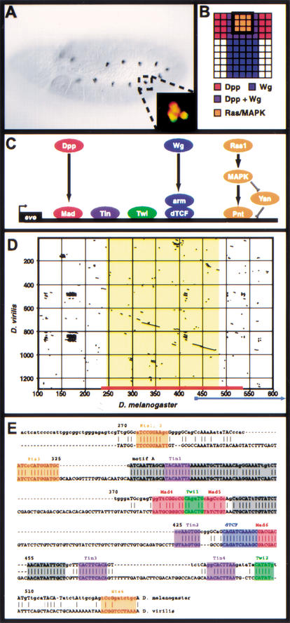
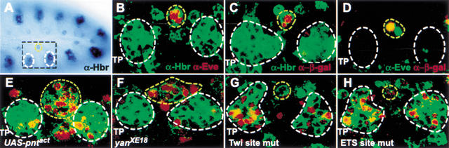
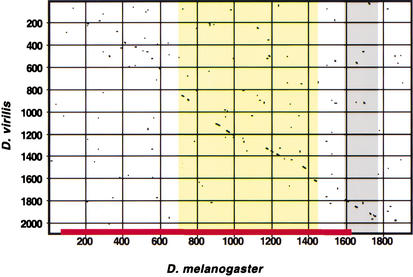
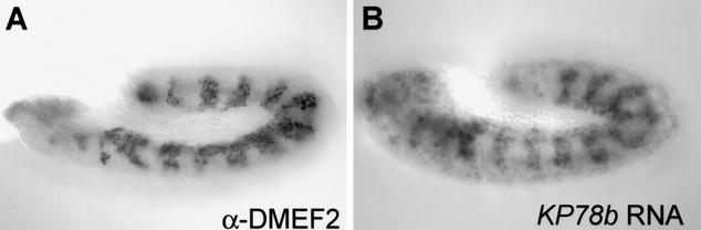
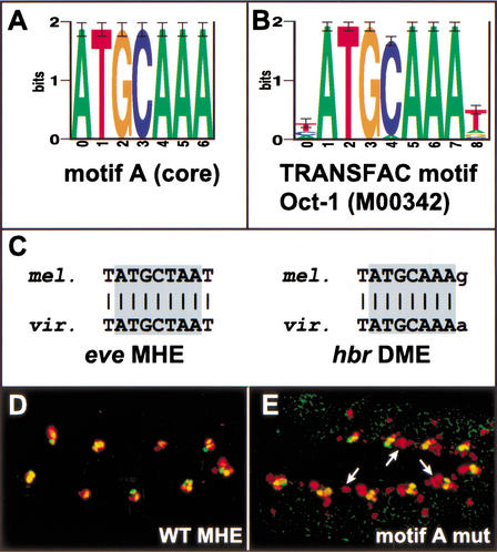
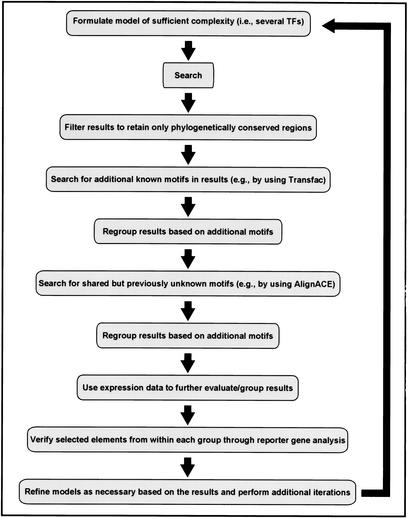
Gene expression is regulated by transcription factors that interact with cis-regulatory elements. Predicting these elements from sequence data has proven difficult. We describe here a successful computational search for elements that direct expression in a particular temporal-spatial pattern in the Drosophila embryo, based on a single well characterized enhancer model. The fly genome was searched to identify sequence elements containing the same combination of transcription factors as those found in the model. Experimental evaluation of the search results demonstrates that our method can correctly predict regulatory elements and highlights the importance of functional testing as a means of identifying false-positive results. We also show that the search results enable the identification of additional relevant sequence motifs whose functions can be empirically validated. This approach, combined with gene expression and phylogenetic sequence data, allows for genome-wide identification of related regulatory elements, an important step toward understanding the genetic regulatory networks involved in development.


Figures

Figure 1
Figure 1
The Eve MHE provides a model for the transcriptional integration of multiple intercellular signals. (A) Stage 11 Drosophila embryo stained with antibodies against Eve. A small cluster of cells in the dorsal mesoderm of each segment, the Eve-positive muscle and cardiac progenitors, express Eve. Anterior is to the left. Inset: Double-staining for Eve (green) and the MHE reporter construct (red) shows that the MHE is sufficient to drive expression in the Eve-positive cells (Halfon et al. 2000). (B) Signaling events required for Eve expression in the dorsal mesoderm. One hemisegment is represented, with dorsal to the top and anterior to the left. Expression of Eve is induced in cells that receive signaling from the Dpp, Wg, and Ras/MAPK pathways (Carmena et al. 1998). (C) The transcriptional code used at the MHE. The signal-responsive TFs, that is, Mad, dTcf, and Pnt bind along with the mesodermal selector proteins Twi and Tin to activate transcription. (D) Dot plot showing that the MHE sequence (x-axis, red bar) is conserved in D. virilis (y-axis). The area of extensive homology (on the diagonal) is shown in yellow; little homology exists flanking this region for several hundred base pairs. An alternate sequence recently proposed to have MHE-like activity (Knirr and Frasch 2001) is indicated by the blue arrow; note that most of this sequence lies in nonconserved regions. (E) Sequence alignment of MHE and vMHE. Known MHE binding sites are shown in color; gray boxes indicate conserved regions where functional sites have not yet been identified. Base pair numbering corresponds to that shown in D.
Figure 2
Figure 2
The Hbr DME. In all panels, white circles mark the developing tracheal cells, and a yellow circle denotes the mesodermal progenitors. (A) Stage 11 embryo stained with antibodies against Hbr. Expression can be seen in the ectodermally derived tracheal pits and the mesodermal progenitors. The portion of each hemisegment shown in the remaining panels is indicated by a black box. (B) Double-labeling for Hbr (green) and Eve (red) shows that the two proteins are found in the same cells in the dorsal mesoderm. Note that Hbr is membrane-associated, whereas Eve is nuclear. Additional mesodermal cells expressing Hbr but not Eve can be seen on both sides of the Eve progenitors. (C) The Hbr DME reporter (nuclear β-galactosidase, red) is expressed in a subset of the Hbr-positive mesodermal progenitors (green). The cells of the developing trachea do not express the reporter. (D) Double-labeling for Eve (green) and the DME reporter (red) show that the DME is expressed in at least one of the Eve progenitors. (E,F) Ectopic activation of the Ras/MAPK pathway through either mesodermal expression of activated Pnt (E) or loss-of-function of the repressor Yan (F) is accompanied by an expanded number of both Hbr- (green) and DME- (red) expressing cells. (G) Mutation of the Twi binding sites in the DME results in a loss of mesodermal reporter gene activity and the acquisition of expression in the developing trachea. (H) Mutation of the Ets binding sites in the DME causes the acquisition of expression in the developing trachea but has only a minimal effect on mesodermal expression. TP, tracheal pits. All panels show anterior to the left and dorsal up.
Figure 3
Figure 3
Conservation of the Hbr DME. Extensive regions of conservation can be detected between D. melanogaster (x-axis) and D. virilis (y-axis) in the region of the DME (red bar). Conservation within the DME is indicated by yellow shading; gray shading indicates a conserved region not required for DME activity that may represent part of a tracheal-specific enhancer (M. Halfon, unpubl.). Little conservation is evident in the adjacent sequences (see Supplemental Fig. 3 for details. Online at http://www.genome.org).
Figure 4
Figure 4
Additional elements identified in the search map in proximity to mesodermally expressed genes. Pictured is antibody staining showing mesodermal expression of DMEF2 (A) and whole mount in situ hybridization of KP78b RNA (B), a gene not previously known to be expressed in the mesoderm.
Figure 5
Figure 5
Identification of a new functional motif using AlignACE. (A) Sequence logo (Schneider and Stephens 1990) showing the motif A core as identified by AlignACE. (B) Sequence logo for the POU/homeodomain TF Oct-1 from TRANSFAC (Wingender et al. 2001; accession #M00342). (C) Alignments of motif A from the Eve MHE and Hbr DME with the corresponding sequences from D. virilis. (D) Wild-type and (E) motif A site mutated Eve MHE reporter gene expression (red) along with endogenous Eve expression (green). The motif A mutation leads to an expansion of reporter gene expression along the anteroposterior axis (arrows).
Figure 6
Figure 6
An iterative search strategy for discovery of cis-regulatory elements based on an experimentally validated model. (See text for details).

Similar articles

Cited by

References

    1. Adams MD, Celniker SE, Holt RA, Evans CA, Gocayne JD, Amanatides PG, Scherer SE, Li PW, Hoskins RA, Galle RF, et al. The genome sequence of Drosophila melanogaster. Science. 2000;287:2185–2195. - PubMed
    1. Arnone MI, Davidson EH. The hardwiring of development: Organization and function of genomic regulatory systems. Development. 1997;124:1851–1864. - PubMed
    1. Berman BP, Nibu Y, Pfeiffer BD, Tomancak P, Celniker SE, Levine M, Rubin GM, Eisen MB. Exploiting transcription factor binding site clustering to identify cis-regulatory modules involved in pattern formation in the Drosophila genome. Proc Natl Acad Sci. 2002;99:757–762. - PMC - PubMed
    1. Blackman RK, Meselson M. Interspecific nucleotide sequence comparisons used to identify regulatory and structural features of the Drosophila hsp82 gene. J Mol Biol. 1986;188:499–515. - PubMed
    1. Bussemaker HJ, Li H, Siggia ED. Regulatory element detection using correlation with expression. Nat Genet. 2001;27:167–171. - PubMed

Publication types

MeSH terms

Associated data

LinkOut - more resources