. 2002 Jul 1;22(13):5300-9.
doi: 10.1523/JNEUROSCI.22-13-05300.2002.

Alternative splicing of an insect sodium channel gene generates pharmacologically distinct sodium channels

Affiliations

Alternative splicing of an insect sodium channel gene generates pharmacologically distinct sodium channels

Jianguo Tan et al. J Neurosci. .

Abstract

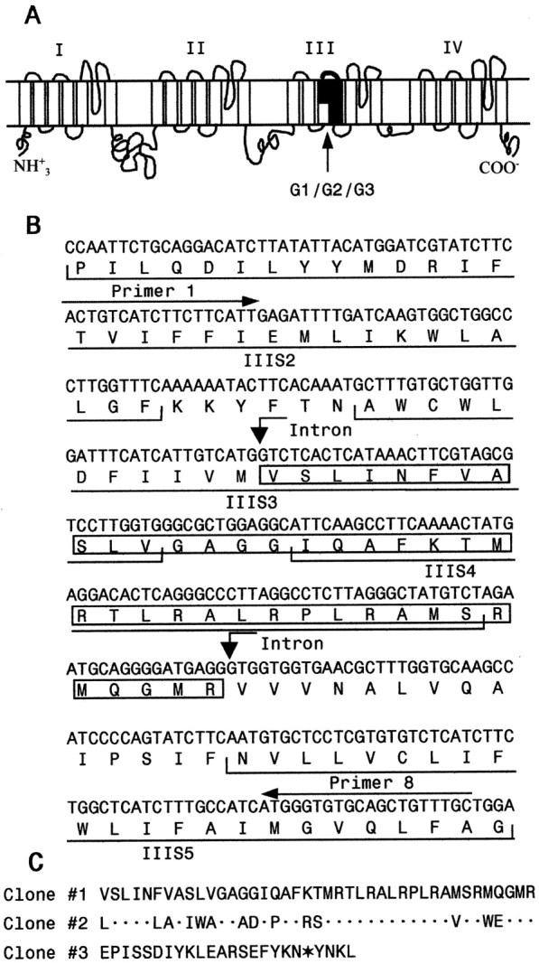
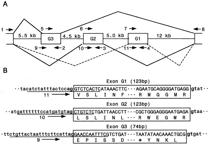
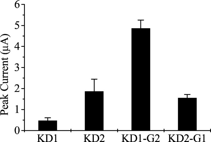
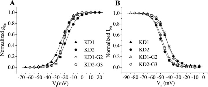
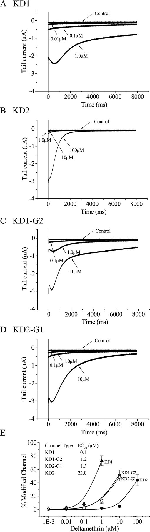
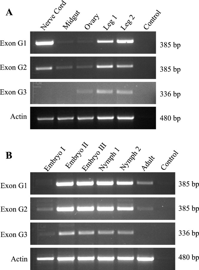
Alternative splicing is a major mechanism by which potassium and calcium channels increase functional diversity in animals. Extensive alternative splicing of the para sodium channel gene and developmental regulation of alternative splicing have been reported in Drosophila species. Alternative splicing has also been observed for several mammalian voltage-gated sodium channel genes. However, the functional significance of alternative splicing of sodium channels has not been demonstrated. In this study, we identified three mutually exclusive alternative exons encoding part of segments 3 and 4 of domain III in the German cockroach sodium channel gene, para(CSMA). The splice site is conserved in the mouse, fish, and human Na(v)1.6 sodium channel genes, suggesting an ancient origin. One of the alternative exons possesses a stop codon, which would generate a truncated protein with only the first two domains. The splicing variant containing the stop codon is detected only in the PNS, whereas the other two full-size variants were detected in both the PNS and CNS. When expressed in Xenopus oocytes, the two splicing variants produced robust sodium currents, but with different gating properties, whereas the splicing variant with the stop codon did not produce any detectable sodium current. Furthermore, these two functional splicing variants exhibited a striking difference in sensitivity to a pyrethroid insecticide, deltamethrin. Exon swapping partially reversed the channel sensitivity to deltamethrin. Our results therefore provide the first evidence that alternative splicing of a sodium channel gene produces pharmacologically distinct channels.


Figures

Fig. 1.
Fig. 1.
Alternatively spliced variants of the cockroachparaCSMA sodium channel gene.A, The schematic diagram of the cockroach sodium channel topology indicating four homologous domains (I–IV), each with six transmembrane segments. The location of three mutually exclusive alternative exons G1/G2/G3 (Fig. 2) is indicated. B, Nucleotide and predicted amino acid sequences of clone 1 encoding IIIS2–5. The amino acid sequence of the variable region in clone 1 (containing exon G1) is boxed. Positions of PCR primers 1 and 8 are indicated above the sequence. C, Alignment of amino acid sequences of the variable region of the three clones.Dots in Clone #2 sequence represent amino acid residues identical to those in Clone #1. The in-frame stop codon in Clone #3 sequence is indicated with an asterisk. The nucleotide sequences of the variable region in clones 2 and 3 are available in GenBank under accession numbers AF478702 and AF478703.
Fig. 2.
Fig. 2.
Identification of three alternatively spliced exons encoding IIIS3–4 in the cockroach ParaCSMA sodium channel protein. A, Schematic representation of the genomic organization. G1, G2, and G3 are the three alternatively spliced exons. Approximate sizes of the introns are indicated. Lines with arrowsindicate the positions of the primers used in PCR analysis of the genomic DNA. Primers 1, 5, 6, 7, 9, 10, and 11 are sense primers, and primers 2, 3, 4, and 8 are antisense primers. Sequences of the primers are presented in Table 1. B, Exon–intron boundaries. The intron sequences are in lowercase letters, and the exon sequences are in capital letters. The sequences inboxes represent exons. The sizes of the exons are indicated in parentheses. The consensus splice donor and acceptor sequences, gt/ag, of each exon/intron border are inbold. Sequences of primers 9, 10, and 11, which cover the intron–exon boundaries, are underlined. The stop codon in Exon G3 is indicated with an asterisk.
Fig. 3.
Fig. 3.
Peak currents of ParaCSMAsodium channel splice variants in oocytes. Amplitude of the maximal peak current was measured during a 20 msec depolarization from −120 to −10 mV 4 d after injection. The error bars indicate the SEM for six oocytes.
Fig. 4.
Fig. 4.
Voltage dependence of activation and inactivation of ParaCSMA splice variants. A, Normalized conductance–voltage curves. Sodium currents were recorded during 14 msec depolarizations ranging from −120 to 60 mV in 5 mV increments. The peak current was converted to conductance as described in Materials and Methods and plotted against the depolarizing voltage. B, Steady-state inactivation curves. The voltage dependence of inactivation was determined using 200 msec inactivating prepulses from a holding potential of −120 to +40 mV in 5 mV increments, followed by test pulses to −5 mV for 5 msec. The peak current amplitude during the test depolarization was normalized to the maximum current amplitude and plotted as a function of the prepulse potential. The smooth curves represent the best fits using a Boltzmann equation, as described in Materials and Methods. Symbolsrepresent means, and error bars indicate the SEM for four oocytes.
Fig. 5.
Fig. 5.
Sensitivity of ParaCSMA splice variants to deltamethrin. Shown are tail currents induced by deltamethrin in oocytes expressing the splice variants KD1 (A) and KD2 (B) and the exon-swapped channels, KD1-G2 (C) and KD2-G1 (D). Tail currents were recorded in response to a 100-pulse train of 5 msec depolarizations from −100 to 0 mV with a 5msec interval between each depolarization.E, The percentage of channel modification by deltamethrin was determined using the equation M = {[Itail/(EhENa)]/[INa/(EtENa)]} × 100, as described in Materials and Methods (Tatebayashi and Narahashi, 1994). The data were fitted with the Hill equation as described in Materials and Methods. EC20 values (inset) were derived from the fitted curves. The Hill coefficients range from 0.8 to 1.4. Symbols represent means, and error bars indicate the SEM for five oocytes.
Fig. 6.
Fig. 6.
RT-PCR analysis of splice variants in five tissues (A) and six developmental stages (B). Equal amounts of total RNA (5 μg) were used in cDNA synthesis, and equal amounts of cDNA templates (1 μl) were used in PCR. Amplification of actin (480 bp) is included to ensure that similar amounts of RNA and cDNA were used in RT-PCR for the various tissues and developmental stages. Each PCR product (10 μl) was separated on a 1.5% agarose gel. The criteria for classification of the developmental stages and tissues are described in Materials and Methods.

Similar articles

Cited by

References

    1. Belcher SM, Zerillo CA, Levinson R, Ritchie JM, Howe JR. Cloning of a sodium channel α-subunit from rabbit Schwann cells. Proc Natl Acad Sci USA. 1995;92:11034–11038. - PMC - PubMed
    1. Bell WL. The laboratory cockroach. Chapman and Hall; New York: 1981.
    1. Bourinet E, Soong TW, Sutton K, Slaymaker S, Mathews E, Monteil A, Zamponi GW, Nargeot J, Snutch TP. Splicing of alpha 1A subunit gene generates phenotypic variants of P- and Q. Nat Neurosci. 1999;2:407–415. - PubMed
    1. Burt PE, Goodchild RE. The site of action of pyrethrin I in the nervous system of the cockroach Periplaneta Americana. Ent Exp Appl. 1971;14:179–189.
    1. Byerly L, Leung H. Ionic currents of Drosophila neurons in embryonic cultures. J Neurosci. 1988;8:4379–4393. - PMC - PubMed

Publication types

MeSH terms

Associated data

LinkOut - more resources