. 2002 Jul 1;22(13):5423-31.
doi: 10.1523/JNEUROSCI.22-13-05423.2002.

In Vivo Delivery of a Bcl-xL Fusion Protein Containing the TAT Protein Transduction Domain Protects against Ischemic Brain Injury and Neuronal Apoptosis

Affiliations

In Vivo Delivery of a Bcl-xL Fusion Protein Containing the TAT Protein Transduction Domain Protects against Ischemic Brain Injury and Neuronal Apoptosis

Guodong Cao et al. J Neurosci. .

Abstract

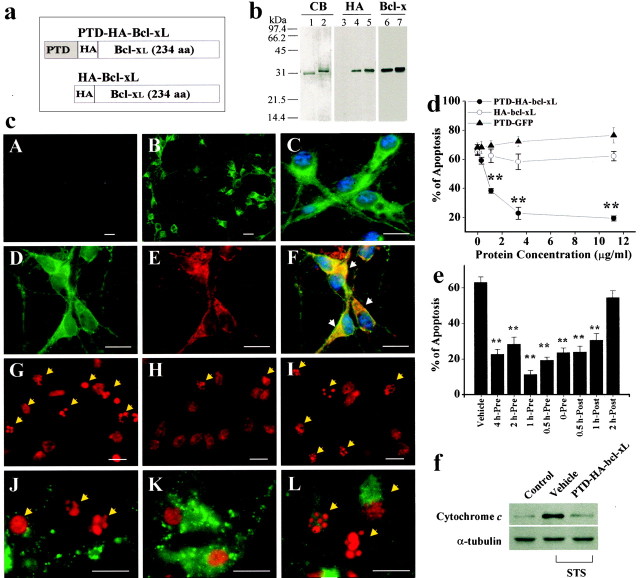
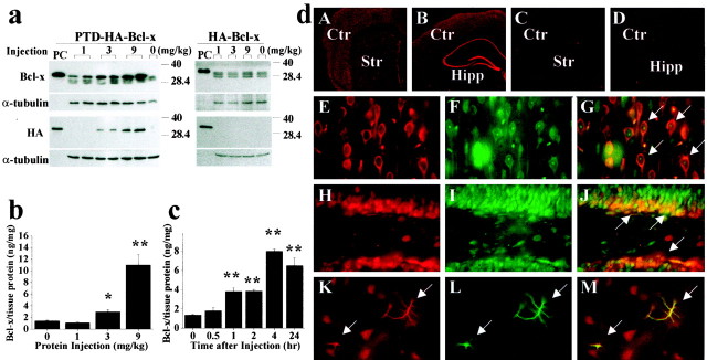
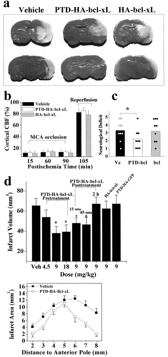
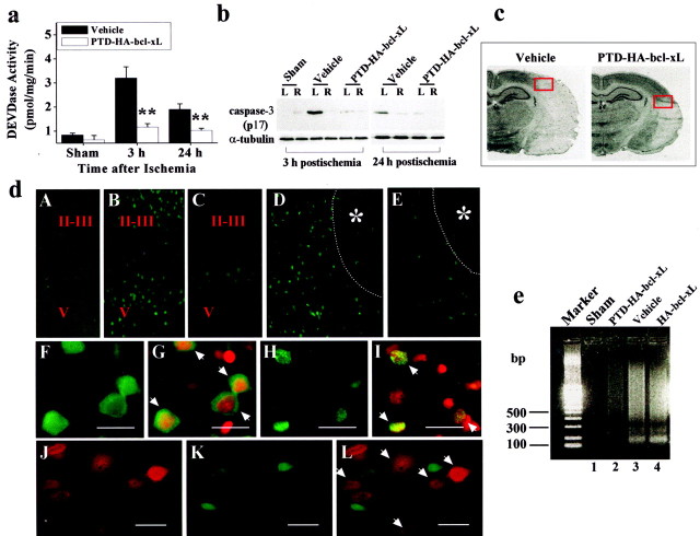
Bcl-xL is a well characterized death-suppressing molecule of the Bcl-2 family. Bcl-xL is expressed in embryonic and adult neurons of the CNS and may play a critical role in preventing neuronal apoptosis that occurs during brain development or results from diverse pathologic stimuli, including cerebral ischemia. In this study, we used a novel approach to study the potential neuroprotective effect of Bcl-xL as a therapeutic agent in the murine model of focal ischemia/reperfusion. We created a Bcl-xL fusion protein, designated as PTD-HA-Bcl-xL, which contains the protein transduction domain (PTD) derived from the human immunodeficiency TAT protein. We demonstrated that this fusion protein is highly efficient in transducing into primary neurons in cultures and potently inhibited staurosporin-induced neuronal apoptosis. Furthermore, intraperitoneal injection of PTD-HA-Bcl-xL into mice resulted in robust protein transduction in neurons in various brain regions within 1-2 hr, and decreased cerebral infarction (up to approximately 40%) in a dose-dependent manner, as determined at 3 d after 90 min of focal ischemia. PTD-HA-Bcl-xL was effective even when it was administered after the completion of ischemia (up to 45 min), and the protective effect was independent of the changes in cerebral blood flow or other physiological parameters. Finally, as shown by immunohistochemistry, Western blotting, and substrate-cleavage assays, PTD-HA-Bcl-xL attenuated ischemia-induced caspase-3 activation in ischemic neurons. These results thus confirm the neuroprotective effect of Bcl-xL against ischemic brain injury and provide the first evidence that the PTD can be used to efficiently transduce a biologically active neuroprotectant in experimental cerebral ischemia.


Figures

Fig. 1.
Fig. 1.
The death-suppressing effect of PTD-HA-Bcl-xL in primary cultures of cortical neurons. a, Structures of the Bcl-xL fusion proteins. PTD-HA-Bcl-xL contains both HA tag and the PTD, whereas HA-Bcl-xL does not contain the PTD; the latter served as the control protein in subsequent studies. b, Verification of the Bcl-xL fusion proteins. Coomassie blue (CB) staining shows that the proteins have been purified to near homogeneity. Western blots show that the proteins could be detected using either anti-HA or anti-Bcl-x antibody.c, Representative immunofluorescent images show the transduction and death-suppressing effect in cortical neurons. PTD-HA-Bcl-xL (B, low power; C, high power) but not HA-Bcl-xL (A) transduces into neurons within 15 min of exposure, as determined using HA immunostaining. Immunofluorescence for HA (D) and CCOX IV (E) are partially colocalized (F, overlay), suggesting that a portion of the transduced PTD-HA-Bcl-xL is associated with the mitochondria. The addition of PTD-HA-Bcl-xL (H) but not HA-Bcl-xL (I) reduces the amounts of nuclei showing apoptotic changes (arrows), compared with the vehicle control (G). Immunofluorescence for cytochrome c is preserved in PTD-HA-Bcl-xL-treated neurons (K) but not in HA-Bcl-xL-treated neurons (L) or vehicle-treated neurons (J), consistent with the speculation that PTD-HA-Bcl-xL inhibits STS-induced cytochrome c release.G–L were obtained 24 hr after STS. Scale bar, 20 μm. d, e, PTD-HA-Bcl-xL but not HA-Bcl-xL or PTD-GFP inhibits STS-induced apoptosis in a dose-dependent manner (d). PTD-HA-Bcl-xL was equally effective when it was added to the cultures before or ≤1 hr after STS (e). Apoptosis was quantified after PI staining at 24 hr after STS exposure (0.3 μm). Data are mean ± SE from three independent experiments. **p < 0.01 versus vehicle controls (ANOVA and post hocScheffe's tests). f, Western blots demonstrate that PTD-HA-Bcl-xL treatment attenuated STS-induced cytochromec release to the cytoplasm in cultured neurons. Eachlane contains 20 μg of cytosolic protein, prepared from noninduced (control) or STS-induced (0.3 μm for 6 hr) neurons, respectively. The blots are representative of three independent experiments with similar results.
Fig. 2.
Fig. 2.
In vivo protein transduction of PTD-HA-Bcl-xL in murine brain. a, Western blotting with either the anti-Bcl-x or anti-HA antibody detects the transduction of PTD-HA-Bcl-xL into the cortex 4 hr after intraperitoneal injection of the protein at various doses (left); the HA-Bcl-xL protein lacking PTD failed to transduce across the blood–brain barrier (right). In all blots, the fusion proteins serve as the positive controls (PC). Immunoblotting of α-tubulin serves as a control for sample loadings (30 μg perlane). Note that the endogenous Bcl-x proteins are detectable in all brains. b, c, Quantitation of PTD-HA-Bcl-xL transduction in the brain by ELISA. Intraperitoneal injection of PTD-HA-Bcl-xL results in dose-dependent (b, 4 hr after injection) and time-dependent (c, injection at the dose of 9 mg/kg) protein transduction into the murine cerebral cortex. Data are mean ± SE (three animals per group). *p < 0.05 versus controls; **p < 0.01 versus controls. d, Immunofluorescent detection (using anti-HA antibody) of PTD-HA-Bcl-xL transduction in the cortex/caudate (A) and hippocampal (Hipp) formation (B) 4 hr after intraperitoneal injection; the immunofluorescence is lost after the primary antibody was preabsorbed with the fusion protein (C). Injection of HA-Bcl-xL serves as the negative control (Ctr) (D). In the cerebral parenchyma, most of the cells transduced with PTD-HA-Bcl-xL (HA immunoreactive; E, H, red) are neurons, being immunoreactive for the neuronal markers NSE (F, arrows in G; cortex) or NeuN (I, arrows in J; hippocampal dentate). Some astrocytes (GFAP-immunoreactive;L, green) in the caudate are also transduced (arrows in K–M).Str, Striatum.
Fig. 3.
Fig. 3.
Systemic delivery of PTD-HA-Bcl-xL protects against focal ischemic infarction. a, Representative photographs of TTC-stained coronal sections of mouse brains recovered from 90 min of focal ischemia and 72 hr of reperfusion. The sections were chosen at the levels of 4 mm (top) and 6 mm (bottom) from the anterior pole. The dotted white lines illustrate the infarct border for each section.b, Changes in cortical blood flow, as determined using laser–Doppler flowmetry, are not different between fusion protein-treated and vehicle-treated brains during or after ischemia (n = 6 per group). CBF, Cerebral blood flow. c, Neurological deficit scores in vehicle-treated (Ve), PTD-HA-Bcl-xL-treated (PTD-bcl), and HA-Bcl-xL-treated (bcl) mice at 24 hr after ischemia.Dots and columns represent the scores of each animal and the mean scores, respectively. The neurological scores were graded according to the scale described previously (Murakami et al., 1998). d, Effects of intraperitoneal injection ofPTD-HA-Bcl-xL on infarct volumes in the brain after 90 min of focal ischemia. Vehicle (Veh), PTD-HA-Bcl-xL, or the control proteins were injected at the indicated doses either 1 hr before or at the indicated time after ischemia, and the infarct volume was measured at 72 hr after ischemia. Data are mean ± SE;n = 6–8 per group. *p < 0.05 versus vehicle treatment (ANOVA and post hoc Scheffe's tests). The graph at the bottomillustrates the comparison of infarct sizes in each brain section between vehicle-treated and PTD-HA-Bcl-xL-treated mice.
Fig. 4.
Fig. 4.
PTD-HA-Bcl-xL inhibits an apoptotic component of ischemic neuronal death. a, Effects of PTD-HA-Bcl-xL on caspase-3-like protease activity in the brain at 3 and 24 hr after ischemia, as determined in the cortical cell extracts using the fluorogenic substrate DEVD-AFC. Data are mean ± SE; n = 5 per group. **p < 0.01 versus vehicle treatment. b, Attenuation of caspase-3 activation after ischemia by PTD-HA-Bcl-xL, as demonstrated by Western blotting. The active (p17) caspase-3 fragment is increased in the ischemic hemisphere (L) compared with the nonischemic side (R) in vehicle-treated but not in PTD-HA-Bcl-xL-treated brains. The blots are representative of three independent experiments with similar results. c, Coronal sections obtained from vehicle-treated and PTD-HA-Bcl-xL-treated brains, respectively, 24 hr after ischemia. The red boxes mark the approximate locations from which the immunofluorescent images in c were obtained.d, Representative immunofluorescent images show that PTD-HA-Bcl-xL attenuates caspase-3 activation after ischemia. Immunostaining was accomplished using the antibody recognizing the active caspase-3. A–E, Low power (25×):A, Nonischemic cortex. B, At 3 hr after ischemia, caspase-3-immunoreactive cells are present primarily in layers I-II and V of the cortex. C, The mounts of caspase-3-immunoreactive cells are decreased in PTD-HA-Bcl-xL-treated brain. D, 24 hr after ischemia, caspase-3-immunoreactive cells are localized in the border zone of the infarction (*). Thedotted lines in D and E mark the estimated infarct border. E, Few caspase-3-immunoreactive cells are present in PTD-HA-Bcl-xL-treated brain. F–I, high power (400×). Scale bar, 20 μm. F, At 3 hr after ischemia (vehicle treated), caspase-3 immunoreactivity primarily shows a cytosolic location. G, As indicated by the arrows, the cells in F counterstained with the DNA dye PI.H, At 24 hr after ischemia, caspase-3 immunoreactivity is localized in the nucleus. I, As indicated by thearrows, the cells in H counterstained with PI. J–L, Double-label immunofluorescence of PTD-HA-Bcl-xL (J) and caspase-3 (K) in the border zone of the cortical infarction at 24 hr after ischemia (PTD-HA-Bcl-xL-treated brain); Lis the overlay of J and K. Note that there is no colocalization of caspase-3 and PTD-HA-Bcl-xL in neurons (L, arrows indicate protein-transduced neurons). e, Apoptotic DNA fragmentation is attenuated by PTD-HA-Bcl-xL after ischemia, as demonstrated by DNA electrophoresis. The DNA was prepared from the cortical tissues 72 hr after ischemia, using the brains pretreated with vehicle, PTD-HA-Bcl-xL, or HA-Bcl-xL. The gel is representative of two independent experiments with similar results.

References

    1. Blomer U, Kafri T, Randolph-Moore L, Verma IM, Gage FH. Bcl-xL protects adult septal cholinergic neurons from axotomized cell death. Proc Natl Acad Sci USA. 1998;95:2603–2608. - PMC - PubMed
    1. Cao G, Minami M, Pei W, Yan C, Chen D, O'Horo C, Graham SH, Chen J. Intracellular Bax translocation after transient cerebral ischemia: implications for a role of the mitochondrial apoptotic signaling pathway in ischemic neuronal death. J Cereb Blood Flow Metab. 2001a;21:321–333. - PubMed
    1. Cao G, Pei W, Lan J, Stetler RA, Luo Y, Nagayama T, Graham SH, Yin XM, Simon RP, Chen J. Caspase-activated DNase/DNA fragmentation factor 40 mediates apoptotic DNA fragmentation in transient cerebral ischemia and in neuronal cultures. J Neurosci. 2001b;21:4678–4690. - PMC - PubMed
    1. Cao G, Luo Y, Nagayama T, Pei W, Stetler RA, Graham SH, Chen J. Cloning and characterization of rat caspase-9: implications for a role in mediating caspase-3 activation and hippocampal cell death after transient cerebral ischemia. J Cereb Blood Flow Metab. 2002;22:534–546. - PubMed
    1. Chen D, Stetler RA, Cao G, Pei W, O'Horo C, Yin XM, Chen J. Characterization of the rat DNA fragmentation factor 35/inhibitor of caspase-activated DNase (short form): the endogenous inhibitor of caspase-dependent DNA fragmentation in neuronal apoptosis. J Biol Chem. 2000;275:38508–38517. - PubMed

Publication types

MeSH terms