. 2002 Jul 1;22(13):5442-51.
doi: 10.1523/JNEUROSCI.22-13-05442.2002.

A third vesicular glutamate transporter expressed by cholinergic and serotoninergic neurons

Affiliations

A third vesicular glutamate transporter expressed by cholinergic and serotoninergic neurons

Christelle Gras et al. J Neurosci. .

Abstract

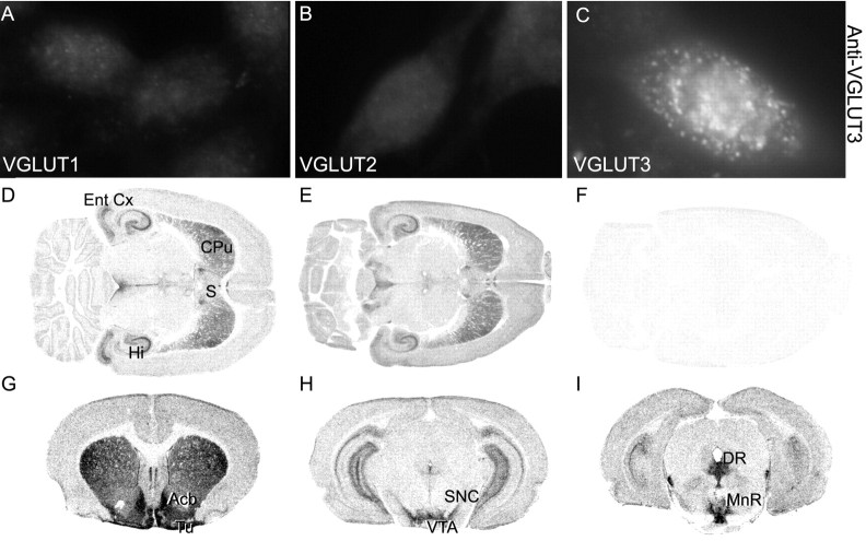
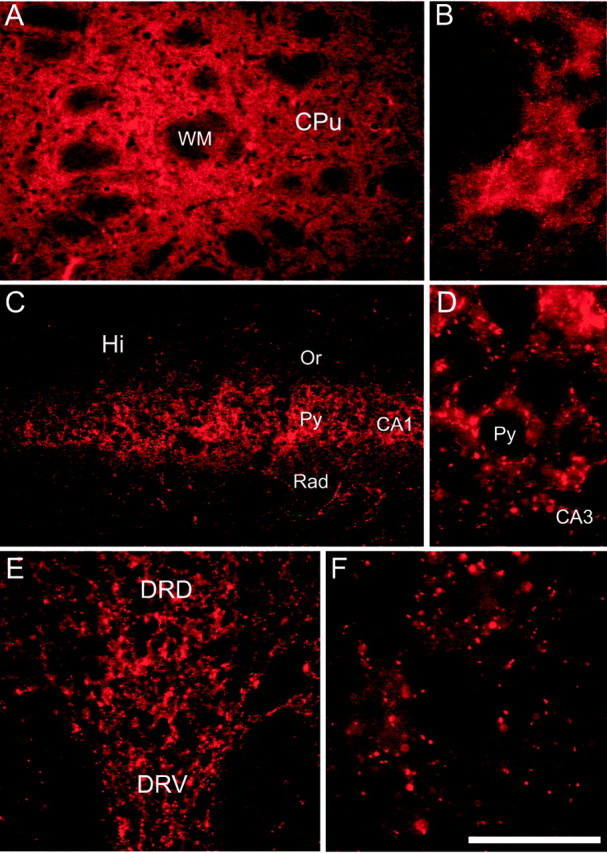
Two proteins previously known as Na(+)-dependent phosphate transporters have been identified recently as vesicular glutamate transporters (VGLUT1 and VGLUT2). Together, VGLUT1 and VGLUT2 are operating at most central glutamatergic synapses. In this study, we characterized a third vesicular glutamate transporter (VGLUT3), highly homologous to VGLUT1 and VGLUT2. Vesicles isolated from endocrine cells expressing recombinant VGLUT3 accumulated l-glutamate with bioenergetic and pharmacological characteristics similar, but not identical, to those displayed by the type-1 and type-2 isoforms. Interestingly, the distribution of VGLUT3 mRNA was restricted to a small number of neurons scattered in the striatum, hippocampus, cerebral cortex, and raphe nuclei, in contrast to VGLUT1 and VGLUT2 transcripts, which are massively expressed in cortical and deep structures of the brain, respectively. At the ultrastructural level, VGLUT3 immunoreactivity was concentrated over synaptic vesicle clusters present in nerve endings forming asymmetrical as well as symmetrical synapses. Finally, VGLUT3-positive neurons of the striatum and raphe nuclei were shown to coexpress acetylcholine and serotonin transporters, respectively. Our study reveals a novel class of glutamatergic nerve terminals and suggests that cholinergic striatal interneurons and serotoninergic neurons from the brainstem may store and release glutamate.


Figures

Fig. 1.
Fig. 1.
Alignment of VGLUT3, VGLUT2, and VGLUT1 amino acid sequences. The three proteins are highly conserved in their central portion. (Black boxing indicates identical residues.)
Fig. 2.
Fig. 2.
Functional characterization of VGLUT3. Membranes from BON cells stably expressing VGLUT3, VGLUT2, or an unrelated VIAAT (mock) were incubated at 30°C for 10 min with 40 μm or 2 μCi of [3H]l-glutamate and the following additives as stated: 50 μm CCCP, 5 μmnigericin, 20 μm valinomycin, and 5 μmEvans Blue, as well as 10 mm (C)l-glutamate (GLU),l-aspartate (ASP), acetylcholine (Ach), or GABA. Representative experiments are shown. The error bars represent the SEM of triplicate determinations.A, Expression of VGLUT2 or VGLUT3 induces a CCCP-sensitive uptake of glutamate. In B–D, only the specific (CCCP-sensitive) component of uptake is shown. InC and D, the subtracted CCCP-resistant component was determined in the presence of an identical concentration of amino acid or Evans Blue. B, VGLUT3-mediated uptake is more sensitive to nigericin than VGLUT2. C, Both transporters prefer l-glutamate over other amino acids or transmitters. D, VGLUT3 is less sensitive to Evans Blue than VGLUT2. The dotted lines in C andD represent the control level (i.e., 100%).
Fig. 3.
Fig. 3.
Saturation kinetics of VGLUT2 and VGLUT3.A, Time course of VGLUT2- and VGLUT3-mediated uptake at a 40 μm [3H]glutamate.B, The initial rate of uptake (at 3 min) into VGLUT2-containing vesicles was determined with increasing concentrations of [3H]glutamate (0.25–3 mm). Specific uptake was determined at each glutamate concentration by subtracting the background uptake observed in the presence of 50 μm CCCP. C, Dependence of the initial rate of VGLUT3-mediated uptake (at 10 min) on glutamate concentration was determined as in B. D, A Lineweaver–Burke plot of the data shown in B andC indicates KM values of 1.27 and 0.56 mm and Vmax values of 152 and 19 pmol · mg−1protein · min−1 for VGLUT2 (closed circles) and VGLUT3 (open circles), respectively. Regression lines for VGLUT2 (r = 0.9993) and VGLUT3 (r = 0.8959) intersect the 1/S axis at distinct −1/KM values (closed and open arrowheads). The results in B–D represent the average of three independent paired analyses of VGLUT2 and VGLUT3, each performed in triplicate. Error bars in A–C correspond to SEM.
Fig. 4.
Fig. 4.
Regional distribution of VGLUT3 mRNA.A, RT-PCR detection of VGLUT3 mRNA in body organs and in different brain areas. G3PDH-specific primers (Clontech) were used to normalize the RNA level in tissue sample extracts. B–H,In situ hybridization analysis of VGLUT3 transcript distribution in the rat brain. Horizontal (B) or coronal (C–H) brain sections were hybridized with antisense 35S-labeled oligonucleotides (B–E) or DIG-UTP-labeled cRNA probes (F–H). CA3, CA3 field of the hippocampus; CPu, caudate-putamen; DR, dorsal raphe; DRD, dorsal part of the dorsal raphe nucleus; DRV, ventral part of the dorsal raphe nucleus;E, ependymal cells; Hi, hippocampus;Hil, hilus of the dentate gyrus; Or, oriens layer of the hippocampus; PMnR, paramedian raphe nuclei; py, pyramidal layer of the hippocampus;Rad, radiatum layer of the hippocampus. Scale bars:F, H, 200 μm; G, 100 μm.
Fig. 5.
Fig. 5.
Antiserum specificity and regional distribution of VGLUT3. Two polyclonal antisera directed against distinct peptides were generated. A–C, Immunofluorescence detection of VGLUT3 in BON stable transfectants (white puncta) with the P45-1 antiserum at a dilution of 1:5000. The serum recognizes VGLUT3 but not VGLUT1 or VGLUT2 in BON stable transfectants. The same result was obtained with the P45-3 antiserum (data not shown).D–I, Localization of VGLUT3 protein by immunoautoradiography. In D and G–I the P45-3 antiserum was used. In E and F, the P45-1 antiserum was used in the presence (F) or absence (E) of its cognate peptide (0.1 mg/ml).Acb, accumbens; CPu, caudate-putamen; DR, dorsal raphe; Ent Cx, entorhinal cortex; Hi, hippocampus;MnR, median raphe nucleus; S, septum; SNC, substantia nigra pars compacta;Tu, olfactory tubercles; VTA, ventral tegmental area. Scale bars: J, L, N, 200 μm;K, M, O, 50 μm.
Fig. 6.
Fig. 6.
Immunofluorescent detection of VGLUT3 in the brain. VGLUT3 immunoreactivity appears as red puncta in the caudate-putamen (A, B), hippocampus (C, D), and dorsal raphe (E, F).CA1, CA1 field of the hippocampus; CA3, CA3 field of the hippocampus; CPu, caudate-putamen;DRD, dorsal part of the dorsal raphe nucleus;DRV, ventral part of the dorsal raphe nucleus;Hi, hippocampus; Or, oriens layer of the hippocampus; py, pyramidal layer of the hippocampus;Rad, radiatum layer of the hippocampus;WM, white matter. Scale bars: A, C, E, 200 μm; B, D, F, 50 μm.
Fig. 7.
Fig. 7.
VGLUT3 is localized in nerve endings in the striatum (A), hippocampus (B, C), and dorsal raphe (D, E). In all three areas, immunoparticles for VGLUT3 are localized in terminals (t). In the striatum, dorsal raphe, and hippocampus (C), the labeled terminals are in close apposition with dendrites (d). In the hippocampus and dorsal raphe, the immunoreactive terminal makes a symmetrical synapse (arrowheads) with a pyramidal cell (Pyr Cell) in B andD; one terminal makes an asymmetrical synapse (arrow) with a dendrite (d) inC and E. Scale bar, 250 nm.
Fig. 8.
Fig. 8.
VGLUT3 is colocalized with ChAT and VAChT in the striatum. A, C, Cold in situhybridization with the ChAT cRNA probe alone (inred). D, Enlargement of the neuron indicated in A by the red arrow.B, D, Double-labeling in situhybridization (ChAT in red and VGLUT3 inblue). D, The double-labeled neuron indicated by a blue arrow in B has been enlarged. E, F, Confocal laser detection of double-immunofluorescence for VGLUT3 (CY3, red fluorescence) and VAChT (fluorescein, green fluorescence) in the striatum (E) or the hippocampus (F). In false colors, overlapping signals appear prominently as yellow-orange.G, double detection of VGLUT3 (immunogold) and VAChT (immunoperoxidase) with the electron microscope in the striatum. Scale bars: A, B, E, F, 50 μm; C, D, 10 μm;G, 0.25 μm.
Fig. 9.
Fig. 9.
VGLUT3 is colocalized with SERT in the dorsal and medial raphe. The SERT probe is shown in red, and the VGLUT3 probe is shown in blue. A shows the dorsal and medial raphe. B, Higher magnification of the dorsal raphe. C, Enlargement of double-labeled neurons (indicated in B by a blue arrow).A and B are taken from different sections. DR, Dorsal raphe; MnR, medial raphe. Scale bars: A, 300 μm; B, 100 μm; C, 10 μm.

References

    1. Aihara Y, Mashima H, Onda H, Hisano S, Kasuya H, Hori T, Yamada S, Tomura H, Yamada Y, Inoue I, Kojima I, Takeda J. Molecular cloning of a novel brain-type Na+-dependent inorganic phosphate cotransporter. J Neurochem. 2000;74:2622–2625. - PubMed
    1. Antunes Bras JM, Epstein AL, Bourgoin S, Hamon M, Cesselin F, Pohl M. Herpes simplex virus 1-mediated transfer of preproenkephalin A in rat dorsal root ganglia. J Neurochem. 1998;70:1299–1303. - PubMed
    1. Bai L, Xu H, Collins JF, Ghishan FK. Molecular and functional analysis of a novel neuronal vesicular glutamate transporter. J Biol Chem. 2001;276:36764–36769. - PubMed
    1. Bellocchio EE, Hu H, Pohorille A, Chan J, Pickel VM, Edwards RH. The localization of the brain-specific inorganic phosphate transporter suggests a specific presynaptic role in glutamatergic transmission. J Neurosci. 1998;18:8648–8659. - PMC - PubMed
    1. Bellocchio EE, Reimer RJ, Fremeau RT, Jr, Edwards RH. Uptake of glutamate into synaptic vesicles by an inorganic phosphate transporter. Science. 2000;289:957–960. - PubMed

Publication types

MeSH terms