Comparative Study
. 2002 Jul 1;22(13):5505-15.
doi: 10.1523/JNEUROSCI.22-13-05505.2002.

Localization of Nogo-A and Nogo-66 receptor proteins at sites of axon-myelin and synaptic contact

Affiliations
Comparative Study

Localization of Nogo-A and Nogo-66 receptor proteins at sites of axon-myelin and synaptic contact

Xingxing Wang et al. J Neurosci. .

Abstract

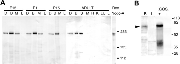
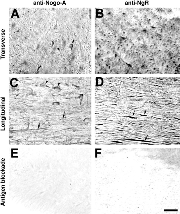
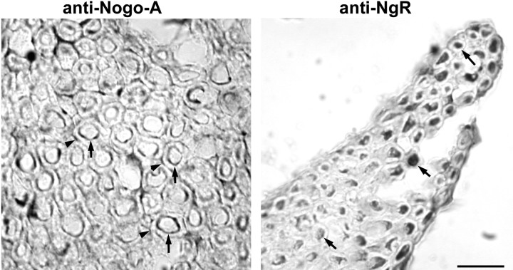
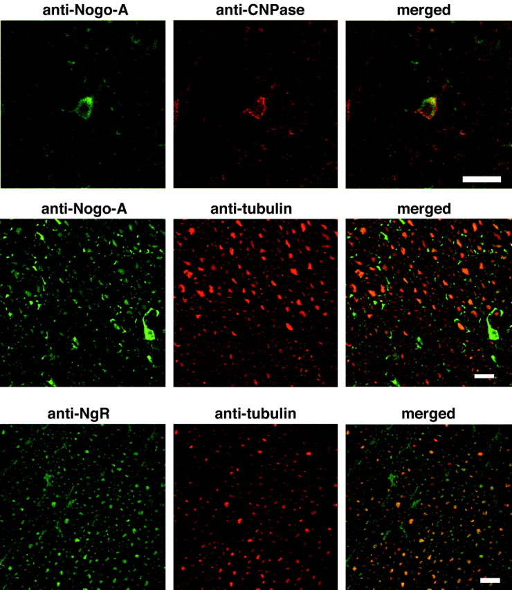
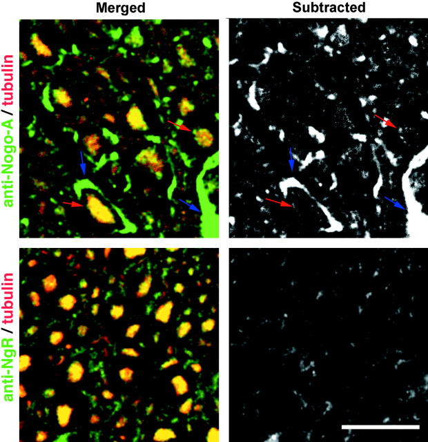
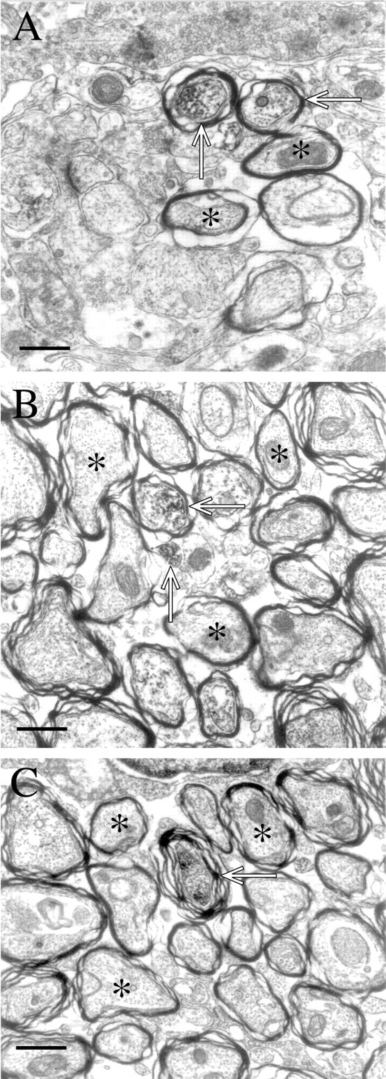
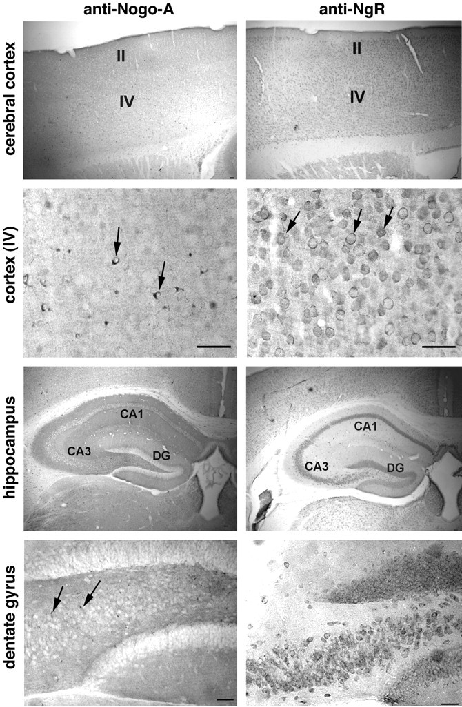
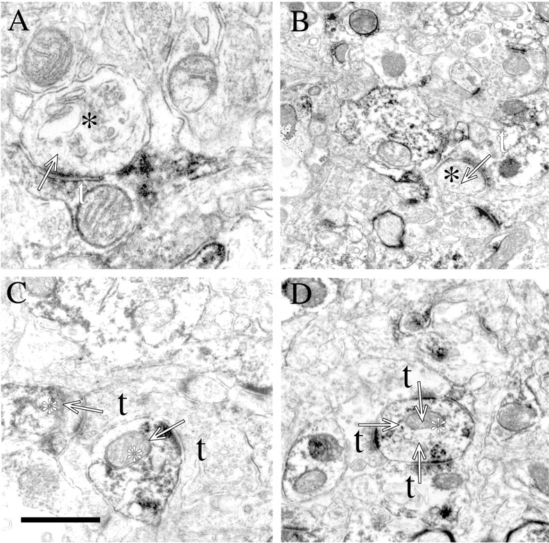
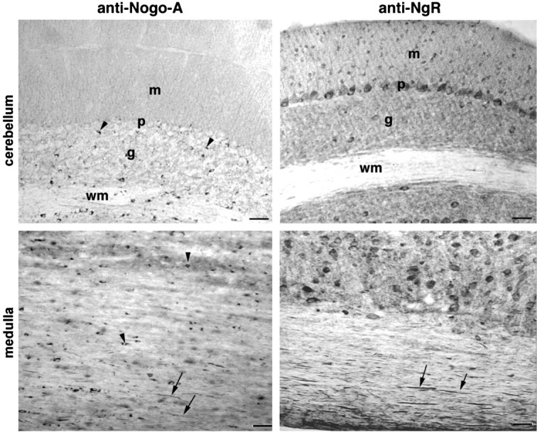
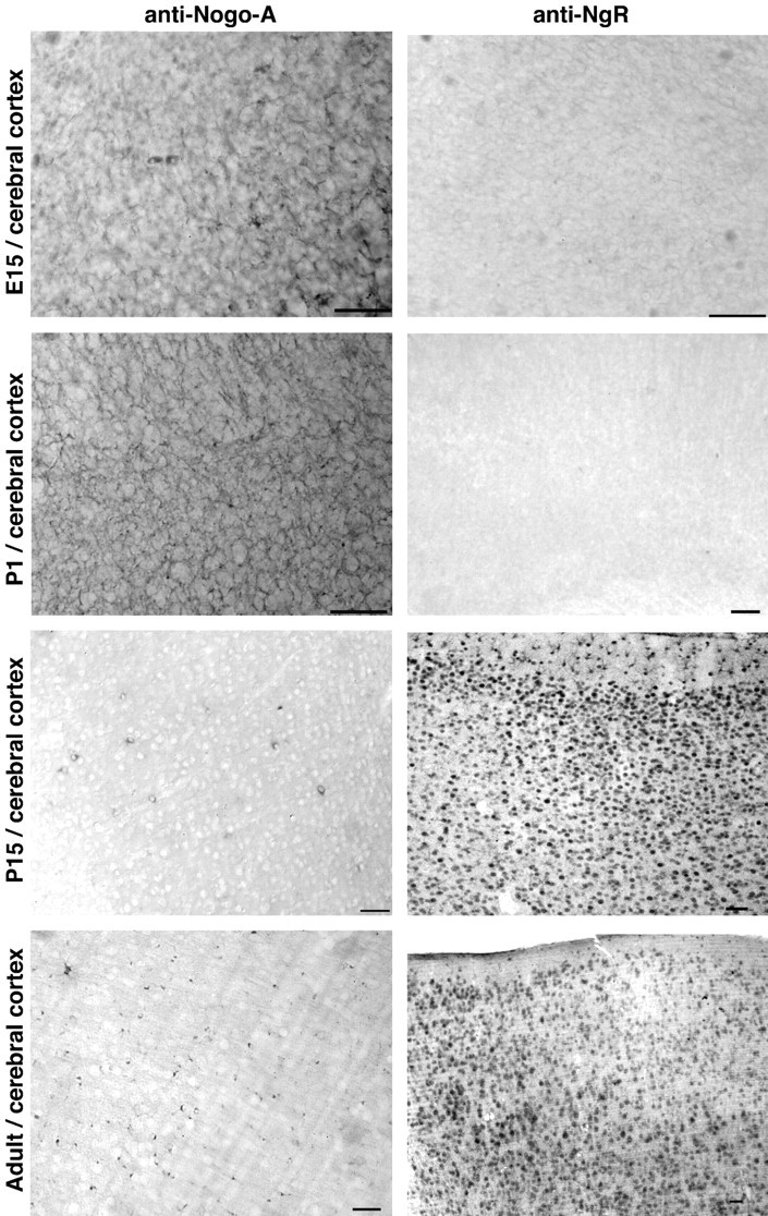
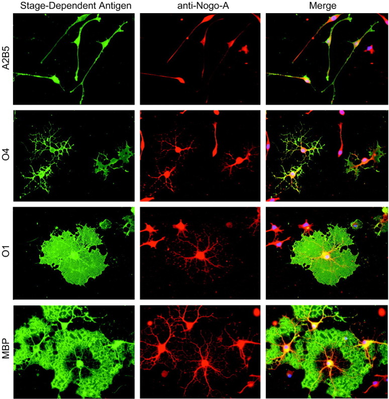
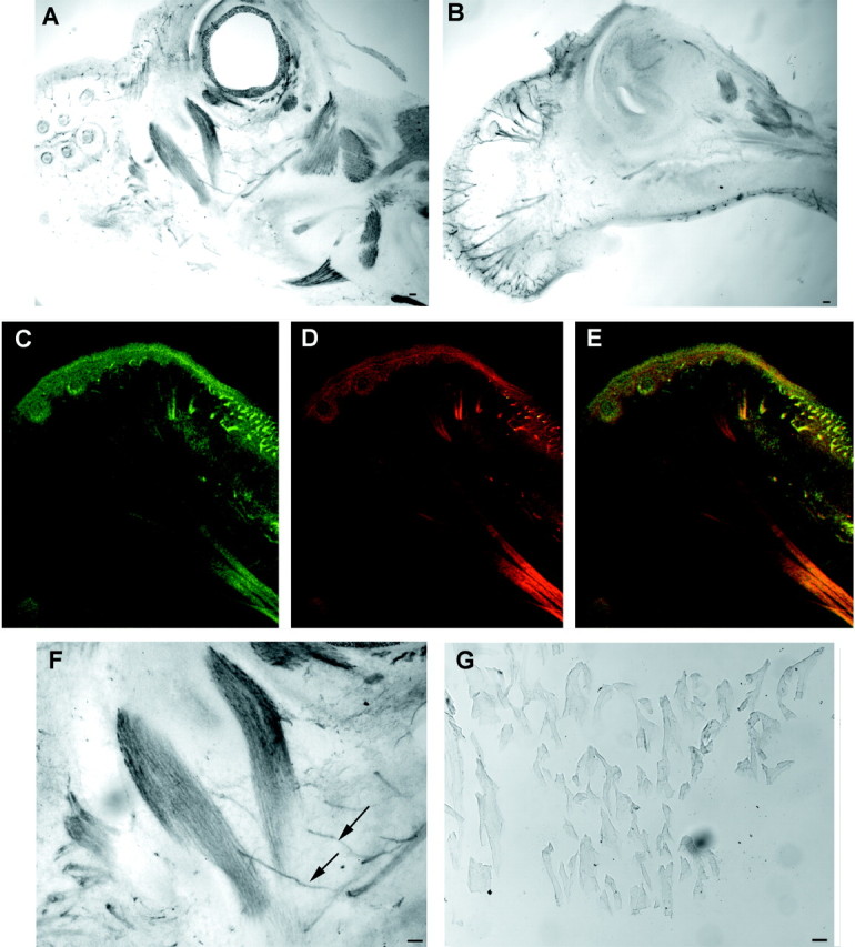
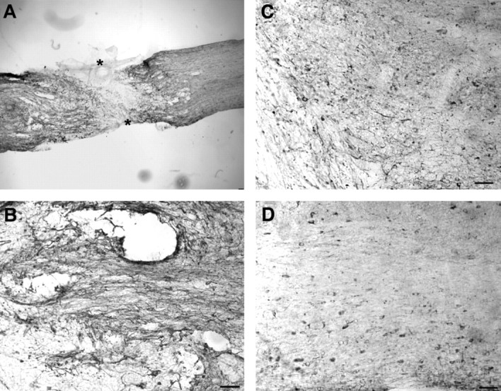
Axon regeneration in the adult CNS is limited by the presence of inhibitory proteins. An interaction of Nogo on the oligodendrocyte surface with Nogo-66 Receptor (NgR) on axons has been suggested to play an important role in limiting axonal growth. Here, we compare the localization of these two proteins immunohistochemically as a test of this hypothesis. Throughout much of the adult CNS, Nogo-A is detected on oligodendrocyte processes surrounding myelinated axons, including areas of axon-oligodendrocyte contact. The NgR protein is detected selectively in neurons and is present throughout axons, indicating that Nogo-A and its receptor are juxtaposed along the course of myelinated fibers. NgR protein expression is restricted to postnatal neurons and their axons. In contrast, Nogo-A is observed in myelinating oligodendrocytes, embryonic muscle, and neurons, suggesting that Nogo-A has additional physiologic roles unrelated to NgR binding. After spinal cord injury, Nogo-A is upregulated to a moderate degree, whereas NgR levels are maintained at constant levels. Taken together, these data confirm the apposition of Nogo ligand and NgR receptor in situations of limited axonal regeneration and support the hypothesis that this system regulates CNS axonal plasticity and recovery from injury.


Figures

Fig. 1.
Fig. 1.
Immunoblot of Nogo-A and NgR expression in mouse tissues. A, Samples (20 μg) from the indicated tissues from mice of the indicated ages were analyzed by anti-Nogo-A immunoblot. D, Dorsal root ganglion; B, brain; S, spinal cord; M, skeletal muscle; H, heart; K, kidney;LU, lung; L, liver. The migration of recombinant Nogo-A is shown at right with molecular weight standards of 233, 135, and 112 kDa. B, Immunoblot for NgR in adult mouse brain (B), adult liver (L), NgR-expressing COS cells (COS, +), or control COS cells (COS, −). Molecular weight standards are indicated at the right, in kilodaltons.
Fig. 2.
Fig. 2.
Localization of Nogo-A and NgR in adult mouse spinal cord. Adult mouse thoracic spinal cord sections were sectioned in the transverse or sagittal plane and stained with anti-Nogo-A or anti-NgR antiserum, as indicated. Note the oligodendrocyte staining (arrowhead) for Nogo-A and the staining along axonal profiles for both proteins (arrows). In some cases, 50 μg/ml of purified antigen peptide or protein was included together with primary antibody (Antigen blockade). Scale bar, 100 μm.
Fig. 3.
Fig. 3.
Nogo–NgR distribution in ventral spinal roots. Cross sections of lumbar ventral roots stained with the anti-Nogo-A and anti-NgR antibodies. Note the axonal staining for NgR (arrows in right panel) and the myelin staining for Nogo-A, detectable at the outer (arrowheads in left panel) and adaxonal (arrows in left panel) surface of the myelin. Scale bar, 100 μm.
Fig. 4.
Fig. 4.
Cellular localization of Nogo-A and NgR. Transverse sections of the adult spinal cord were processed for anti-Nogo-A/anti-CNPase (top panels), anti-Nogo-A/anti-β-III tubulin (middle panels), or anti-NgR/anti-β-III tubulin staining (bottom panels) and examined by confocal microscopy. Note the colocalization of Nogo-A with CNPase and NgR with β-III tubulin. Strong oligodendrocyte Nogo-A immunoreactivity surrounds axonal staining. Scale bars, 100 μm.
Fig. 5.
Fig. 5.
Relationship of Nogo-A and NgR immunoreactivity to axons. Sections stained with anti-Nogo-A or anti-NgR (green) plus anti-β-III tubulin (red) as in Figure 4 are presented as higher magnification merged images (merged) or with the β-III tubulin signal subtracted from the Nogo-A or NgR signal (subtracted). For Nogo-A, but not for NgR, note the rim of staining at the inner (red arrow) and outer (blue arrow) circumference of the myelin. Scale bar, 100 μm.
Fig. 6.
Fig. 6.
Ultrastructural localization of NgR to axons. InA, labeled axons in cortex, apparent because of the accumulation of reaction product within the axon, are shown (open arrows). Myelin membranes are dense in these pictures because of the osmium treatment, and this density is present in samples not stained with anti-NgR. It is the electron-dense reaction product within the axons that is NgR-selective. Unlabeled axons are also within the field of view (asterisks). InB and C, both labeled (arrows) and unlabeled (asterisks) are seen. Scale bars: A,B, 5 μm;C, 6 μm.
Fig. 7.
Fig. 7.
Nogo-A and NgR in the adult forebrain. Coronal sections of adult mouse brain were examined for anti-Nogo-A and anti-NgR staining. Note the small oligodendrocyte cell bodies (arrows) and neuropil staining for Nogo-A, and the multiple neuronal cell bodies (arrows) and neuropil staining for NgR. The abbreviations are: II andIV, layers II and IV of the cortex; CA1and CA3, pyramidal cell layers of the hippocampus;DG, dentate gyrus of the hippocampus. Scale bars, 100 μm.
Fig. 8.
Fig. 8.
NgR localizes to both presynaptic and postsynaptic profiles in cortex. In A and B, labeled presynaptic terminals (white t) are seen making asymmetric synapses (arrows) onto unlabeled processes (black asterisks). In C andD, unlabeled presynaptic terminals (black t) are seen making asymmetric synapses (arrows) onto labeled processes (white asterisk). In all examples, small accumulations of spherical vesicles are observed in the presynaptic terminal while a thick postsynaptic specialization characterizes the postsynaptic element. Scale bar (shown inC): A, 0.5 μm; B, 1.3 μm; C, 0.75 μm; D, 1.0 μm.
Fig. 9.
Fig. 9.
Presence of Nogo and NgR protein in the hindbrain Sagittal sections of the cerebellum and medulla oblongata were analyzed with anti-Nogo-A and anti-NgR antibodies. Oligodendrocyte cell bodies (arrowheads) and myelinated fibers (arrows) contain Nogo-A, whereas NgR immunoreactivity is present in numerous neuronal cell bodies, including Purkinje cells and myelinated fibers (arrows). m, Molecular layer; p, Purkinje cell layer; g, granule cell layer; wm, deep white matter layer. Scale bars, 100 μm.
Fig. 10.
Fig. 10.
Developmental pattern of Nogo-A and NgR expression in the forebrain. Sections of the indicated tissues from mice of the indicated ages were stained for Nogo-A or NgR protein. Note the shift of Nogo-A immunoreactivity from a diffuse reticular pattern at E15 to a predominantly oligodendrocyte pattern in the adult. NgR is present only at low levels before birth but is widely expressed in neurons postnatally. Scale bars, 100 μm.
Fig. 11.
Fig. 11.
Nogo-A expression during oligodendrocyte differentiation. Oligodendrocyte precursors isolated from rat forebrain were differentiated in vitro and stained for the stage-dependent antigens recognized by A2B5, O4, O1, and anti-MBP antibodies (green). Nogo-A immunoreactivity is observed in double-stained cells from each stage of oligodendrocyte differentiation (red). The merged images include nuclear staining (blue) by propidium iodide.
Fig. 12.
Fig. 12.
Peripheral expression of Nogo-A in E15 mouse embryo. A, B, Parasagittal sections of the head of the E15 mouse embryo were stained for Nogo-A expression. Note the strong skeletal muscle expression of Nogo-A inA and the nerve fiber expression in B. Anterior is to the left, dorsal is up.C–E, Nogo-A (C, E, green) and neurofilament (D, E, red) are colocalized in a section similar to that in B. The presence of Nogo-A in neurofilament-positive structures is clear. F, Higher magnification of A reveals a Nogo-A-positive nerve fiber innervating a Nogo-A-positive muscle. G, Adult skeletal muscle expresses little or no Nogo-A protein as compared with the high level E15 expression in F. Scale bars, 100 μm.
Fig. 13.
Fig. 13.
Nogo-A in transected adult spinal cord. Parasagittal sections of the thoracic spinal cord were processed for Nogo-A (A) immunohistology 1 week after complete spinal cord transection (asterisk). Note the moderate increase of Nogo-A in the region immediately surrounding the lesion site and the low level within the glial scar. Higher magnification of the perilesional site stained for Nogo-A at 1 week (B), 2 weeks (C), or 1 month after injury (D). Scale bars, 100 μm.

References

    1. Benfey M, Aguayo AJ. Extensive elongation of axons from rat brain into peripheral nerve grafts. Nature. 1982;296:150–152. - PubMed
    1. Bregman BS, Kunkel-Bagden E, Schnell L, Dai HN, Gao D, Schwab ME. Recovery from spinal cord injury mediated by antibodies to neurite growth inhibitors. Nature. 1995;378:498–501. - PubMed
    1. Brittis PA, Flanagan JG. Nogo domains and a Nogo receptor: implications for axon regeneration. Neuron. 2001;30:11–14. - PubMed
    1. Cai D, Qiu J, Cao Z, McAtee M, Bregman BS, Filbin MT. Neuronal cyclic AMP controls the developmental loss in ability of axons to regenerate. J Neurosci. 2001;21:4731–4739. - PMC - PubMed
    1. Chen MS, Huber AB, van der Haar ME, Frank M, Schnell L, Spillmann AA, Christ F, Schwab ME. Nogo-A is a myelin-associated neurite outgrowth inhibitor and an antigen for monoclonal antibody IN-1. Nature. 2000;403:434–439. - PubMed

Publication types

MeSH terms

LinkOut - more resources