. 2002 Jul 1;22(13):5572-80.
doi: 10.1523/JNEUROSCI.22-13-05572.2002.

Enhanced learning and memory and altered GABAergic synaptic transmission in mice lacking the alpha 5 subunit of the GABAA receptor

Affiliations

Enhanced learning and memory and altered GABAergic synaptic transmission in mice lacking the alpha 5 subunit of the GABAA receptor

Neil Collinson et al. J Neurosci. .

Abstract

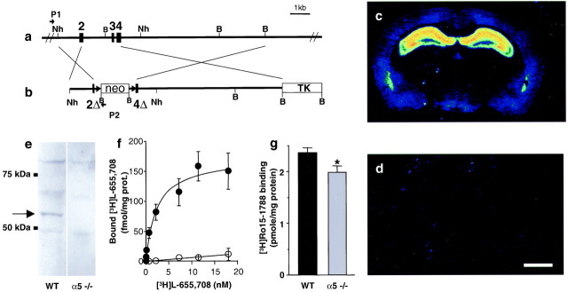
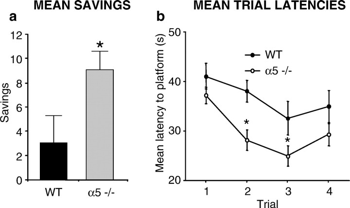
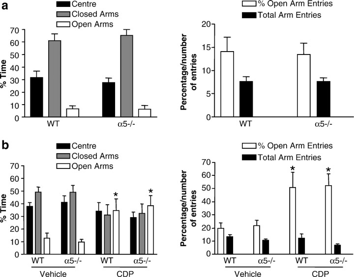
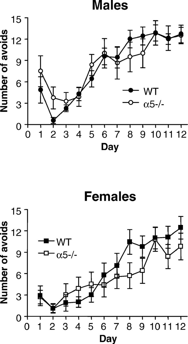
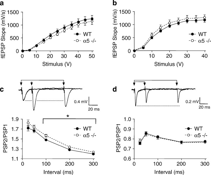
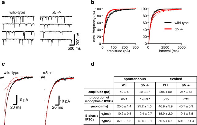
The alpha5 subunit of the GABA(A) receptor is localized mainly to the hippocampus of the mammalian brain. The significance of this rather distinct localization and the function of alpha5-containing GABA(A) receptors has been explored by targeted disruption of the alpha5 gene in mice. The alpha5 -/- mice showed a significantly improved performance in a water maze model of spatial learning, whereas the performance in non-hippocampal-dependent learning and in anxiety tasks were unaltered in comparison with wild-type controls. In the CA1 region of hippocampal brain slices from alpha5 -/- mice, the amplitude of the IPSCs was decreased, and paired-pulse facilitation of field EPSP (fEPSP) amplitudes was enhanced. These data suggest that alpha5-containing GABA(A) receptors play a key role in cognitive processes by controlling a component of synaptic transmission in the CA1 region of the hippocampus.


Figures

Fig. 1.
Fig. 1.
Generation and validation of α5 −/− mice.a, b, Schematic representation of the WT α5 allele of the GABAA receptor and the targeting vector, respectively.2, 3,4, Exons 2, 3, and 4; 2Δ,4Δ, partial deletion of exons 2 and 4; neo, neomycin resistance gene;TK, thymidine kinase gene; Nh,NheI restriction site; B,BamHI restriction site; P1,P2, PCR primers. c–g, Pharmacological and biochemical characterization of α5-deficient mice. c, d, Color-coded autoradiograms for [3H]L-655,708 binding to mouse brain sections reveals binding to the hippocampus in the WT (c) and absence of signal in the α5 −/− mice (d). Scale bar, 1 mm. e, Western blot shows the absence of a specific α5 subunit band at 55 kDa in membranes from α5 −/− hippocampus. f, Saturation experiment demonstrating the absence of high-affinity [3H]L-655,708 binding sites in α5 −/− mice (open circles) compared with WT mice (closed circles). g, Total number of [3H] BZ sites labeled by [3H]Ro15–1788 is reduced (−16%) in α5 −/− mice compared with WT controls.
Fig. 2.
Fig. 2.
Enhanced performance of α5 −/− mice in the matching-to-place version of the water maze test. Eighteen WT and 20 α5 −/− mice were used in this test. a, The difference in time taken between trial 1 and trial 2 (savings) to find the hidden platform over the 10 d testing period is shown. α5 −/− mice made significantly higher savings compared with WT mice.b, α5 −/− mice were significantly quicker (*) at finding the hidden platform for both trial 2 and trial 3.
Fig. 3.
Fig. 3.
Normal performance of the α5 −/− mice in the elevated plus maze. a, Data shown are mean (±SEM) values for percentage time spent in (left panel) and number of entries made to (right panel) different areas of the elevated plus-maze by WT and α5 −/− mice (n = 20–23). There were no significant differences between WT and α5 −/− mice on any of the measures. b, Data shown are mean (± SEM) values for percentage time spent in (left panel) and number of entries made to (right panel) different areas of the elevated plus-maze by 10.0 mg/kg CDP or vehicle-treated WT and α5 −/− mice (n = 13–20). CDP had a significant anxiolytic-like effect of the same magnitude in both WT and α5 −/− mice (*). This is indicated by the increase in the percentage of time spent in (p < 0.01) and percentage of entries made to (p < 0.01) the open arms of the plus-maze in comparison with vehicle-treated WT and α5 −/− mice.
Fig. 4.
Fig. 4.
Normal performance of the α5 −/− mice in the non-hippocampal-dependent two-way active avoidance. Data shown are mean (± SEM) avoid responses recorded on each of 12 d by untreated male (top) and female (bottom) wild-type (WT) and GABAA receptor α5 knock-out (α5 −/−) mice (n = 7–10). There was no significant difference between WT and α5 −/− mice but a significant effect of sex (p < 0.05).
Fig. 5.
Fig. 5.
Synaptic strength in hippocampal brain slices of α5 −/− mice compared with its effect on paired-pulse facilitation in CA1 region and paired-pulse depression in the dentate gyrus. In all figures the filled circles represent data from age-matched WT mice, and open circles are from α5 −/− mice. No significant difference was found in the maximal fEPSP amplitude in the CA1 region. a, Forty-six slices from 16 WT controls and 48 slices from 16 α5 −/− mice or dentate gyrus.b, Forty-one slices from 16 WT controls and 42 slices from 16 α5 −/− mice. In the CA1 region the amplitude of fEPSPs was significantly enhanced during paired-pulse stimulus intervals of 100–300 msec. c, Filled circles, 46 slices from 16 WT controls; open circles, 48 slices from 16 α5 −/− mice. In comparison, paired-pulse depression of fEPSPs in the dentate gyrus remained unaffected. d, Filled circles, 41 slices from 16 WT controls; open circles, 42 slices from 16 α5 −/− mice.
Fig. 6.
Fig. 6.
Long-term potentiation in CA1 region of hippocampal brain slices from α5 −/− mice. LTP was induced by theta burst (4 stimuli at 100 Hz repeated 10 times every 200 msec). The amount of LTP was not different between WT and α5 −/− mice (filled circles, 32 slices from 8 WT mice;open circles, 38 slices from α5 −/− mice) at 1 hour after the theta burst (p = 0.18;F(1,68) = 1.82).
Fig. 7.
Fig. 7.
Characteristics of IPSCs recorded from CA1 pyramidal neurons in hippocampal slices in vitroprepared from WT and α5 −/− mice. a, Examples of recordings of membrane current from CA1 neurons in WT and α5 −/− mice at a holding potential of −60 mV. The regular downward deflections are sIPSCs. b, Cumulative frequency distributions of the sIPSC amplitude and interevent interval in WT (black;n = 15 slices) and α5 −/− mice (red;n = 13 slices), showing a clear reduction in IPSC amplitude but no change in IPSC frequency in the α5 −/− mice. c, Representative sIPSCs recorded from CA1 pyramidal neurons in hippocampal brain slices from WT and α5 −/− mice. In the examples shown, the decay of the sIPSC recorded from the WT mouse was best fitted to a double-exponential function, whereas the decay of the sIPSC recorded from the α5 −/− mouse was best fitted to a monoexponential function (decay fits superimposed in red).d, Summary of the mean data of spontaneous and evoked IPSC mean peak amplitude and decay kinetics from WT and α5 −/− mice.

References

    1. Abeliovich A, Paylor R, Chen C, Kim JJ, Wehner JM, Tonegawa S. PKC gamma mutant mice exhibit mild deficits in spatial and contextual learning. Cell. 1993;75:1263–1271. - PubMed
    1. Barnard EA, Skolnick P, Olson RW, Möhler H, Sieghart W, Biggio G, Braestrup C, Bateson AN, Langer SZ. International Union of Pharmacology. XV. Subtypes of γ-aminobutyric acidA receptors: classification on the basis of subunit structure and receptor function. Pharmacol Rev. 1998;50:291–313. - PubMed
    1. Brickley SG, Revilla V, Cull-Candy SG, Wisden W, Farrant M. Adaptive regulation of neuronal excitability by a voltage-independent K+ conductance. Nature. 2001;409:88–92. - PubMed
    1. Crestani F, Lorez M, Baer K, Essrich C, Benke D, Laurent JP, Belzung C, Fritschy J-M, Luscher B, Möhler H. Decreased GABAA receptor clustering results in enhanced anxiety and a bias for threat cues. Nat Neurosci. 1999;2:833–839. - PubMed
    1. Culiat CT, Stubbs L, Woychik RP, Russell LB, Johnson DK, Rinchik EM. Deficiency of the β3 subunit of the type A γ-aminobutyric acid receptor causes cleft palate in mice. Nat Genet. 1995;11:344–346. - PubMed