. 2002 Jul 1;22(13):5769-76.
doi: 10.1523/JNEUROSCI.22-13-05769.2002.

mu-Opioid receptors: Ligand-dependent activation of potassium conductance, desensitization, and internalization

Affiliations

mu-Opioid receptors: Ligand-dependent activation of potassium conductance, desensitization, and internalization

Veronica A Alvarez et al. J Neurosci. .

Abstract

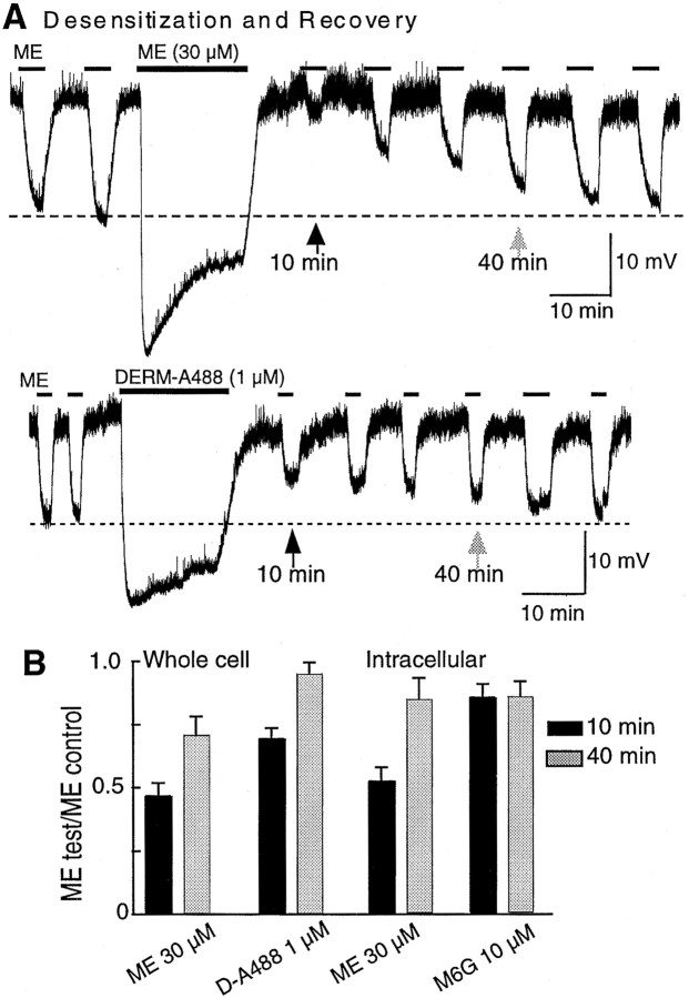
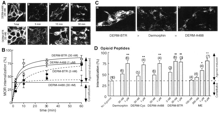
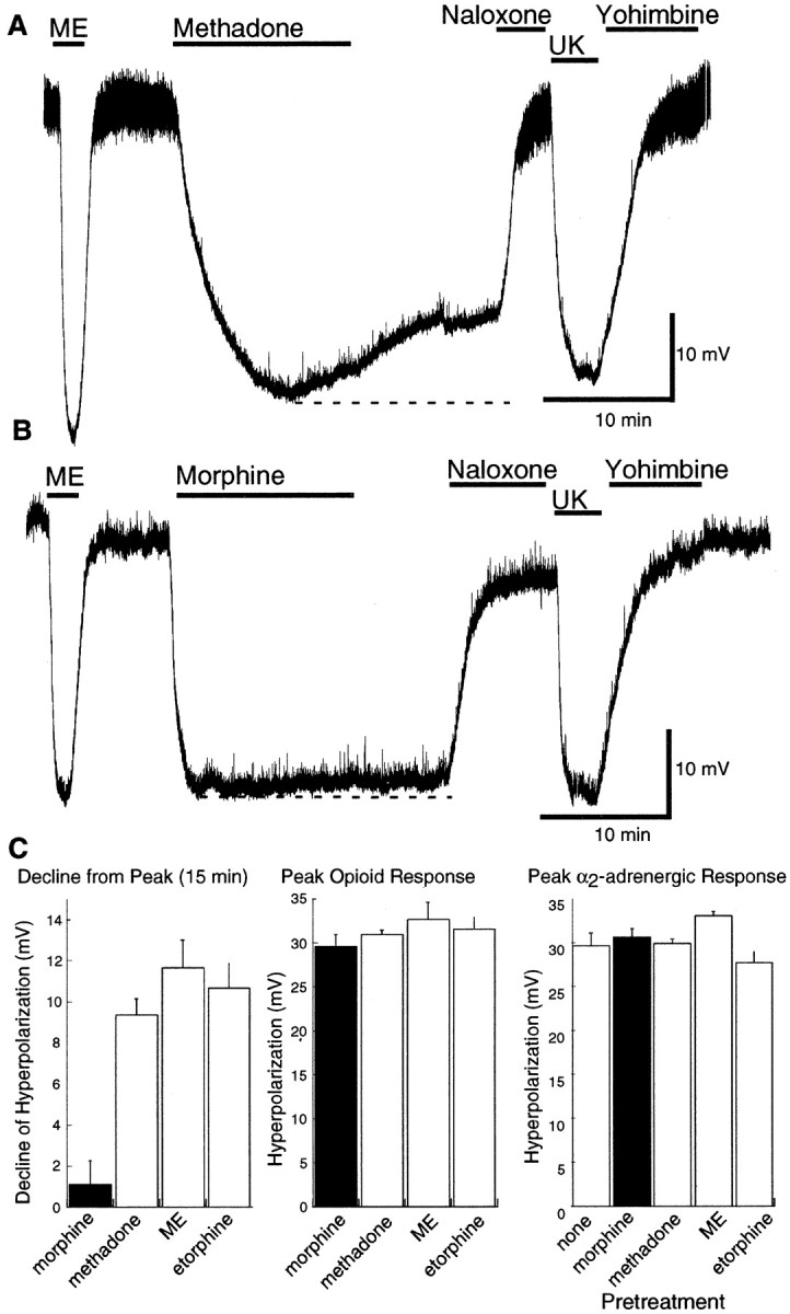
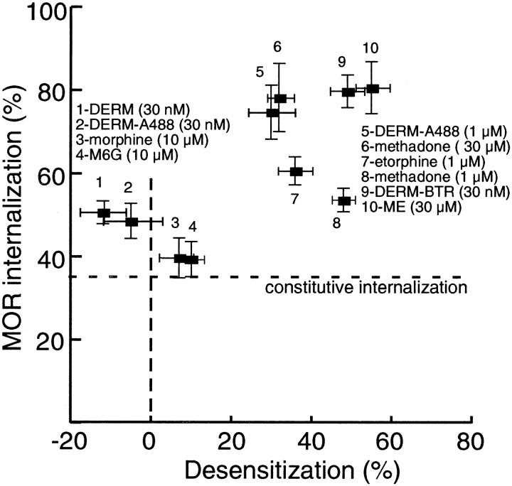
micro-Opioid receptor (MOR) desensitization and endocytosis have been implicated in tolerance and dependence to opioids. The efficiency of each process is known to be agonist dependent; however, it is not known what determines the relative efficiency of various agonists at either process. In the present study, homologous MOR desensitization in locus ceruleus (LC) neurons and MOR internalization in HEK293 cells were examined using a series of agonists. The results show that the rank order of this series of agonists was different when comparing the magnitude of hyperpolarization and the ability to cause desensitization in LC neurons. Endocytosis of MOR was also examined in HEK293 cells using the same agonists. The relative ability to cause endocytosis in HEK293 cells correlated with the degree of desensitization in LC cells. This strong correlation suggests that the two processes are closely linked. The results also suggest that agonist efficacy is not necessarily a predictor of the ability to cause MOR desensitization or endocytosis. Identification and characterization of the biophysical properties of agonists that favor desensitization and internalization of receptors will lead to a better understanding of opioid signaling.


Figures

Fig. 1.
Fig. 1.
Protocols used in recordings from LC neurons to measure desensitization. A, An EC50concentration of ME (300 nm) was used as a test before (1) and after (2) treating the tissue with a maximal concentration of agonist. The decrease in amplitude of the hyperpolarization immediately after washout was taken as a measure of desensitization. Over time, the amplitude of the hyperpolarization in response to the EC50 test returned toward control values. B, The hyperpolarization induced by a maximal concentration of ME (10 μm) was tested before (1) and during (2) the continuous superfusion with an EC50 concentration of opioid agonists. The decline in the peak hyperpolarization (3) was taken as a sign of desensitization. The hyperpolarization induced by the EC50 concentration of agonist was reversed with naloxone (1 μm). The hyperpolarization induced by a maximal concentration of UK14304 (3 μm) was determined for each experiment. C, A maximal concentration of opioid agonist was applied, and the difference between the peak hyperpolarization (1) and the amplitude of the hyperpolarization after 15 min (2) was taken as a sign of desensitization. In each experiment, naloxone was used to reverse the opioid-induced hyperpolarization, and the hyperpolarization induced by UK14304 was determined.
Fig. 2.
Fig. 2.
Induction of and recovery from desensitization. In this and other figures, the recordings are of membrane potential. In some recordings, the presence of spontaneous oscillations in membrane potential resulted in “noise” in the trace. The oscillations and thus the noise was abolished in the presence of opioid agonists.A, An EC50 concentration of ME (300 nm) was tested every 5 min before and after the application of a maximal concentration of ME (30 μm; top trace) and DERM-A488 (1 μm; bottom trace; applied by pressure ejection). The amplitude of the EC50 MOR response was significantly decreased after the desensitizing treatment with ME (top trace) but only slightly reduced after DERM-A488 (bottom trace). Desensitization recovered over 30–40 min. B, Summarized data from desensitization experiments done with whole-cell (left) and intracellular (right) recordings. The decrease in amplitude of the hyperpolarization after desensitization is presented as a fraction of the amplitude of the initial response to ME (300 nm). Black barsare the ratio obtained after 10 min application, and gray bars are the ratio after 40 min.
Fig. 3.
Fig. 3.
DERM-BTR induced desensitization in LC.A, Examples of experiments done with the low-dose protocol using DERM-A488 (30 nm; top) and DERM-BTR (30 nm; bottom). The maximum hyperpolarization induced by ME (10 μm) was not changed during treatment with DERM-A488, whereas the maximum hyperpolarization was reduced in the presence of DERM-BTR. B, Summarized data showing the amplitude of the ME-induced hyperpolarization during the treatment of slices with dermorphin, DERM-A488, and DERM-BTR (n = 5 for each experiment). The amplitude is plotted as a fraction of that observed during the first application of ME in the presence of the low concentration of each dermorphin analog.C, A summary of the acute effects of dermorphin, DERM-A488, and DERM-BTR, all applied at 30 nm(left). Right side indicates that the maximal hyperpolarization induced by DERM-A488 (1 μm; applied by pressure ejection) and DERM-BTR (1 μm; applied by pressure ejection) is the same as that induced by ME (10 μm).
Fig. 4.
Fig. 4.
Endocytosis of flag-tagged MOR in HEK293 cells by opioid peptide agonists. A, Examples of experiments examining the endocytosis induced by DERM-A488 (top) and DERM-BTR (bottom). The far left images(a, e) show total receptor binding using anti-flag antibodies. In b–d and f–h, the cells were incubated with DERM-A488 and DERM-BTR for the period indicated. At each time point, the anti-flag antibody remaining on the plasma membrane was stripped off so that only internalized label remained. The results show more internalization during treatment with DERM-BTR. B, Summarized data showing the time course of internalization induced by different concentrations of DERM-A488 and DERM-BTR. C, Examples of the maximal internalization caused by DERM-BTR, dermorphin, and DERM-A488 all applied at 30 nm for 30 min. D, Summary of many experiments with the dermorphin analogs and ME after a 30 min incubation period at 37°C. Each of the opioid peptides caused internalization that was dependent on the concentration. Also shown is constitutive internalization, which is the amount of receptor that was internalized in 30 min in the absence of any opioid agonist (No Agonist). ∗p < 0.05; ∗∗p < 0.001; one-way ANOVA; Tukey's multiple comparison test.
Fig. 5.
Fig. 5.
Methadone but not morphine caused endocytosis of flag-tagged MOR in HEK293 cells. A, Representative images from endocytosis experiments. Fluorescence stains internalized receptor after 30 min exposure to 30 μm methadone (left), morphine (middle), and M6G (right). B, Summarized data from several endocytosis experiments testing exposure to alkaloid opioids for 30 min at 37°C. Normorphine, methadone, and etorphine caused internalization that was dependent on the concentration. Morphine and M6G did not cause MOR endocytosis above the constitutive level (dotted lines), even at high concentrations. ∗∗p < 0.05; one-way ANOVA; Tukey's multiple comparison test.
Fig. 6.
Fig. 6.
Methadone-induced desensitization using the low-dose protocol. A, Examples of experiments using the low-dose protocol with morphine and methadone. The amplitude of the ME-induced hyperpolarization was not changed during treatment with morphine (3 μm; top trace) but decreased progressively during superfusion with methadone (3 μm;bottom trace). B, Summarized data showing the decline in the ME-induced hyperpolarization in the presence of methadone and the lack of any change in the presence of morphine (3 μm), M6G (3 μm), and UK14304 (100 nm).
Fig. 7.
Fig. 7.
Methadone-induced desensitization using the high-dose protocol. A, An example of the desensitization caused by a high concentration of methadone (30 μm). The peak hyperpolarization caused by methadone was approximately the same as the hyperpolarization induced by ME (1 μm) and UK14304 (3 μm). The decline in the hyperpolarization during the 15 min application was taken as a sign of desensitization.B, An example of the same experiment using a high concentration of morphine (30 μm). There is little or no decline in the hyperpolarization induced by morphine. C,Left, Summarized data showing the amplitude of the decline in hyperpolarization during a 15 min application of several agonists. This decline was ∼10 mV for methadone, ME, and etorphine but <2 mV for morphine. Middle shows the peak amplitude of the hyperpolarization caused by each agonist. Rightshows the amplitude of the hyperpolarization induced by UK14304 (3 μm) before (none) and after treatment with each of the indicated opioid agonists. There is no sign of heterologous desensitization induced by any of the opioid agonists.
Fig. 8.
Fig. 8.
A comparison of MOR endocytosis and desensitization. Summarized data from all endocytosis and desensitization experiments. The percentage of the total MOR internalized by a given concentration of agonist (taken from Figs. 4,5) was plotted as a function of amount of desensitization achieved when tested in LC neurons (expressed as percentage of the maximal opioid hyperpolarization). A significant correlation was found (nonparametric Spearman's test; r = 0.77; two-tailedp = 0.01269). The lowest possible value on they-axis (35%) is the level of constitutive endocytosis.

References

    1. Arttamangkul S, Alvarez-Maubecin V, Thomas G, Williams JT, Grandy DK. Binding and internalization of fluorescent opioid peptide conjugates in living cells. Mol Pharmacol. 2000;58:1570–1580. - PubMed
    1. Ferguson SS. Evolving concepts in G protein-coupled receptor endocytosis: the role in receptor desensitization and signaling. Pharmacol Rev. 2001;53:1–24. - PubMed
    1. Finn AK, Whistler JL. Endocytosis of the mu opioid receptor reduces tolerance and a cellular hallmark of opiate withdrawal. Neuron. 2001;32:829–839. - PubMed
    1. Fiorillo CD, Williams JT. Opioid desensitization: interactions with G-protein-coupled receptors in the locus coeruleus. J Neurosci. 1996;16:1479–1485. - PMC - PubMed
    1. Gomes BA, Shen J, Stafford K, Patel M, Yoburn BC. μ-Opioid receptor down-regulation and tolerance are not equally dependent upon G-protein signaling. Pharmacol Biochem Behav. 2002;72:273–278. - PubMed