. 2002 Aug;76(15):7385-97.
doi: 10.1128/jvi.76.15.7385-7397.2002.

RNA editing in hepatitis delta virus genotype III requires a branched double-hairpin RNA structure

Affiliations

RNA editing in hepatitis delta virus genotype III requires a branched double-hairpin RNA structure

John L Casey. J Virol. 2002 Aug.

Abstract

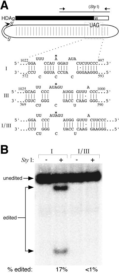
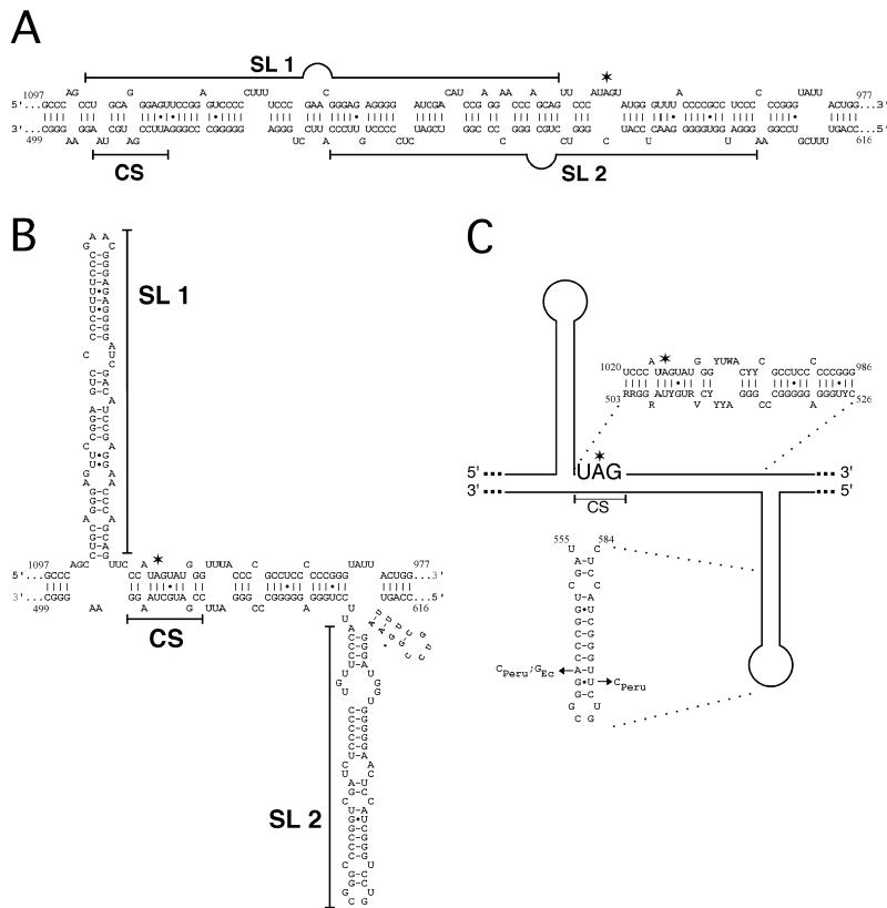
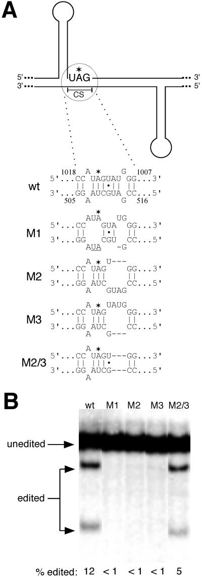
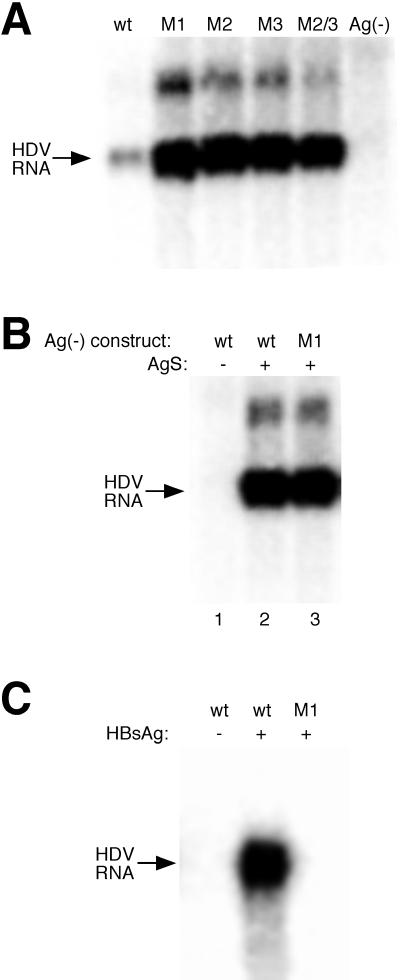
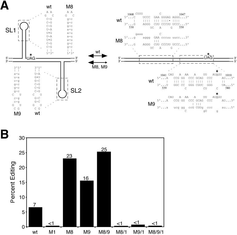
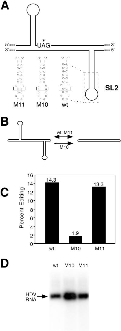
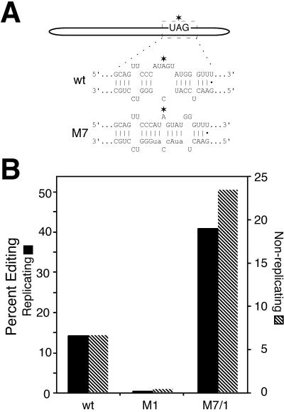
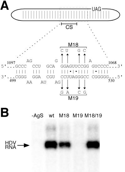
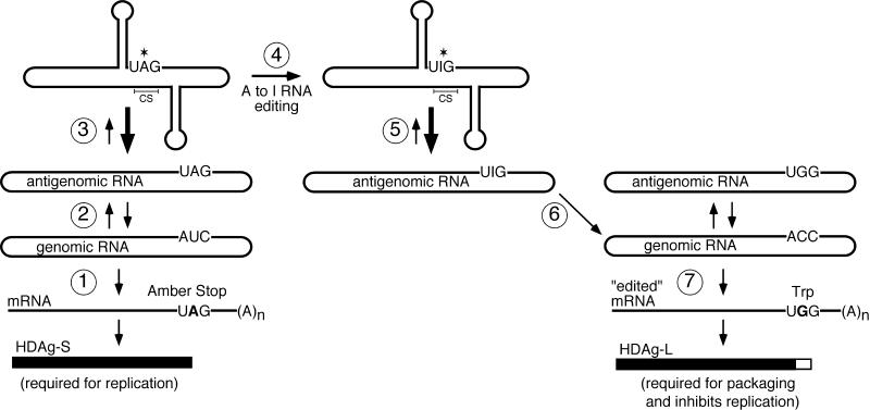
RNA editing at the amber/W site plays a central role in the replication scheme of hepatitis delta virus (HDV), allowing the virus to produce two functionally distinct forms of the sole viral protein, hepatitis delta antigen (HDAg), from the same open reading frame. Editing is carried out by a cellular activity known as ADAR (adenosine deaminase), which acts on RNA substrates that are at least partially double stranded. In HDV genotype I, editing requires a highly conserved base-paired structure that occurs within the context of the unbranched rod structure characteristic of HDV RNA. This base-paired structure is disrupted in the unbranched rod of HDV genotype III, which is the most distantly related of the three known HDV genotypes and is associated with the most severe disease. Here I show that RNA editing in HDV genotype III requires a branched double-hairpin structure that deviates substantially from the unbranched rod structure, involving the rearrangement of nearly 80 bp. The structure includes a UNCG RNA tetraloop, a highly stable structural motif frequently involved in the folding of large RNAs such as rRNA. The double-hairpin structure is required for editing, and hence for virion formation, but not for HDV RNA replication, which requires the unbranched rod structure. HDV genotype III thus relies on a dynamic conformational switch between the two different RNA structures: the unbranched rod characteristic of HDV RNA and a branched double-hairpin structure that is required for RNA editing. The different mechanisms of editing in genotypes I and III underscore their functional differences and may be related to pathogenic differences as well.


Figures

FIG. 1.
FIG. 1.
The unbranched rod structure is not involved in HDV genotype III amber/W site RNA editing. (A) Predicted secondary structure around the amber/W site in HDV genotype I, genotype III, and genotype I/III chimera RNAs. Oval, HDV RNA antigenome; light vertical bars, base pairs that form the unbranched rod structure; the curved arrow indicates the orientation of the HDV antigenome. The coding region for HDAg-S is indicated by the solid bar above the oval; the UAG (amber) stop codon is indicated. Open bar, C-terminal extension of HDAg-L, in which the UAG terminator has been changed to a Trp (W) codon. The horizontal line above the HDAg bar indicates the PCR product used to analyze editing. Leftward- and rightward-pointing arrows above this line represent PCR primers. The location ofthe StyI restriction site created as a result of amber/W site editing is indicated. Predicted RNA secondary structures (29, 54) surrounding the amber/W site, indicated by asterisks, are shown for genotype I (I), genotype III (III), and the genotype I/III chimera in construct pHDV•I/IIIΔ1-NR (I/III). Vertical lines, Watson-Crick base pairs; dots, GU pairs. (B) RT-PCR-restriction digestion assay of amber/W editing in nonreplicating HDV RNA isolated from Huh-7 cells transfected with pHDV•IΔ1-NR (lanes I) or pHDV•I/IIIΔ1-NR (lanes I/III). RT-PCR products, either uncut (lanes −) or cut with StyI (lanes +), were analyzed by polyacrylamide gel electrophoresis. PCR products derived from edited RNAs contain a StyI restriction site (CCATGG) that is not present in products derived from unedited RNAs (CCATAG). Because only a fraction of HDV RNAs are edited, PCR products are a mixture of those derived from edited and unedited RNAs. Restriction digestion products from edited and unedited RNAs are indicated by arrows. Percent editing is determined by dividing the sum of the two bands due to editing by the sum of the edited and unedited bands.
FIG. 2.
FIG. 2.
Predicted secondary structures of a segment of the HDV genotype III RNA antigenome that includes the amber/W site. Structures were obtained with the mfold program (29, 54). (A) Unbranched rod structure. Sequences that would be involved in the stem-loops and CS of a double-hairpin structure are indicated. The amber/W site is indicated by an asterisk. (B) Double-hairpin branched structure including two stem-loops, SL1 and SL2, and a segment complementary to the amber/W site (CS). (C) Sequence conservation in the double-hairpin structure among six independent genotype III isolates. Variable positions in the central base-paired region are indicated by standard IUPAC (International Union of Pure and Applied Chemistry) abbreviations (R = A or G; Y = C or U; W = A or U; V = A, C, or G). Positions marked CPeru or GEc are unique to the Peru-1 (10) or Ecuador (28) isolate, respectively.
FIG. 3.
FIG. 3.
The CS in the central base-paired region of the double-hairpin structure is required for editing in replicating HDV genotype III RNA. (A) Schematic of the predicted double-hairpin RNA structure and predicted effects of mutations on base pairing between the CS and the amber/W site. The amber/W site is indicated by an asterisk.Base substitutions are underlined, and deletions are represented by dashes. (B) RT-PCR restriction digestion assay of HDV amber/W RNA editing in Huh-7 cells 9 days after transfection with replicating genotype III constructs containing the indicated site-directed mutations.
FIG. 4.
FIG. 4.
Effects of genotype III amber/W editing mutations on HDV RNA replication and virion production. (A) Northern blot analysis of HDV genomic RNA from cells 9 days after transfection with replication-competent genotype III constructs that produce antigenomic RNA containing the indicated site-directed mutations. Lane Ag(−), RNA from cells transfected with pHDV•III(+)Ag(−), a genotype III HDV construct defective in HDAg production. wt, wild type. (B) Northern blot analysis of HDV genomic RNA from cells 6 days after cotransfection with pHDAg-S•III and either wild-type or M1 mutant pHDV•III(+)Ag(−) constructs (lanes 2 and 3) or with pHDV•III(+)Ag(−) alone (lane 1). (C) Northern blot analysis of HDV virion RNA isolated from the culture medium of cells 9 days after transfection with pHDV•III(+) containing either wild-type sequence or the M1 mutation (M1). Cells were also cotransfected with either a hepatitis B surface antigen expression construct (HBsAg, +), or an equivalent amount of a negative plasmid control, (HBsAg, −).
FIG. 5.
FIG. 5.
(A) Schematic diagram of the double-hairpin and unbranched rod structures, indicating locations of mutations M8 and M9, and the effects of these mutations on base pairing in these structures. Mutations are indicated by lowercase letters. The horizontal double arrow indicates that the two RNA structures may normally interconvert; the shortened rightward-pointing arrow for M8 and M9 indicates that these mutations likely shift the distribution between these two structures to the left by destabilizing the unbranched rod structure. wt, wild type. (B) Amber/W editing in nonreplicating HDV RNAs isolated from Huh-7 cells 3 days after transfection with the nonreplicating RNA expression construct pHDV•III-NR containing the indicated mutations. Mutant constructs denoted by two or more numbers separated by a slash (e.g., M8/9, M8/9/1) contain all indicated mutations (e.g., M8/9 contains both M8 and M9; M8/9/1 contains M8, M9, and M1). Editing was determined by RT-PCR and StyI digestion as in Fig. 1 and 3.
FIG.6.
FIG.6.
A UNCG tetraloop in SL2 is essential for maximal amber/W site editing but is not required for type III RNA replication. (A) Schematic of the double-hairpin structure showing base pairing in SL2 and the effects of the M10 and M11 mutations (lowercase letters). (B) Potential effects of the M10 and M11 mutations on the relative abundance of the double-hairpin and unbranched rod structures. The shortened leftward-pointing arrow for M10 indicates that this mutation likely shifts the distribution between these two structures to the right, while the M11 mutation should restore the distribution to near-wild-type levels. wt, wild type. (C) Amber/W editing in HDV RNAs isolated from Huh-7 cells 9 days after transfection of Huh-7 cells with pHDV•III(+) constructs containing the indicated mutations. Editing was determined by RT-PCR and StyI digestion as for Fig. 1 and 3. (D) Northern blot of HDV genomic RNAs from panel C. Arrow indicates HDV genomic RNA.
FIG. 7.
FIG. 7.
Creation of a functional amber/W editing site in the unbranched rod structure of HDV genotype III RNA. (A) Schematic of the unbranched rod of HDV genotype III. In the M7 mutation, base pairing in the vicinity of the amber/W site is modified to resemble the structure required for amber/W site editing in HDV genotype I. Mutated positions are indicated by lowercase letters. (B) Amber/W editing in replicating (solid bars) and nonreplicating (diagonally striped bars) HDV RNAs from cells transfected with either wild-type (wt), M1 mutant, or M7/1 combination mutant expression constructs. Replicating RNAs were harvested 9 days after transfection with pHDV•III(+) or the indicated mutant derivative; nonreplicating RNAs were harvested 3 days after transfection with pHDV•III-NR and the indicated mutant derivative.
FIG. 8.
FIG. 8.
The unbranched rod structure is required for replication of HDV genotype III RNA. (A) Schematic of unbranched rod structure of HDV antigenomic RNA. Mutants M18 and M19 each introduce four transversion mutations that are predicted to destabilize a 10-bp segment that includes the CS. The introduced mutations are complementary to each other; base pairing is predicted to be restored by combining M18 with M19. (B) Northern blot analysis of HDV genomic RNA from cells 6 days after transfection with pHDAg-S•III and pHDV•III(+)Ag(−) constructs containing either the wild-type (wt) or the indicated mutant sequence. Lane −AgS, transfection with pCI and pHDV•III(+)Ag(−).
FIG. 9.
FIG. 9.
Schematic of HDV genotype III replication cycle, including secondary-structure changes required for HDV genotype III amber/W RNA editing and RNA replication. Steps: 1, transcription of HDV genomic RNA to produce mRNA that is translated to yield HDAg-S, which is required for RNA replication; 2, rolling-circle replication of genomic and antigenomic RNA; 3, conformational change from unbranched rod to double-hairpin structure; 4, amber/W RNA editing by host RNA ADAR; 5, conformational change from double-hairpin to unbranched rod structure; 6, replication of antigenome to genome (I created by editing is copied to C); 7, transcription from genomic RNA with C at position 1012 (in the genome) yields mRNA with UGG (tryptophan) codon; production of HDAg-L, which is required for virion production and inhibits RNA replication.

Similar articles

Cited by

References

    1. Abdelkafi, M., N. Leulliot, V. Baumruk, L. Bednarova, P. Y. Turpin, A. Namane, C. Gouyette, T. Huynh-Dinh, and M. Ghomi. 1998. Structural features of the UCCG and UGCG tetraloops in very short hairpins as evidenced by optical spectroscopy. Biochemistry 37:7878-7884. - PubMed
    1. Baumstark, T., and D. Riesner. 1995. Only one of four possible secondary structures of the central conserved region of potato spindle tuber viroid is a substrate for processing in a potato nuclear extract. Nucleic Acids Res. 23:4246-4254. - PMC - PubMed
    1. Baumstark, T., A. R. Schroder, and D. Riesner. 1997. Viroid processing: switch from cleavage to ligation is driven by a change from a tetraloop to a loop E conformation. EMBO J. 16:599-610. - PMC - PubMed
    1. Been, M. D., and G. S. Wickham. 1997. Self-cleaving ribozymes of hepatitis delta virus RNA. Eur. J. Biochem. 247:741-753. - PubMed
    1. Bergmann, K. F., and J. L. Gerin. 1986. Antigens of hepatitis delta virus in the liver and serum of humans and animals. J. Infect. Dis. 154:702-706. - PubMed

Publication types

MeSH terms