. 2002 Aug;76(15):7453-9.
doi: 10.1128/jvi.76.15.7453-7459.2002.

Hepatitis C virus subgenomic replicons induce endoplasmic reticulum stress activating an intracellular signaling pathway

Affiliations

Hepatitis C virus subgenomic replicons induce endoplasmic reticulum stress activating an intracellular signaling pathway

Keith D Tardif et al. J Virol. 2002 Aug.

Abstract

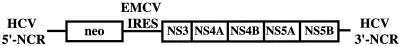
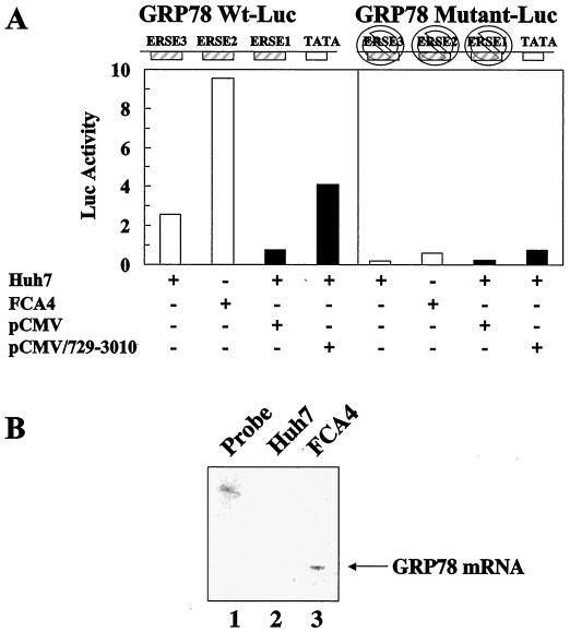
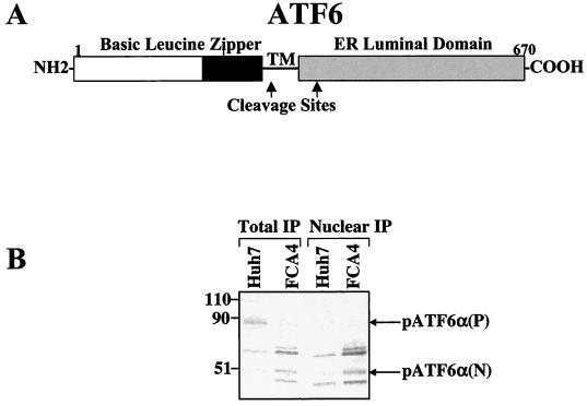
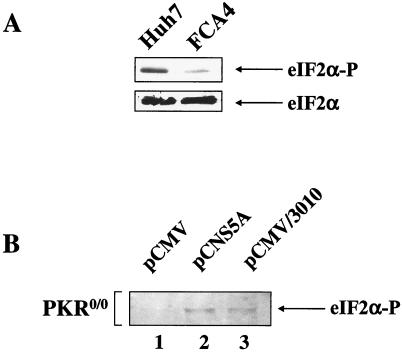
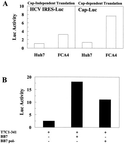
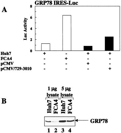
Hepatitis C virus (HCV) replicates from a ribonucleoprotein (RNP) complex that is associated with the endoplasmic reticulum (ER) membrane. The replication activities of the HCV subgenomic replicon are shown here to induce ER stress. In response to this stress, cells expressing HCV replicons induce the unfolded protein response (UPR), an ER-to-nucleus intracellular signaling pathway. The UPR is initiated by the proteolytic cleavage of a transmembrane protein, ATF6. The resulting cytoplasmic protein fragment of ATF6 functions as a transcription factor in the nucleus and activates selective genes required for an ER stress response. ATF6 activation leads to increased transcriptional levels of GRP78, an ER luminal chaperone protein. However, the overall level of GRP78 protein is decreased. While ER stress is also known to affect translational attenuation, cells expressing HCV replicons have lower levels of phosphorylation of the alpha subunit of eukaryotic initiation factor 2. Interestingly, cap-independent internal ribosome entry site-mediated translation directed by the 5' noncoding region of HCV and GRP78 is activated in cells expressing HCV replicons. These studies provide insight into the effects of HCV replication on intracellular events and the mechanisms underlying liver pathogenesis.


Figures

FIG. 1.
FIG. 1.
Structure of the HCV subgenomic replicon, I377/NS3-3′. The structure of the replicon is composed of the HCV 5′-NCR (solid line), the neo gene (open box), encephalomyocarditis virus IRES (solid line), coding region of NS3 to NS5B (open box), and the HCV 3′-NCR. The neomycin resistance marker allows the establishment of a transformed cell line that stably expresses replicons in RNA form.
FIG. 2.
FIG. 2.
GRP78 gene expression is stimulated by HCV replicons and HCV nonstructural proteins. (A) HCV replicons and HCV nonstructural proteins activate GRP78 transcription in an ERSE-dependent manner. Huh7 and FCA4 cells were transfected with GRP78 wild-type (left panel) or mutant (right panel) luciferase reporter plasmids (white). Huh7 cells were also cotransfected with pCMV/729-3010 or pCMV and wild-type (left panel) or mutant (right panel) GRP78-Luc (black). The GRP78 luciferase reporter has three ERSE sequences. The mutant GRP78-Luc has all three of these ERSE sequences eliminated. (B) RNase protection assay indicating an increase in GRP78 mRNA steady-state levels in FCA4 cells. RNA was extracted from Huh7 and FCA4 cells and analyzed using a probe specific to GRP78. Lane 1, probe alone; lane 2, Huh7 GRP78 mRNA analysis; lane 3, FCA4 GRP78 mRNA analysis.
FIG. 3.
FIG. 3.
HCV replicons activate ATF6. (A) Structure of ATF6. ATF6 is an ER transmembrane protein. Upon ER stress, the cytoplasmic N-terminal domain of ATF6 is cleaved at the cytoplasmic face of the ER membrane. The cleaved cytoplasmic domain is translocated to the nucleus where it activates the transcription of ER chaperone genes containing the ERSE. TM is the region of the protein that spans the ER membrane. The ATF6 cleavage sites are indicated by arrows (45). (B) HCV replicons induce the cleavage of endogenous p90ATF6 to p50ATF6. Total and nuclear Huh7 and FCA4 cell lysates were immunoprecipitated with anti-ATF6 antibody. Molecular masses (in kilodaltons) are indicated at left.
FIG. 4.
FIG. 4.
Effects of HCV replicons or HCV nonstructural proteins on eIF-2α phosphorylation levels in cells with or without PKR. (A) FCA4 cells with HCV replicons have lower levels of eIF-2α phosphorylation. Top panel, Huh7 and FCA4 cell lysates were analyzed by Western blot analysis using anti-eIF-2α serum specific to phosphoserine-51. Bottom panel, Western blot analysis of Huh7 and FCA4 cell lysates with antiserum reactive with all forms of eIF-2α. (B) NS5A stimulates eIF-2α phosphorylation in PKR knockout cells (PKR0/0). Western blot analysis of eIF-2α phosphoprotein from PKR0/0 transfected with pCMV (lane 1), pCNS5A (lane 2), and pCMV/3010 (lane 3) expression vectors.
FIG. 5.
FIG. 5.
HCV IRES-directed translation and cap-dependent translation increases in cells with HCV replicons. (A) HCV replicons induce HCV IRES translation and cap-dependent translation. Huh7 and FCA4 cells were transfected with a dual luciferase reporter plasmid containing the HCV IRES linked to Renilla luciferase (left panel). Cap-dependent translation activity was measured in Huh7 and FCA4 cells from firefly luciferase activity using the same dual luciferase reporter plasmid (right panel). (B) Wild-type HCV replicons enhance HCV IRES activity more than replication defective HCV replicons. Huh7 cells were transfected with an HCV IRES RNA luciferase reporter alone, T7C1-341, and a wild-type HCV replicon (BB7) or a replication-defective mutant HCV replicon (BB7 pol) was transfected into Huh7 cells along with the luciferase reporter, T7C1-341.
FIG. 6.
FIG. 6.
HCV replicons induce GRP78 translation while lowering overall levels of GRP78 protein. (A) HCV replicons and HCV nonstructural proteins activate GRP78 IRES-mediated translation. Huh7 and FCA4 cells were transfected with an GRP78 IRES-linked luciferase reporter plasmid (white). Huh7 cells were also cotransfected with GRP78 IRES-Luc and pCMV/729-3019 or pCMV (black). (B) GRP78 protein levels in Huh7 and FCA4 cells. Shown is a Western blot analysis of Huh7 and FCA4 cell lysates with anti-GRP78 antibody. Lanes 1 and 2, 1 μg of Huh7 and FCA4 cell lysates, respectively; lanes 3 and 4, 5 μg of Huh7 and FCA4 cell lysates, respectively.

Similar articles

Cited by

References

    1. Abraham, V., D. F. Stojkl, P. I. Duncan, N. Methot, T. Ishii, M. Dube, B. C. Vanderhyden, H. L. Atkins, D. A. Gray, M. W. McBurney, A. E. Koromilas, E. G. Brown, N. Sonenberg, and J. C. Bell. 1999. Characterization of transgenic mice with targeted disruption of the catalytic domain of the double-stranded RNA-dependent protein kinase, PKR. J. Biol. Chem. 274:5953-5962. - PubMed
    1. Bartenschlager, R., and V. Lohmann. 2000. Replication of hepatitis C virus. J. Gen. Virol. 81:1631-1648. - PubMed
    1. Berlanga, J. J., J. Santoyo, and C. De Haro. 1999. Characterization of a mammalian homologue of the GCN2 eukaryotic initiation factor 2 alpha kinase. Eur. J. Biochem. 265:754-762. - PubMed
    1. Berry, M. J., G. S. Knutson, S. R. Lasky, S. M. Munemitsu, and C. E. Samuel. 1985. Mechanism of interferon from untreated and interferon-treated mouse fibroblasts. J. Biol. Chem. 270:11240-11247. - PubMed
    1. Bertolotti, A., Y. Zhang, L. M. Hendershot, H. P. Harding, and D. Ron. 2000. Dynamic interaction of BiP and ER stress transducers in the unfolded-protein response. Nat. Cell Biol. 2:326-332. - PubMed

Publication types

MeSH terms

LinkOut - more resources