. 2002 Aug;76(15):7495-505.
doi: 10.1128/jvi.76.15.7495-7505.2002.

Analysis of deletion mutants indicates that the 2A polypeptide of hepatitis A virus participates in virion morphogenesis

Affiliations

Analysis of deletion mutants indicates that the 2A polypeptide of hepatitis A virus participates in virion morphogenesis

Lisette Cohen et al. J Virol. 2002 Aug.

Abstract

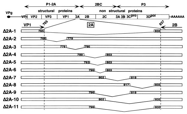
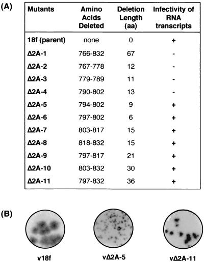
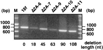
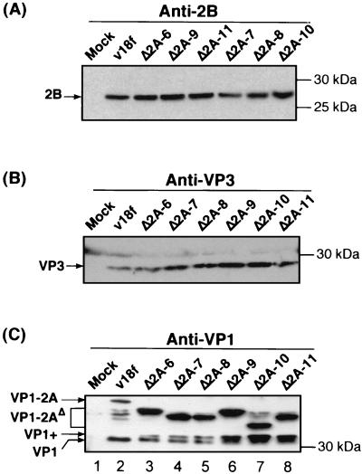
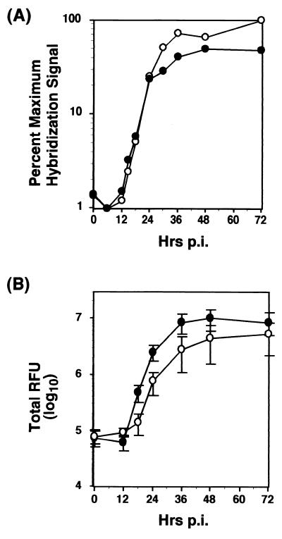
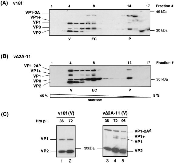
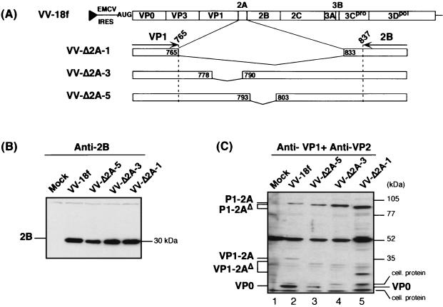
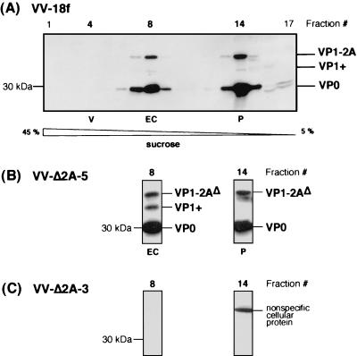
Unlike all other picornaviruses, the primary cleavage of the hepatitis A virus (HAV) polyprotein occurs at the 2A/2B junction and is carried out by the only proteinase encoded by the virus, 3C(pro). The resulting P1-2A capsid protein precursor is subsequently cleaved by 3C(pro) to generate VP0, VP3, and VP1-2A, which associate as pentamers. An unidentified cellular proteinase acting at the VP1/2A junction releases the mature capsid protein VP1 from VP1-2A later in the morphogenesis process. Although these aspects of polyprotein processing are well characterized, the function of 2A is unknown. To study its role in the viral life cycle, we assessed the infectivity of synthetic, genome-length RNAs containing 11 different in-frame deletions in the 2A region. Deletions in the N-terminal 40% of 2A abolished infectivity, whereas deletions in the C-terminal 60% resulted in viruses with a small-focus replication phenotype. C-terminal deletions in 2A had no effect on RNA replication kinetics under one-step growth conditions, nor did they have an effect on capsid protein synthesis and 3C(pro)-mediated processing. However, C-terminal deletions in 2A altered the VP1/2A cleavage, resulting in accumulation of uncleaved VP1-2A precursor in virions and possibly accounting for a delay in the appearance of infectious particles with these mutants, as well as a fourfold decrease in specific infectivity of the virus particles. When the capsid proteins were expressed from recombinant vaccinia viruses, the N-terminal part of 2A was required for efficient cleavage of the P1-2A precursor by 3C(pro) and assembly of structural precursors into pentamers. These data indicate that the N-terminal domain of 2A must be present as a C-terminal extension of P1 for folding of the capsid protein precursor to allow efficient 3C(pro)-mediated cleavages and to promote pentamer assembly, after which cleavage at the VP1/2A junction releases the mature VP1 protein, a process that appears to be necessary to produce highly infectious particles.


Figures

FIG. 1.
FIG. 1.
Schematic representation of HAV cDNAs with deletions in 2A. The C terminus of VP1 and N terminus of 2B are indicated at the top of the 2A enlargement, with the amino acid residues within the HAV polyprotein numbered according to p5′P2P3-18f cDNA (38). Internal 2A deletions are indicated by broken lines. The positions of amino acids framing each deletion within the HAV polyprotein are indicated in boxes.
FIG. 2.
FIG. 2.
Infectivity of full-length HAV RNA transcripts with deletions in 2A. (A) For each genome-length RNA transcript, the extent of the deletion is indicated, both by the positions within the polyprotein of the corresponding deleted amino acids and by the number of amino acid residues deleted. The infectivity of deleted transcripts is indicated (+, infectious; −, lethal), as determined by RIFA in BS-C-1 cells inoculated with FRhK-4 cell lysates harvested 2 weeks posttransfection of RNA. (B) RIFA autoradiograms for selected progeny viruses.
FIG. 3.
FIG. 3.
Retention of engineered deletions in the genomes of viable 2A mutants. The 2A coding region of the mutant viral RNA isolated from passage 1 virions was amplified by RT-PCR, and the resulting fragments were run on a 1% agarose gel. Positions of the DNA molecular weight markers are indicated on both sides of the gel. The length of the corresponding 2A deletion, in terms of the number of nucleotides deleted, is indicated for each mutant below the lanes.
FIG. 4.
FIG. 4.
Polyprotein synthesis and 3Cpro processing of 2A deletion mutants. FRhK-4 cells were mock infected (Mock) or infected with the parent virus (v18f) or the indicated mutant at an MOI of 1 RFU/cell. Proteins from cytoplasmic extracts prepared at 72 hpi were separated by SDS-10% PAGE and identified by immunoblot with anti-2B (A), anti-VP3 (B), or anti-VP1 (C) antibodies. HAV polypeptides and molecular mass markers, are indicated on the left and right sides of each panel, respectively.
FIG. 5.
FIG. 5.
Single-cycle growth and RNA replication kinetics of vΔ2A-11 variant. FRhK-4 cells were infected at an MOI of 4 RFU/cell, either with the virus carrying the most-extensive deletion (vΔ2A-11 [open circles]) or with the parent virus (v18f [closed circles]). (A) Cytoplasmic RNAs were harvested at the indicated times p.i., denatured, immobilized onto a nylon membrane, and hybridized to an HAV-specific, 32P-labeled, negative-strand riboprobe. HAV-specific positive-strand RNA was quantified by PhosphorImager analysis. Results of one experiment are represented and expressed as the relative percentage of the maximum hybridization signal obtained in the experiment. (B) Virus present in the supernatant and within the cells was harvested at the indicated times p.i. and titrated by RIFA. Each point represents the mean of three independent experiments. Error bars indicate the standard deviation.
FIG. 6.
FIG. 6.
vΔ2A-11 capsid protein assembly. FRhK-4 cells were infected with the parent virus (v18f) (A) or the vΔ2A-11 mutant (B) at an MOI of 1 RFU/cell. Morphogenesis intermediates present in cytoplasmic cell extracts harvested at 72 hpi were fractionated on 5 to 45% sucrose gradients. Each fraction was loaded on an SDS-3.5 M urea-12% polyacrylamide gel. The HAV polypeptide content of each fraction was determined by immunoblotting using a mixture of anti-VP1 and anti-VP2 antibodies. (C) FRhK-4 cells were infected with the parent virus (v18f) or the vΔ2A-11 mutant at an MOI of 4 RFU/cell, and cytoplasmic cell extracts were harvested at the indicated times p.i. (36, 72, or 96 hpi). Morphogenesis intermediates were separated on a 5 to 45% sucrose gradient, and the polypeptide content of fraction 4, containing virus particles (V), was analyzed by immunoblotting with anti-VP1 and anti-VP2 antibodies. HAV polypeptides, as well as molecular mass markers, are indicated on each side of the panels.
FIG. 7.
FIG. 7.
Effect of N-terminal 2A deletions on 3Cpro-mediated processing of the capsid protein precursors expressed by recombinant VVs. (A) Schematic showing HAV polyprotein expressed by the recombinant VV containing the open reading frame, as well as the 3′ nontranslated region, of the v18f genome downstream of the EMCV internal ribosome entry site and the T7 RNA polymerase promoter (black triangle). Enlargements are shown for the 2A deletions introduced into the polyproteins of the indicated mutants. The positions within the HAV polyprotein of amino acids framing each deletion are indicated in boxes. FRhK-4 cells were infected with vTF7-3 (Mock) or coinfected with vTF7-3 and either the parent virus (VV-18f) or the indicated mutant virus (VV-Δ2A-1, -3, or -5), each at an MOI of 5 PFU/cell. HAV proteins from cytoplasmic extracts prepared at 20 hpi were separated by SDS-10% PAGE and identified by immunoblotting using anti-2B antibodies (B) or a mixture of anti-VP1 and anti-VP2 antibodies (C). HAV polypeptides, as well as molecular mass markers, are indicated on each side of the panels.
FIG. 8.
FIG. 8.
Effect of N-terminal 2A deletions on assembly of capsid proteins expressed by recombinant VVs. FRhK-4 cells were coinfected with vTF7-3 and virus expressing either the parent polyprotein, VV-18f (A), or the indicated mutant polyproteins, VV-Δ2A-5 (B) or VV-Δ2A-3 (C), each at an MOI of 5 PFU/cell. Morphogenesis intermediates were fractionated on 5 to 45% sucrose gradients and loaded on an SDS-3.5 M urea-12% polyacrylamide gel, and their polypeptide composition was determined by immunoblot analysis using a mixture of anti-VP1 and anti-VP2 antibodies. All fractions are shown for the gradient containing the parent VV-18f (A), whereas only fractions in which pentamers (fraction 14 [P]) and empty capsids (fraction 8 [EC]) were expected are shown for the mutant viruses (B and C).

References

    1. Ansardi, D. C., and C. D. Morrow. 1993. Poliovirus capsid proteins derived from P1 precursors with glutamine-valine cleavage sites have defects in assembly and RNA encapsidation. J. Virol. 67:7284-7297. - PMC - PubMed
    1. Beard, M. R., L. Cohen, S. M. Lemon, and A. Martin. 2001. Characterization of recombinant hepatitis A virus genomes containing exogenous sequences at the 2A/2B junction. J. Virol. 75:1414-1426. - PMC - PubMed
    1. Bishop, N. E., and D. A. Anderson. 2000. Uncoating kinetics of hepatitis A virus virions and provirions. J. Virol. 74:3423-3426. - PMC - PubMed
    1. Boege, U., and D. G. Scraba. 1989. Mengo virus maturation is accompanied by C-terminal modification of capsid protein VP1. Virology 168:409-412. - PubMed
    1. Borovec, S. V., and D. A. Anderson. 1993. Synthesis and assembly of hepatitis A virus-specific proteins in BS-C-1 cells. J. Virol. 67:3095-3102. - PMC - PubMed

Publication types

MeSH terms

LinkOut - more resources