. 2002 Aug;76(15):7777-89.
doi: 10.1128/jvi.76.15.7777-7789.2002.

Similarities in the induction of post-Golgi vesicles by the vaccinia virus F13L protein and phospholipase D

Affiliations

Similarities in the induction of post-Golgi vesicles by the vaccinia virus F13L protein and phospholipase D

Matloob Husain et al. J Virol. 2002 Aug.

Abstract

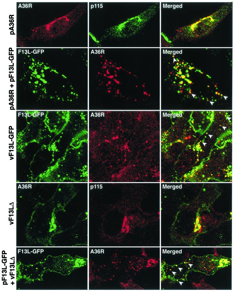
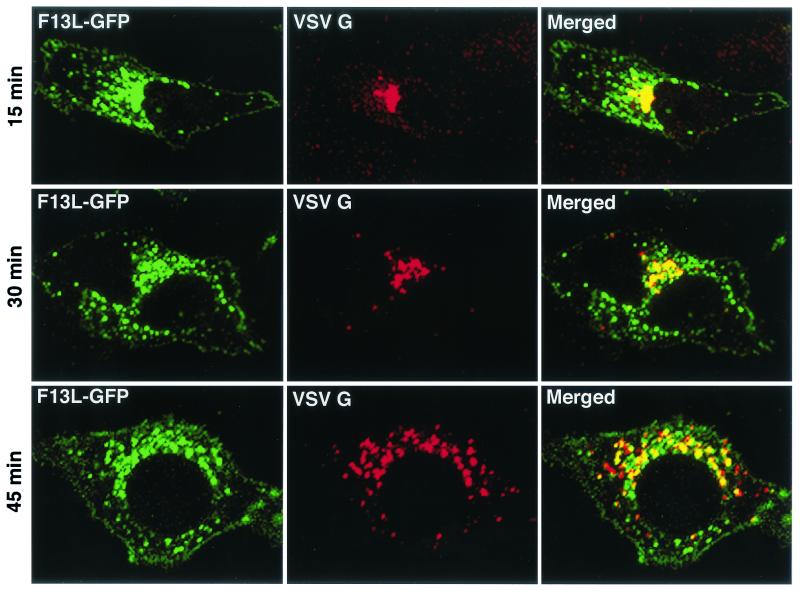
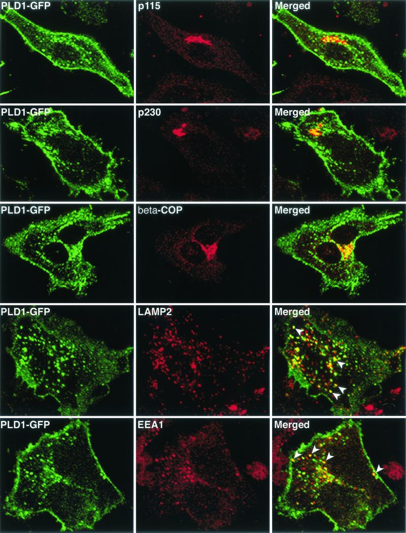
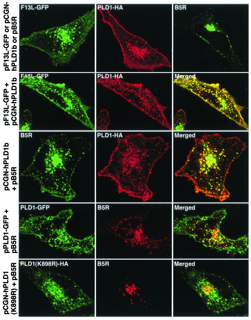
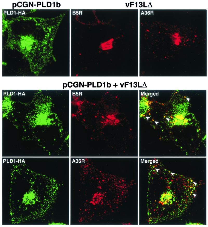
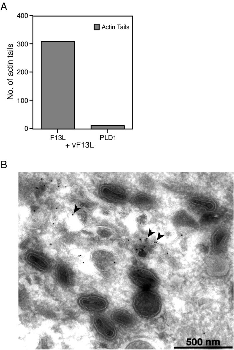
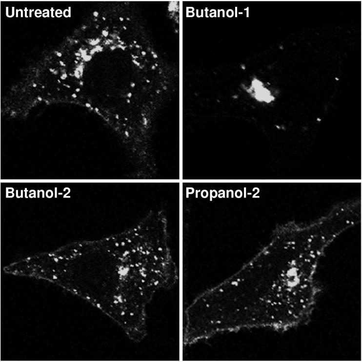
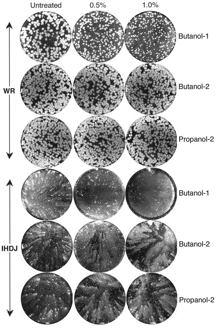
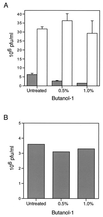
Intracellular mature vaccinia virions are wrapped by cisternae, derived from virus-modified trans-Golgi or endosomal membranes, and then transported via microtubules to the cell periphery. Two viral proteins, encoded by the F13L and B5R open reading frames, are essential for the membrane-wrapping step. Previous transfection studies indicated that F13L induces the formation of post-Golgi vesicles that incorporate the B5R protein and that this activity depends on an intact F13L phospholipase motif. Here we show that the F13L protein has a general effect on the trafficking of integral membrane proteins from the Golgi apparatus, as both the vaccinia virus A36R protein and the vesicular stomatitis virus G protein also colocalized with the F13L protein in vesicles. In addition, increased expression of cellular phospholipase D, which has a similar phospholipase motif as, but little amino acid sequence identity with, F13L, induced post-Golgi vesicles that contained B5R and A36R proteins. Butanol-1, which prevents the formation of phosphatidic acid by phospholipase D and specifically inhibits phospholipase D-mediated vesicle formation, also inhibited F13L-induced vesicle formation, whereas secondary and tertiary alcohols had no effect. Moreover, inhibition of phospholipase activity by butanol-1 also reduced plaque size and decreased the formation of extracellular vaccinia virus without affecting the yield of intracellular mature virus. Phospholipase D, however, could not complement a vaccinia virus F13L deletion mutant, indicating that F13L has additional virus-specific properties. Taken together, these data support an important role for F13L in inducing the formation of vesicle precursors of the vaccinia virus membrane via phospholipase activity or activation.


Figures

FIG. 1.
FIG. 1.
Effect of F13L-GFP on the intracellular localization of A36R in transfected or infected cells. HeLa cells were transfected or infected for 24 or 18 h, respectively, with the name of the plasmid or virus indicated at the left of each row. First row, cells transfected with pA36R were stained with anti-p115 MAb followed by indodicarbocyanine (Cy5)-conjugated anti-mouse immunoglobulin antibody and then with anti-A36R polyclonal antibody followed by Texas red-conjugated anti-rabbit immunoglobulin antibody. Second row, cells were cotransfected with pA36R and pF13L-GFP and stained for A36R as in the first row. Third row, cells infected with vF13L-GFP (1 PFU/cell) were stained with anti-A36R polyclonal antibody followed by tetramethyl rhodamine isothiocyanate-conjugated anti-rabbit immunoglobulin antibody. Fourth row, cells infected with vF13LΔ (1 PFU/cell) were stained with anti-p115 MAb followed by tetramethyl rhodamine isothiocyanate-conjugated anti-mouse immunoglobulin antibody and then stained with anti-A36R polyclonal antibody followed by Alexa 488-conjugated anti-rabbit immunoglobulin antibody. Fifth row, cells were transfected with plasmid pF13L-GFP; after 24 h the cells were infected with vF13LΔ as in the fourth row and stained with anti-A36R polyclonal antibody followed by tetramethyl rhodamine isothiocyanate-conjugated anti-rabbit immunoglobulin antibody. Cells were analyzed by confocal microscopy. Green, GFP or Alexa 488; red, Texas red or tetramethyl rhodamine isothiocyanate; yellow, overlap of green and red. White arrowheads show the colocalization of F13L-GFP with A36R in vesicular structures.
FIG. 2.
FIG. 2.
Time course of VSV G colocalization with F13L-GFP. HeLa cells were cotransfected with plasmids pF13L-GFP and pVSVGts045 and incubated at 39°C. After 24 h, the cells were shifted to 31°C and chased for the time indicated on the left of each row. Cells were fixed, permeabilized, stained with anti-VSV G MAb followed by rhodamine red-conjugated anti-mouse immunoglobulin antibody, and analyzed by confocal microscopy. Green, GFP; red, rhodamine red; yellow, overlap of green and red.
FIG. 3.
FIG. 3.
Colocalization of PLD1-GFP with cellular markers. HeLa cells were transfected with plasmid pPLD1-GFP for 24 h and then fixed, permeabilized, and stained with the indicated antibodies. Transfected HeLa cells were stained with mouse MAbs to p115, p230, LAMP2, or EEA1 or rabbit polyclonal anti-β-COP antibodies followed by rhodamine red-conjugated anti-mouse immunoglobulin antibody or tetramethyl rhodamine isothiocyanate-conjugated anti-rabbit immunoglobulin antibody, respectively. Cells were analyzed by confocal microscopy. Green, GFP; red, tetramethyl rhodamine isothiocyanate; yellow, overlap of green and red. White arrowheads indicate vesicles containing PLD1-GFP and endosomal marker LAMP2 or EEA1.
FIG.4.
FIG.4.
Colocalization of PLD1 with F13L-GFP and B5R in transfected cells. HeLa cells were transfected with plasmids indicated at the left of each row and incubated for 24 h. First row, cells transfected with pF13L-GFP alone (left), pCGN-hPLD1b alone (middle), or pB5R alone (right) were unstained or stained with mouse anti-HA MAb or rat anti-B5R MAb followed by rhodamine red-conjugated anti-mouse immunoglobulin antibody or fluorescein isothiocyanate-conjugated anti-rat immunoglobulin antibody. Second row, cells cotransfected with plasmid pF13L-GFP and pCGN-hPLD1b were stained with mouse anti-HA followed by rhodamine red-conjugated immunoglobulin antibody. Third row, cells cotransfected with plasmids pCGN-hPLD1b and pB5R were stained for HA and then for B5R as in the first row. Fourth and fifth rows, cells cotransfected with pPLD1-GFP and pB5R or pPLD1(K898R)-HA and pB5R, respectively, were stained for B5R or HA and examined by confocal microscopy as in the third row. Green, GFP or fluorescein isothiocyanate; red, rhodamine red; yellow, overlap of green and red.
FIG. 5.
FIG. 5.
Intracellular localization of B5R and A36R in vF13LΔ-infected cells expressing PLD1-HA. First row, HeLa cells were transfected with plasmid pCGN-PLD1b or infected with vF13LΔ. Second and third rows, transfected cells were infected with vF13LΔ and after 18 h were fixed, permeabilized, and stained with anti-HA MAb followed by Alexa 488-conjugated anti-mouse immunoglobulin antibody. Cells were then stained with either anti-B5R MAb or anti-A36R followed by tetramethyl rhodamine isothiocyanate-conjugated anti-rat or anti-rabbit immunoglobulin antibody, respectively. Stained cells were analyzed by confocal microscopy. Green, Alexa 488; red, tetramethyl rhodamine isothiocyanate; yellow, overlap of green and red. White arrowheads show the colocalization of PLD1 with B5R and A36R in vesicular structures.
FIG. 6.
FIG. 6.
Failure of PLD1 to complement an F13L deletion mutant. (A) Quantitation of actin tail formation in vF13LΔ-infected cells transfected with either pGF13L or pGPLD1-LR. HeLa cells were infected with vF13LΔ for 2 h and then transfected with plasmid pGF13L or pGPLD1-LR. At 24 h after transfection, the cells were fixed and permeabilized. Cells transfected with plasmid pGPLD1-LR were stained with anti-HA MAb followed by Alexa 488-conjugated anti-mouse immunoglobulin antibody. Plasmid pGF13L has GFP at the C terminus of F13L. Data shown are the averages of three separate experiments. (B) vPLD1-GFP does not produce IEV particles. A confluent monolayer of RK13 cells was infected with vPLD1-GFP. After 24 h, cells were fixed, cryosectioned, and probed with rabbit anti-GFP polyclonal antibodies followed by protein A conjugated to 10-nm colloidal gold particles. Arrowheads point to PLD1-GFP fusion protein on membranes close to IMV particles.
FIG. 7.
FIG. 7.
Effects of butanol-1, butanol-2, and propanol-2 on intracellular localization of F13L-GFP. HeLa cells were transfected with plasmid pF13L-GFP. After 4 h, 1% concentrations of the indicated alcohols were added to the media and the incubations were continued for a total of 24 h at 37°C. Cells were fixed and analyzed by confocal microscopy.
FIG. 8.
FIG. 8.
Effects of butanol-1, butanol-2, and propanol-2 on plaque size of vaccinia virus. BS-C-1 cells, in six-well tissue culture plates, were infected with 50 to 100 PFU of vaccinia virus strain WR or IHD-J/well. After 2 h of adsorption of IHD-J, the virus inocula were replaced with liquid media supplemented with 0.5 or 1.0% concentrations of the indicated alcohol and the incubation was continued for 2 days. For vaccinia virus WR, methylcellulose was included in the overlay and the cells were incubated for 3 days. Plaques were visualized by staining with crystal violet.
FIG. 9.
FIG. 9.
Effect of butanol-1 on the production of IMV and EEV particles. (A) BS-C-1 cells were infected with vaccinia virus strain IHD-J. After 2 h, the inoculum was replaced with medium supplemented with 0.5 or 1.0% butanol-1. After 48 h at 37°C, the medium and cells were harvested separately in similar volumes and analyzed by plaque assay. Plaque numbers are presented as the averages of three separate counts from two different experiments with standard deviations. Filled and unfilled bars refer to extracellular and cell-associated virus, respectively. (B) BS-C-1 cells were infected with vaccinia virus strain IHD-J, and after adsorption, inoculum was replaced with normal medium. After 48 h, the medium was collected; cleared by low-speed centrifugation; and incubated with 0, 0.5, or 1.0% butanol-1 at 37°C for 48 h. Infectious virus was quantified by plaque assay.

References

    1. Baek, S.-H., J.-Y. Kwak, S. H. Lee, T. Lee, S. H. Ryu, D. J. Uhlinger, and J. D. Lambeth. 1997. Lipase activities of p37, the major envelope protein of vaccinia virus. J. Biol. Chem. 272:32042-32049. - PubMed
    1. Bednarek, S. Y., L. Orici, and R. Schekman. 1996. Traffic COPS and the formation of vesicle coats. Trends Cell Biol. 6:468-473. - PubMed
    1. Blasco, R., and B. Moss. 1991. Extracellular vaccinia virus formation and cell-to-cell virus transmission are prevented by deletion of the gene encoding the 37,000-Dalton outer envelope protein. J. Virol. 65:5910-5920. - PMC - PubMed
    1. Blasco, R., and B. Moss. 1992. Role of cell-associated enveloped vaccinia virus in cell-to-cell spread. J. Virol. 66:4170-4179. - PMC - PubMed
    1. Brown, F. D., N. Thompson, K. M. Saqib, J. M. Clark, D. Powner, N. T. Thompson, R. Solari, and M. J. Wakelam. 1998. Phospholipase D1 localizes to secretory granules and lysosomes and is plasma-membrane translocated on cellular stimulation. Curr. Biol. 8:835-838. - PubMed

MeSH terms

LinkOut - more resources