

Oligomeric structure of the human immunodeficiency virus type 1 envelope protein on the virion surface
- PMID: 12097599
- PMCID: PMC136379
- DOI: 10.1128/jvi.76.15.7863-7867.2002
Oligomeric structure of the human immunodeficiency virus type 1 envelope protein on the virion surface
Abstract
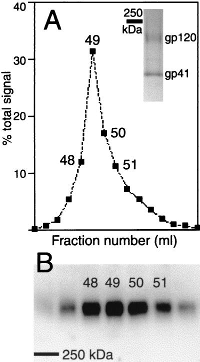
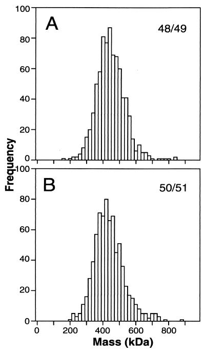
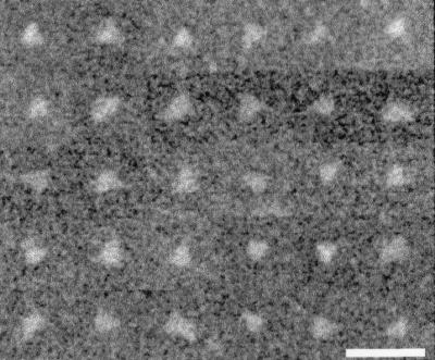
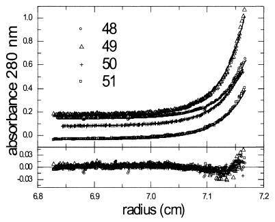
The envelope protein (Env) of human immunodeficiency virus type 1 forms homo-oligomers in the endoplasmic reticulum. The oligomeric structure of Env is maintained after cleavage in a Golgi compartment and transport to the surfaces of infected cells, where incorporation into budding virions takes place. Here, we use biophysical techniques to assess the oligomeric valency of virion-associated Env prior to fusion activation. Virion-associated Env oligomers were stabilized by chemical cross-linking prior to detergent extraction and were purified by immunoaffinity chromatography. Gel filtration revealed a single predominant oligomeric species, and sedimentation equilibrium analysis-derived mass values indicated a trimeric structure. Determination of the masses of individual Env molecules by scanning transmission electron microscopy demonstrated that virion-associated Env was trimeric, and a triangular morphology was observed in 20 to 30% of the molecules. These results, which firmly establish the oligomeric structure of human immunodeficiency virus virion-associated Env, parallel those of our previous analysis of the simian immunodeficiency virus Env.
Figures
References
-
- Abacioglu, Y. H., T. R. Fouts, J. D. Laman, E. Claassen, S. H. Pincus, J. P. Moore, C. A. Roby, R. Kamin-Lewis, and G. K. Lewis. 1994. Epitope mapping and topology of baculovirus-expressed HIV-1 gp160 determined with a panel of murine monoclonal antibodies. AIDS Res. Hum. Retrovir. 10:371-381. - PubMed
-
- Baker, K. A., R. E. Dutch, R. A. Lamb, and T. S. Jardetzky. 1999. Structural basis for paramyxovirus-mediated membrane fusion. Mol. Cell 3:309-319. - PubMed
-
- Borchers, C., and K. B. Tomer. 1999. Characterization of the noncovalent complex of human immunodeficiency virus glycoprotein 120 with its cellular receptor CD4 by matrix-assisted laser desorption/ionization mass spectrometry. Biochemistry 38:11734-11740. - PubMed
-
- Bullough, P. A., F. M. Hughson, J. J. Skehel, and D. C. Wiley. 1994. Structure of influenza hemagglutinin at the pH of membrane fusion. Nature 371:37-43. - PubMed
Publication types
MeSH terms
Substances
LinkOut - more resources
Full Text Sources
Other Literature Sources
