

doi: 10.1128/jvi.76.15.7890-7896.2002.
Kaposi's sarcoma-associated herpesvirus (human herpesvirus 8) contains two functional lytic origins of DNA replication
Affiliations
- PMID: 12097603
- PMCID: PMC136389
- DOI: 10.1128/jvi.76.15.7890-7896.2002
Item in Clipboard
Kaposi's sarcoma-associated herpesvirus (human herpesvirus 8) contains two functional lytic origins of DNA replication
J Virol.
2002 Aug.
Abstract
We used a transient-transfection replication assay to identify two functional copies of the human herpesvirus 8 (HHV8) lytic origin of DNA replication (oriLyt). BCLB-1 cells were transfected with HHV8 subgenomic fragments containing the putative lytic origin along with a plasmid expressing viral transactivator open reading frame (ORF) 50. The HHV8 left-end oriLyt (oriLyt-L) lies between ORFs K4.2 and K5 and is composed of a region encoding various transcription factor binding sites and an A+T-rich region and a G+C repeat region. The right-end oriLyt (oriLyt-R) maps between ORF 69 and vFLIP, a region similar to the RRV oriLyt, and is an inverted duplication of oriLyt-L.
Figures
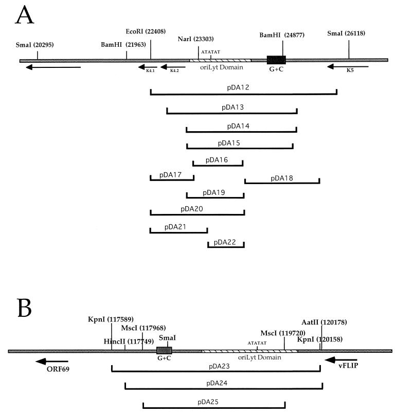
Schematic representation of HHV8 oriLyt regions and subclones used in the transient-transfection replication assay. Shown are the relative positions of ORFs. Hatched regions indicate the oriLyt domain sequences, and sold blocks indicate G+C repeat regions. Shown below are each oriLyt region and the locations of oriLyt subclones. Nucleotide coordinates for each subclone and other details are given in Table 1. (A) HHV8 oriLyt-L; (B) HHV8 oriLyt-R.
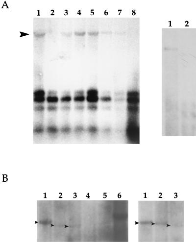
Identification of HHV8 oriLyt. (A, left side) Autoradiogram of a Southern blot of EcoRI- and DpnI-cleaved total cellular DNA from BCBL-1 cells transfected by electroporation with pDA12 with or without a plasmid expressing HHV8 ORF 50 (pEXP50). Blots were hybridized with pGEM7zf(−). The arrowhead indicates the replicated product. Lanes: 1, transfection of pDA12 plus pEXP50 (5 μg); 2, pDA12 plus pGEM7zf(−) (5 μg); 3, pDA12 plus pEXP50 (0.5 μg); 4, pDA12 plus pEXP50 (2 μg); 5, pDA12 plus pEXP50 (5 μg); 6, pDA12 plus TPA treatment for 4 days; 7, pDA12 plus TPA treatment for 2 days; 8, pDA12 plus pEXP50 (5 μg) incubated with PFA (300 μg/ml). (A, right side) Replication products are susceptible to cleavage by the restriction enzyme MboI. Cells were transfected with pDA12 and pEXP50 (5 μg), and DNA was extracted and treated with EcoRI and DpnI (lane 1) or EcoRI, DpnI, and MboI (lane 2). (B) Both the oriLyt domain and the G+C repeat regions are required for replication and identification of the OriLyt-R sequence. Transfection of HHV8 oriLyt-L and oriLyt-R subclones was done to identify essential DNA elements. All transfections contained 20 μg of oriLyt-containing plasmids plus 5 μg of pEXP50. Arrowheads indicate replicated plasmids. Refer to Table 1 and Fig. 1 for details of subclones. (B, left side) Transfection of oriLyt-L subclones. Lanes: 1, pDA13; 2, pDA14; 3, pDA15; 4, pDA16; 5, pDA18; 6, pDA12. (B, right side) Transfection of oriLyt-R subclones. Lanes: 1, pDA23; 2, pDA24; 3, pDA25.
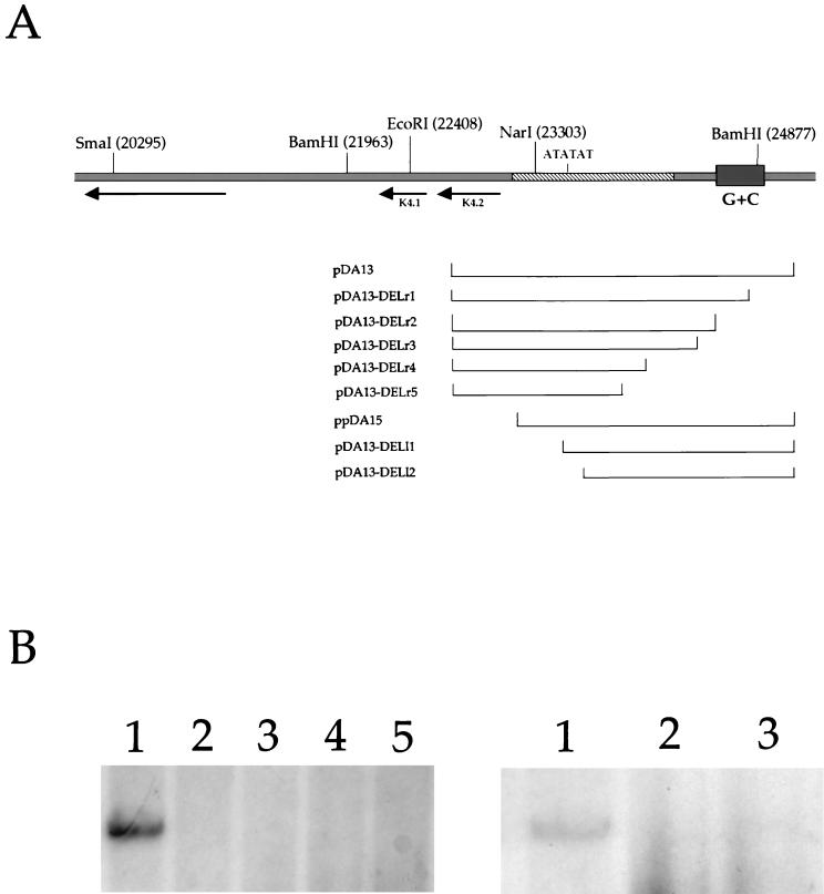
Mapping of the right- and left-hand boundaries of HHV8 oriLyt-R. (A) Exonuclease cleavage constructs used to define the 3′ and 5′ boundaries of the HHV8 oriLyt. Nucleotide coordinates for cleavage constructs are listed in Table 1. (B) 5′ and 3′ boundaries of the HHV8 oriLyt-R. BCBL-1 cells were transfected with a series of deletion constructs in which a DNA sequence from either the 5′ or the 3′ end of oriLyt subclone pDA13 was removed by exonuclease cleavage. An autoradiogram of a Southern blot of DNA from the transient-transfection replication assay is shown. (B, left side) 3′-end deletions. Lanes: 1, pDA13-DELr1; 2, pDA13-DELr2; 3, pDA13-DELr3; 4, pDA13-DELr4; 5, pDA13-DELr5. (B, right side) 5′-end deletions. Lanes: 1, pDA15; 2, pDA13-DELl1; 3, pDA13-DELl2.
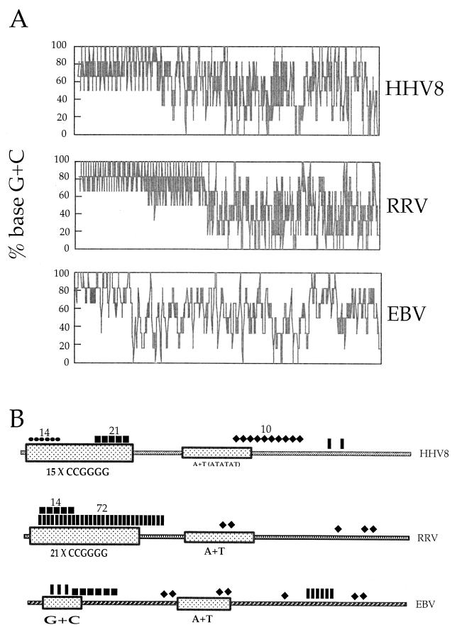
Sequence comparison of core oriLyt regions from HHV8, RRV, and EBV. (A) Comparison of the core oriLyt regions with respect to percent G+C content. (B) Identification of various transcription factor binding sites within HHV8, RRV, and EBV oriLyt regions. GCF, GC factor; JCV, pentanucleotide repeat sequence; bHLH, basic helix-loop-helix; AP2, activator protein 2.
References
-
- Birley, H. D., and T. F. Schultz. 1997. Kaposi's sarcoma and the new herpesvirus. J. Med. Microbiol. 46:433-435. - PubMed
-
- Cesarman, E., Y. Chang, P. S. Moore, J. W. Said, and D. M. Knowles. 1995. Kaposi's sarcoma-associated herpesvirus-like DNA sequences in AIDS-related body-cavity-based lymphomas. N. Engl. J. Med. 332:1186-1191. - PubMed
-
- Cesarman, E., P. S. Moore, P. H. Rao, G. Inghirami, D. M. Knowles, and Y. Chang. 1995. In vitro establishment and characterization of two acquired immunodeficiency syndrome-related lymphoma cell lines (BC-1 and BC-2) containing Kaposi's sarcoma-associated herpesvirus-like (KSHV) DNA sequences. Blood 86:2708-2714. - PubMed
-
- Chang, Y., E. Cesarman, M. S. Pessin, F. Lee, J. Culpepper, D. M. Knowles, and P. S. Moore. 1994. Identification of herpesvirus-like DNA sequences in AIDS-associated Kaposi's sarcoma. Science 266:1865-1869. - PubMed
Publication types
MeSH terms
Substances
Grants and funding
LinkOut - more resources
Full Text Sources
Research Materials
