. 2002 Jul 23;99(15):9840-5.
doi: 10.1073/pnas.152588599. Epub 2002 Jul 3.

Intercellular calcium signaling mediated by point-source burst release of ATP

Affiliations

Intercellular calcium signaling mediated by point-source burst release of ATP

Gregory Arcuino et al. Proc Natl Acad Sci U S A. .

Abstract

Calcium signaling, manifested as intercellular waves of rising cytosolic calcium, is, in many cell types, the result of calcium-induced secretion of ATP and activation of purinergic receptors. The mechanism by which ATP is released has hitherto not been established. Here, we show by real-time bioluminescence imaging that ATP efflux is not uniform across a field of cells but is restricted to brief, abrupt point-source bursts. The ATP bursts emanate from single cells and manifest the transient opening of nonselective membrane channels, which admits fluorescent indicators of < or = 1.5 kDa. These observations challenge the existence of regenerative ATP release, because ATP efflux is finite and restricted to a point source. Transient efflux of cytosolic nucleotides from a subset of cells may represent a conserved pathway for coordinating local activity of electrically nonexcitable cells, because identical patterns of ATP release were identified in human astrocytes, endothelial cells, and bronchial epithelial cells.


Figures

Figure 1
Figure 1
Astrocytic calcium signaling is associated with a transient increase in membrane permeability and uptake of propidium. (a) Sequence of images (in s) demonstrating the time course of propidium uptake in the cell that triggers a calcium wave. Propidium uptake is superimposed upon a time series of confocal images displaying relative increases in fluo-3 emission (ΔF). A single cell located in the center of the calcium wave displays uptake of propidium (red arrowhead). (b) Histogram summarizing the fraction of cells in the center of calcium waves (n = 21) and cells exhibiting calcium oscillations (n = 221) with uptake of propidium (see supporting information, which is published on the PNAS web site, www.pnas.org).
Figure 2
Figure 2
ATP is released as point-source bursts. ATP release was detected as light emissions produced by ATP-triggered luciferase breakdown of luciferin. A series of bioluminescence images visualize the stochastic distribution of ATP burst releases from C6-Cx43 cells during a 3-h recording period. Bioluminescence signal is displayed in arbitrary units (see supporting information).
Figure 3
Figure 3
ATP release is associated with a transient increase in membrane permeability and uptake of fluorescent indicators. (A Upper) Bioluminescence signal from three ATP burst events recorded from rat astrocytes during a 5-min period; calcein AM (1 μM) loading in the same field as b is shown in Lower. Cells in the center of ATP burst events consistently admitted propidium, indicative of a transient increase in membrane permeability. The ability of propidium+ cells (red arrows) to load and retain calcein after ATP release suggests that membrane permeability was only increased transiently in cells releasing ATP. (b) Human astrocytes. An ATP burst event (Top) from a single propidium+ cell (Middle, red arrow). The ATP-releasing cell loaded and retained calcein to a similar extent as surrounding cells (Bottom, red arrow). (c) ATP efflux (Left) and corresponding fields of propidium/calcein uptake in cultures of human bronchial epithelial cells, human umbilical vein endothelial cells (HUVEC), and C6 glioma cells overexpressing connexin 43 (C6-Cx43 cells). Bioluminescence signal is displayed in arbitrary units and scaled as in Fig. 1. (d) Comparison of the frequency of ATP burst events in C6-mock cells (Cx deficient), C6-Cx43, and C6-Cx32 cells. The frequency of ATP release is mapped as burst per min per 100 cells.
Figure 4
Figure 4
Propidium influx is associated with a transient activation of an inward current in C6-Cx43 cells. (a) Propidium uptake and whole-cell recordings in a responding cell. A single cell was patched in the voltage-clamp configuration with a holding potential of −60 mV (right pipette). Another pipette (left) was used to apply a Ca2+-free solution locally (10 mM EGTA, 2 mM propidium, 10-s pulse). (Upper) Sequence of images collected at indicated time displays cellular uptake of propidium evoked by local application of the Ca2+-free solution. (Lower) Relative changes in fluorescence as a function of time (F) and whole-cell recordings (I) in the same cell. Solid bars indicate that the Ca2+-free solution was applied. Uptake of propidium is detected as a relative increase in fluorescence after the first stimulation (ΔF). The first challenge with a Ca2+-free solution activated an inward current, whereas neither inward current nor further propidium uptake was elicited when the stimulation was repeated. (b) Sequence of images illustrating a nonresponding cell. The cell excluded propidium and did not increase membrane permeability during repeated exposures to a Ca2+-free solution.
Figure 5
Figure 5
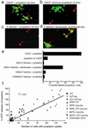
The increase in membrane permeability is transient and allows influx of both cations and anions below 1.5 kDa. (a) CDCF (green arrowhead, 445 Da) and propidium (red arrowhead, 562 Da) were both taken up by the same cells during a 6-min exposure to a Ca2+-free solution containing both indicators. CDCF is gap junction permeable, and clusters of cells with CDCF surround single cells with uptake of both indicators (red and green arrowheads). (b) Separate populations of cells took up the two indicators if first exposed to a Ca2+-free solution containing CDCF (3 min) followed by a second exposure to a Ca2+-free solution containing propidium (3 min). (c) Fluorescein dextran (>1.5 kDa) was excluded by cells with propidium uptake (6 min). (d) Dextranase digestion of the 3-kDa fluorescein-dextran conjugate resulted in uptake of the indicator in propidium+ cells (yellow arrowhead; 6 min). (e) Histogram summarizing the percentage of propidium+ cells with uptake of CDCF or fluorescein dextran. (f) ATP release as a function of the total number of cells with propidium uptake in the same culture. ATP release is plotted as a percentage of baseline (unstimulated) release from the same culture. Each culture contained a total of ≈16,000 cells.
Figure 6
Figure 6
The effect of a cell-free lane on mechanically induced astrocytic calcium signaling. (a) Sequential expansion of a calcium wave (1–44 s). The calcium wave was evoked on the left side of a cell-free lane (*). Relative increase in Ca2+i (ΔF) was superimposed upon a fluo-3 image collected before stimulation. (b) Grid used for quantification of ΔF. The grid was positioned with its center on top of the stimulation site and consisted of concentric rings each with a width of 50 μm. ΔF was calculated in these radially defined sections (a, b, c, d) and compared with matching regions on the opposite side of the cell-free lane (A, B, C, D). (c) Histogram comparing ΔF in matched sections at increasing distance from the point of stimulation (n = 44–67). “All regions” indicates ΔF (mean ± SEM) in a total of 242 matched regions analyzed. No difference between regions on either side of the cell-free lane was evident.

References

    1. Berridge M J. J Physiol. 1997;499:291–306. - PMC - PubMed
    1. Cornell-Bell A H, Finkbeiner S M, Cooper M S, Smith S J. Science. 1990;247:470–473. - PubMed
    1. Nedergaard M. Science. 1994;263:1768–1771. - PubMed
    1. Patel S, Robb-Gaspers L D, Stellato K A, Shon M, Thomas A P. Nat Cell Biol. 1999;1:467–471. - PubMed
    1. Kaneko T, Tanaka H, Oyamada M, Kawata S, Takamatsu T. Circ Res. 2000;86:1093–1099. - PubMed

Publication types

Substances

LinkOut - more resources