

The strict regulation of lymphocyte migration to splenic white pulp does not involve common homing receptors
- PMID: 12100717
- PMCID: PMC1782723
- DOI: 10.1046/j.1365-2567.2002.01443.x
The strict regulation of lymphocyte migration to splenic white pulp does not involve common homing receptors
Abstract
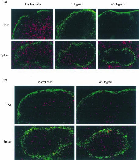
Although the spleen is the largest secondary lymphoid organ, little is known about the regulation of lymphocyte migration towards its different compartments of red and white pulp, in contrast to the well-studied mechanisms of lymphocyte homing to lymph nodes. Here we show that short-term trypsin treatment of lymphocytes cleaved off molecules involved in entry into lymph nodes, while homing to the splenic white pulp was unaltered. Prolonged trypsin treatment also abolished the ability of lymphocytes to enter the white pulp. Analysis of affected cell surface molecules and adoptive transfer studies in combination with blocking antibodies revealed that l-selectin, CD44, PSGL-1 and the alpha4 integrins are not required for migration to the white pulp. Although lymphocyte function-associated antigen-1 (LFA-1) is critical for entry into lymph nodes, we show here that in the absence of functional LFA-1 molecules, lymphocytes can still enter the white pulp, in spite of the high expression of intercellular adhesion molecule-1 on sinus lining cells in the marginal zone. The data indicate that adhesion molecules involved in lymphocyte homing to lymph nodes are not essential for migration towards the splenic white pulp, but that additional, trypsin-sensitive, and so far unidentified, molecules are required.
Figures


Similar articles
-
In vivo function of homing receptors participating in lymphocyte recirculation: transfer analysis in SCID mice.Pathobiology. 1995;63(6):305-13. doi: 10.1159/000163966. Pathobiology. 1995. PMID: 8738469
-
Clever-1 contributes to lymphocyte entry into the spleen via the red pulp.Sci Immunol. 2019 Mar 29;4(33):eaat0297. doi: 10.1126/sciimmunol.aat0297. Sci Immunol. 2019. PMID: 30926591
-
Integrin-dependence of lymphocyte entry into the splenic white pulp.J Exp Med. 2003 Feb 3;197(3):353-61. doi: 10.1084/jem.20021569. J Exp Med. 2003. PMID: 12566419 Free PMC article.
-
The role of the spleen in lymphocyte migration.Scanning Microsc. 1991 Dec;5(4):1075-9; discussion 1079-80. Scanning Microsc. 1991. PMID: 1687991 Review.
-
Physiologic migration of lymphocytes to lymph nodes following bone marrow transplantation: role in immune recovery.Semin Oncol. 1993 Oct;20(5 Suppl 6):34-9. Semin Oncol. 1993. PMID: 7692606 Review.
Cited by
-
Pak1 Kinase Promotes Activated T Cell Trafficking by Regulating the Expression of L-Selectin and CCR7.Front Immunol. 2019 Mar 5;10:370. doi: 10.3389/fimmu.2019.00370. eCollection 2019. Front Immunol. 2019. PMID: 30891040 Free PMC article.
-
Pathogenic Potential of Fresh, Frozen, and Thermally Treated Anisakis spp. Type II (L3) (Nematoda: Anisakidae) after Oral Inoculation into Wistar Rats: A Histopathological Study.J Nematol. 2017 Dec;49(4):427-436. J Nematol. 2017. PMID: 29353932 Free PMC article.
-
Sodium fluoride (NaF) induces the splenic apoptosis via endoplasmic reticulum (ER) stress pathway in vivo and in vitro.Aging (Albany NY). 2016 Dec 27;8(12):3552-3567. doi: 10.18632/aging.101150. Aging (Albany NY). 2016. PMID: 28039491 Free PMC article.
-
Intrinsic T- and B-cell defects impair T-cell-dependent antibody responses in mice lacking the actin-bundling protein L-plastin.Eur J Immunol. 2013 Jul;43(7):1735-44. doi: 10.1002/eji.201242780. Epub 2013 May 18. Eur J Immunol. 2013. PMID: 23589339 Free PMC article.
-
Subtotal splenectomy for splenomegaly in cirrhotic patients.Int J Clin Exp Pathol. 2014 Jul 15;7(8):4981-90. eCollection 2014. Int J Clin Exp Pathol. 2014. PMID: 25197369 Free PMC article.
References
-
- Kraal G, Mebius RE. High endothelial venules: Lymphocyte traffic control and controlled traffic. Adv Immunol. 1997;65:347–95. - PubMed
-
- Campbell JJ, Butcher EC. Chemokines in tissue-specific and microenvironment-specific lymphocyte homing. Curr Opin Immunol. 2000;12:336–41. - PubMed
-
- Kraal G. Cells in the marginal zone of the spleen. Int Rev Cytol. 1992;132:31–73. - PubMed
-
- Nolte MA, ′t Hoen ENM, Van Stijn A, Kraal G, Mebius RE. Isolation of the intact white pulp. Quantitative and qualitative analysis of the cellular composition of the splenic compartments. Eur J Immunol. 2000;30:626–34. - PubMed
Publication types
MeSH terms
Substances
LinkOut - more resources
Full Text Sources
Miscellaneous
