Cross-reactivity of the anti-La monoclonal antibody SW5 with early endosome antigen 2
- PMID: 12100721
- PMCID: PMC1782732
- DOI: 10.1046/j.1365-2567.2002.01432.x
Cross-reactivity of the anti-La monoclonal antibody SW5 with early endosome antigen 2
Abstract
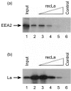
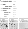
Coimmunoprecipitation studies with SW5, a frequently used and specific mouse monoclonal antibody (mAb) directed against the human La autoantigen, led to the identification of a functionally unrelated 80 000 MW protein, designated early endosome antigen 2 (EEA2). EEA2 appeared to be directly targeted by mAb SW5. Because an RNA-binding domain, a structural element of La containing the SW5-epitope, was not discernable in the primary structure of EEA2, the SW5-epitope on EEA2 was determined. Coiled-coil region 3 of EEA2 appeared to contain the epitope recognized by SW5. The SW5 epitope regions of La and EEA2 share a limited sequence homology and probably share a higher degree of structural similarity at the tertiary level. Most likely, the most critical determinants for recognition by SW5 reside in elements adopting alpha-helical conformations. These data indicate that the application of specific mAbs to purify and characterize (functionally) interacting proteins can be severely obscured by the cross-reactivity of mAbs with structurally, but not functionally, similar proteins.
Figures


Similar articles
-
The human La (SS-B) autoantigen interacts with DDX15/hPrp43, a putative DEAH-box RNA helicase.RNA. 2002 Nov;8(11):1428-43. doi: 10.1017/s1355838202021076. RNA. 2002. PMID: 12458796 Free PMC article.
-
Anti-La monoclonal antibodies recognizing epitopes within the RNA-binding domain of the La protein show differential capacities to immunoprecipitate RNA-associated La protein.Eur J Biochem. 1995 Sep 1;232(2):611-9. doi: 10.1111/j.1432-1033.1995.611zz.x. Eur J Biochem. 1995. PMID: 7556214
-
Identification of a human-specific epitope in a conserved region of the La/SS-B autoantigen.J Clin Invest. 1993 Aug;92(2):1104-8. doi: 10.1172/JCI116617. J Clin Invest. 1993. PMID: 7688758 Free PMC article.
-
Autoantigenic epitopes of the B and D polypeptides of the U1 snRNP. Analysis of domains recognized by the Y12 monoclonal anti-Sm antibody and by patient sera.J Immunol. 1993 Apr 15;150(8 Pt 1):3592-601. J Immunol. 1993. PMID: 7682245
-
Identification of GRASP-1 as a novel 97 kDa autoantigen localized to endosomes.Clin Immunol. 2005 Aug;116(2):108-17. doi: 10.1016/j.clim.2005.03.021. Clin Immunol. 2005. PMID: 15897011
Cited by
-
T Cell Mediated Conversion of a Non-Anti-La Reactive B Cell to an Autoreactive Anti-La B Cell by Somatic Hypermutation.Int J Mol Sci. 2021 Jan 26;22(3):1198. doi: 10.3390/ijms22031198. Int J Mol Sci. 2021. PMID: 33530489 Free PMC article.
-
Rabip4' is an effector of rab5 and rab4 and regulates transport through early endosomes.Mol Biol Cell. 2004 Feb;15(2):611-24. doi: 10.1091/mbc.e03-05-0343. Epub 2003 Nov 14. Mol Biol Cell. 2004. PMID: 14617813 Free PMC article.
-
The human La (SS-B) autoantigen interacts with DDX15/hPrp43, a putative DEAH-box RNA helicase.RNA. 2002 Nov;8(11):1428-43. doi: 10.1017/s1355838202021076. RNA. 2002. PMID: 12458796 Free PMC article.
References
-
- Pruijn GJM. The La (SS-B) antigen. In: Van Venrooij WJ, Maini RN, editors. Manual of Biological Markers of Disease. Dordrecht: Kluwer Academic Publishers; 1994. pp. B4.1/1–B4.1/14.
-
- Van Venrooij WJ, Slobbe RL, Pruijn GJ. Structure and function of La and Ro RNPs. Mol Biol Rep. 1993;18:113–9. - PubMed
-
- Scherly D, Stutz F, Lin MN, Clarkson SG. La proteins from Xenopus laevis. cDNA cloning and developmental expression. J Mol Biol. 1993;231:196–204. - PubMed
Publication types
MeSH terms
Substances
LinkOut - more resources
Full Text Sources
Other Literature Sources

