. 2002 Jul;106(3):343-53.
doi: 10.1046/j.1365-2567.2002.01433.x.

Circulating immunoglobulin A- and immunoglobulin G-secreting hybridoma cells in peripheral blood preferably migrate to female genital tracts. The role of sex hormones

Affiliations

Circulating immunoglobulin A- and immunoglobulin G-secreting hybridoma cells in peripheral blood preferably migrate to female genital tracts. The role of sex hormones

Xiaolei Wang et al. Immunology. 2002 Jul.

Abstract

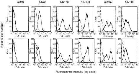
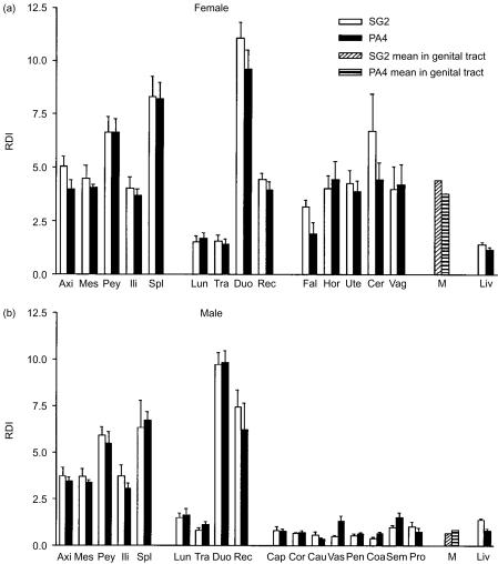
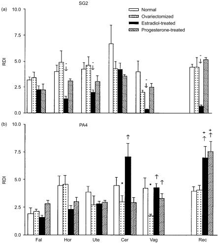
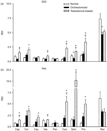
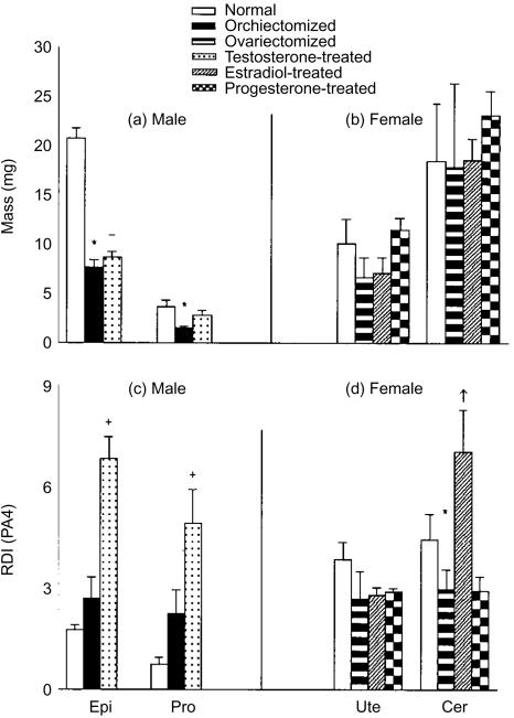
Antigen-specific circulating immunoglobulin-secreting cells (ISC) migrate to various secondary and tertiary lymphoid tissues. To understand the migration of the cells into the genital tract and its regulation by sex hormones, spleen-derived SG2 hybridoma cells secreting immunoglobulin G2b (IgG2b) and Peyer's patch-derived PA4 hybridoma cells secreting polymer IgA were labelled with 3H-TdR, and intravenously injected into syngeneic mice of both sexes. Using flow cytometry, surface molecular markers of plasma cells, CD38 and CD138, and adhesion molecules, CD49d, CD162, and CD11a were found to be positive in SG2 and PA4 cells, but CD62L, alpha4beta7 and CD44 were not expressed on these cells. The relative distribution indexes (RDIs) of the cells in genital tract and other tissues were measured. The means of RDIs of SG2 and PA4 cells in female genital tissues were 6.5 and 4.5 times as many as the means in male genital tissues, respectively. The treatment of ovariectomized mice with beta-oestradiol significantly increased the RDIs of PA4 cells in cervix and vagina, but decreased the RDIs of SG2 cells in vagina, horn of uterus, uterus and rectum (P<0.05). Progesterone treatment increased the RDIs of PA4 cells in vagina and rectum (P<0.05). The treatment with testosterone significantly increased the RDIs of SG2 and PA4 cells in epididymis and accessory sex glands (P<0.05). These results demonstrate that the female genital tract is the preferable site for the migration of circulating hybridoma cells to the male genital tract, and sex hormones play an important role in regulation of the migration of circulating ISC to genital tracts.


Figures

Figure 1
Figure 1
Expression patterns right of the surface molecular markers CD19, CD38, CD138, and adhesion molecules CD49d, CD162 and CD11a on the SG2 (top panels) and PA4 (bottom panels) cells. FL1-Height showed the fluorescence intensity of FITC, FL2-Height showed the fluorescence intensity of PE. Histograms in dotted line represent negative controls, histograms in solid line represent the staining with the corresponding fluorochrome-conjugated antibody. Data are representative of three experiments.
Figure 2
Figure 2
CXCR5 mRNA expression in the representative cells and tissues of mice was detected by RT–PCR. The molecular weight standard is GeneRuler™ 100 bp DNA Ladder plus (MBI). Specific bands of CXCR5 (400 bp) or GAPDH (180 bp) are indicated to the right of the panel. Lanes 1–4, respectively, represent PA4 and SG2 cells, spleen and mesenteric lymph nodes.
Figure 3
Figure 3
The RDIs of SG2 and PA4 cells in various examined organs or tissues of the normal female (a) and male (b) BALB/c mice. Each bar represents the mean of each group, and the line on each bar represents SEM. M, the mean of RDIs of SG2 or PA4 cells in all examined organs of genital tract. Axi, axillary lymph nodes; Mes, mesenteric lymph nodes; Pey, Peyer's patches; Ili, iliac lymph nodes; Spl, spleen; Lun, lung; Tra, trachea; Duo, duodenum; Rec, rectum; Fal, fallopian tube; Hor, horn of uterus; Ute, uterus; Cer, cervix; Vag, vagina; Cap, caput of epididymis; Cor, corpus of epididymis; Cau, cauda of epididymis; Pen, penis; Coa, coagulating gland; Sem, seminal vesicle; Vas, vas defens; Pro, prostate; Liv, liver.
Figure 4
Figure 4
Effects of β-oestradiol and progesterone on the RDIs of SG2 (a) and PA4 (b) cells in female BALB/c mice. Each bar represents the mean of each group, and the line on each bar represents SEM. Marker (*) indicates the RDI of the cells in ovariectomized group significantly decreased compared to that in normal group (P<0·05). +or − indicates that the RDIs of the cells in β-oestradiol- or progesterone-treated group were significantly higher (+) or lower (−) than those in the normal group (P<0·05). ↑↓ indicates the RDIs of the cells in the β-oestradiol- or progesterone-treated group were significantly higher (↑) or lower (↓) than those in the ovariectomized group (P<0·05). Fal, fallopian tube; Hor, horn of uterus; Ute, uterus; Cer, cervix; Vag, vagina; Rec, rectum.
Figure 5
Figure 5
Effects of testosterone on the RDIs of SG2 (a) and PA4 (b) cells in male BALB/c mice. Each bar represents the mean of each group, and the line on each bar represents SEM. * indicates the RDI of the cells in orchiectomized group significantly decreased compared to that in normal group (P<0·05). + indicates the RDIs of the cells in testosterone-treated group significantly increased compared to those in normal group (P<0·05). ↑↓ indicates the RDIs of the cells in the testosterone-treated group were significantly higher (↑) or lower (↓) than those in the orchiectomized group (P<0·05). Cap, caput of epididymis; Cor, corpus of epididymis; Cau, cauda of epididymis; Vas, vas defens; Pen, penis; Coa, coagulating gland; Sem, seminal vesicle; Pro, prostate gland; Rec, rectum.
Figure 6
Figure 6
Changes in the masses and RDIs of PA4 cells in epididymis and prostate in male BALB/c mice (a and c) and in uterus and cervix of female mice (b and d) after treatments. Each bar represents the mean of each group, and the line on each bar represents SEM. * indicates the mass of the organs or RDI of PA4 cells in the ovariectomized or orchiectomized group significantly decreased compared to that in the normal group (P<0·05). + or − indicates the mass of the organs or RDI of PA4 cells in the sex hormone-treated group significantly increased (+) or decreased (−) compared to that in the normal group (P<0·05). ↑ indicates the RDIs of PA4 cells in the sex hormone-treated group were significantly increased compared to those in the ovariectomized group (P<0·05).
Figure 7
Figure 7
The localization of the migrated cells labelled with fluorochrome H33342 in the representative tissues of the normal BALB/c mice 24 hr after intravenous injection. The fluorescent cells were observed under fluorescence microscope (left panels) in conjunction with their phase contrast field (right panels). The upper photomicrographs (a and b) demonstrated the labelled cells appeared in the lamina propria of the duodenal villi. The middle photomicrographs (c and d) demonstrated the labelled cells located in the lamina propria of the vagina. The bottom photomicrographs (e and f) demonstrated the labelled cells in the pseudostratified ciliated columnar epithelium of the epididymis caput. Original magnification: ×132.

Similar articles

References

    1. Cohen MS, Anderson DJ. Sexually Transmitted Diseases. 4. New York: McGraw-Hill Companies, Inc.; 1999. Genitourinary mucosal defenses; pp. 173–90.
    1. Mestecky J, Fultz PN. Mucosal immune system of human genital tract. J Infect Dis. 1999;179(Suppl. 3):S470–4. - PubMed
    1. Frayne J, Hall L. The potential use of sperm antigens as targets for immunocontraception: past, present and future. J Reprod Immunol. 1999;43:1–33. - PubMed
    1. Ogra PL, Mestecky J, Lamm ME, Strober W, Bienenstock J, McGhee JR, editors. Mucosal Immunology. San Diego: Academic Press; 1999.
    1. Mostad SB, Kreiss JK. Shedding of HIV-1 in genital tract. AIDS. 1996;10:1305–12. - PubMed

Publication types

MeSH terms