. 2002 Jul;106(3):404-11.
doi: 10.1046/j.1365-2567.2002.01421.x.

Herpes simplex virus 1 infected neuronal and skin cells differ in their susceptibility to complement attack

Affiliations

Herpes simplex virus 1 infected neuronal and skin cells differ in their susceptibility to complement attack

Riina Rautemaa et al. Immunology. 2002 Jul.

Abstract

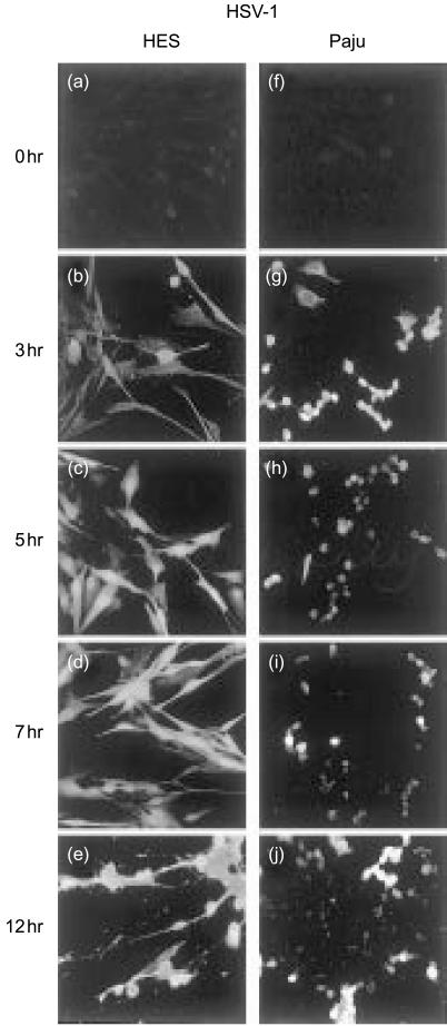
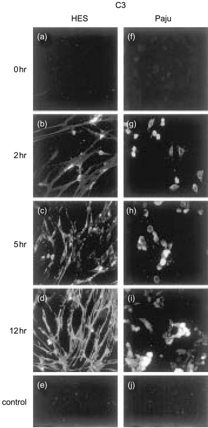
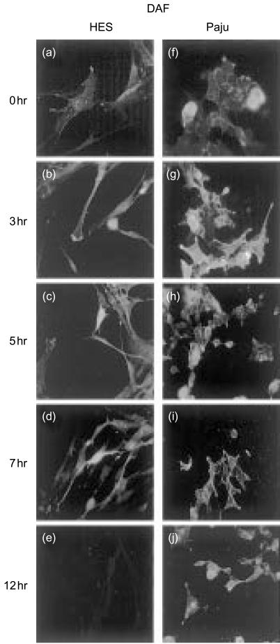
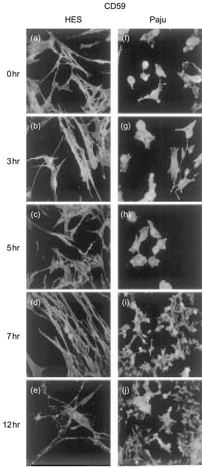
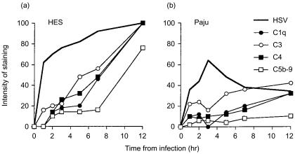
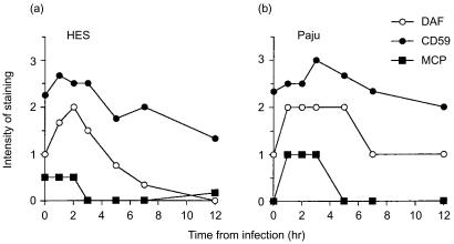
Herpes simplex virus type 1 (HSV-1) infection in neurons is lifelong and generally asymptomatic. Reactivation of this latent infection results in skin blistering whereas the respective peripheral neurons are rarely affected. Why the neuronal cells are spared while the skin cells are sacrificed is not well understood. In the present study our aim was to study whether neuronal and skin cells differ in their ability to control complement attack during HSV-1 infection. Human embryonal skin (HES) cells and neuronal Paju cells were infected by HSV-1 in vitro. Both types of infected cells activated complement but were initially resistant to membrane attack complex (MAC) deposition. During the first hours of infection the expression of the endogenous complement regulators decay accelerating factor (DAF) and CD59 increased on both HES and Paju cells. By 12 hr the infected HES cells had lost their ability to control complement attack. The expression of DAF and CD59 decreased and the cells became targets for MAC attack. In contrast, complement regulator expression on the Paju cells did not decrease below the initial level and complement C5b-9 deposition was found only on 10% of the Paju cells at 12 hr. The results suggest that HSV-infected neuronal cells are better than skin cells in protecting themselves against complement attack. This may contribute to the persistence of a latent HSV-1 infection in neuronal cells for prolonged periods.


Figures

Figure 1
Figure 1
Expression of HSV-1 antigens on skin HES (b–e) and neuronal Paju (g–j) cells at various time points after HSV-1-infection. Immunofluorescence microscopy analysis shows positive staining of the cell nuclei for HSV-1 antigens on both cell types at 3 hr post infection (b, g). At later time points the staining of the HES cells intensifies further (c–e). In contrast, the staining intensity and the percentage of Paju cells positive for HSV-antigens decreases by 5 hr and at 12 hr post infection only 40% of the Paju cells express HSV-antigens (j). In controls, non-infected cells were stained with HSV-1 antibody (a, f) (original magnifications, 400×).
Figure 2
Figure 2
Immunofluorescence microscopy analysis of C3 deposition on non-infected (a, f) and HSV-1-infected HES (b–d) and Paju (g–i) cells after exposure to immune human serum. Two hours post infection positive C3 staining of individual HES cells is mainly located on the contracted cells and cytoplasm adjacent to the nucleus (b) but by 12 hr (d) all the cells stain positively throughout. The shapes of the cells change from leaf-like to spindle-shaped during the infection. HSV-1-infected Paju cells also deposit C3 at 2 hr post infection. By 12 hr 40% of the cells, on the average, were positive (i). The non-infected, serum treated cells showed no staining for C3 (a, f). Controls: FITC-conjugated antibody against mouse IgG alone (e), and irrelevant mouse mAb against Helicobacter pylori as the primary antibody (j) (original magnifications, 400×).
Figure 4
Figure 4
Expression of the membrane regulator DAF (CD55) on non-infected (a, f) or HSV-1 infected HES (b–e) and Paju (g–j) cells. The glycolipid-tailed complement inhibitor DAF is expressed on the non-infected HES (a) and Paju (f) cells. The staining is markedly intensified during the first 3–5 hr of HSV-1 infection (b, c, g, h). The staining is located on cell membranes (c) and Paju (f) cells and on the shrunk extensions of neural Paju cells (g, h). By 7 hr post infection the staining intensity for DAF decreases in both cell types and is totally lost in HES cells at 12 hr (e). Paju cells maintain DAF expression at all time points studied but the individual Paju cells differ in their expression (j) (original magnifications 600×).
Figure 5
Figure 5
Expression of the glycolipid-tailed complement inhibitor CD59 on non-infected (a, f) and HSV-1-infected HES (b–e) and Paju (g–j) cells. CD59 is strongly expressed on non-infected HES (a) and Paju (f) cells. The overall staining intensity for CD59 is stable in both cell types during the first 5–7 hr of infection. The sprouting extensions of the Paju cells stain strongly positive for CD59 (g, h). By 12 h the expression of CD59 has decreased in both cell types (e, j). While in HES cells CD59 staining is homogenously decreased in Paju cells CD59 is lost only from a proportion of the cells (j) (original magnifications a–h, 600×; i–j, 400×).
Figure 3
Figure 3
Complement activation by HES (a) and Paju (b) cells after infecting with HSV-1. The infected cells exposed to human serum (containing antibodies against HSV-1) were stained for complement components C1q, C3, C4 and for the C5b-9 neoepitope. The proportions of cells staining positively were counted from 50 evenly distributed representative cells. The averages of two independently prepared slides are shown. The results shown are averages of two parallel series performed in one experiment.
Figure 6
Figure 6
A summary of complement regulator expression by HES (a) and Paju (b) cells after infecting with HSV-1. The intensity of the staining for the complement regulators studied was estimated semiquantitatively and scored (0–3) under constant, standard settings. The results show average scores from two independently prepared glasses where 50 evenly distributed representative cells were evaluated. The averages of two independently prepared slides are shown. The results shown are averages of two parallel series performed in one experiment.

References

    1. Brown EJ, Joiner KA, Frank MM. The role of complement in host resistance to bacteria. Springer Semin Immunopathol. 1983;6:349–60. - PubMed
    1. Joiner KA. Complement evasion by bacteria and parasites. Annu Rev Microbiol. 1988;42:201–30. - PubMed
    1. Da Costa XJ, Brockman MA, Alicot E, Ma M, Fischer MB, Zhou X, Knipe DM, Carroll MC. Humoral response to herpes simplex virus is complement-dependent. Proc Natl Acad Sci USA. 1999;96:12708–12. - PMC - PubMed
    1. Born J, Bhakdi S. Does complement kill E. coli by producing transmural pores? Immunology. 1986;59:139–45. - PMC - PubMed
    1. Grewal AS, Rouse BT. Destruction of virus infected cells by neutrophils and complement. Experientia. 1980;36:352–4. - PubMed

Publication types