. 2002 Jul;3(7):646-51.
doi: 10.1093/embo-reports/kvf129.

Intranuclear degradation of nonsense codon-containing mRNA

Affiliations

Intranuclear degradation of nonsense codon-containing mRNA

Marc Bühler et al. EMBO Rep. 2002 Jul.

Abstract

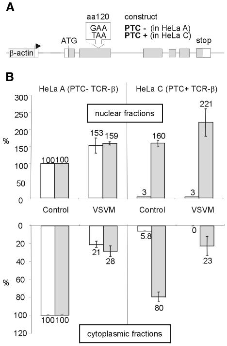
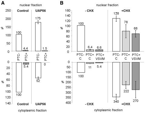
Most vertebrate mRNAs with premature termination codons (PTCs) are specifically recognized and degraded by a process referred to as nonsense-mediated mRNA decay (NMD) while still associated with the nucleus. However, it is still a matter of debate whether PTCs can be identified by intranuclear scanning or only by ribosomes on the cytoplasmic side of the nuclear envelope. Here we show that inhibition of mRNA export by two independent approaches does not affect the downregulation of PTC-containing T-cell receptor beta transcripts in the nuclear fraction of mammalian cells, providing strong evidence for intranuclear NMD. Our results are fully consistent with recently reported evidence for nuclear translation and suggest that an important biological role for nuclear ribosomes is the early elimination of nonsense mRNA during a pioneer round of translation.


Figures

None
Fig. 1. Expression of VSV M inhibits nuclear export of PTC– and PTC+ TCR-β mRNA, but does not affect the extent of NMD in the nuclear fraction. (A) Schematic representation of the PTC– and PTC+ TCR-β constructs stably expressed in HeLa A and C, respectively, under the control of the human β-actin promotor (Muhlemann et al., 2001). (B) Relative amounts of TCR-β (white bars) and endogenous GAPDH mRNA (gray bars) in the nuclear (upper histogram) and in the cytoplasmic fractions (lower histogram) of cells expressing a VSV M–GFP fusion protein (VSVM) or GFP (Control) were determined by real-time RT–PCR. The indicated values are normalized to 18S rRNA levels and corrected for background from untransfected cells (see Methods). Average values of three real-time PCR runs are shown.
None
Fig. 2. Reduction of poly(A) RNA in the cytoplasm of VSV M–GFP-expressing HeLa C cells detected by FISH. Cells were transfected with a plasmid encoding GFP (A and B) or VSV M–GFP (C and D), fixed after 24 h and hybridized with a Cy3-labeled oligo (dT)70 probe to analyze the subcellular distribution of poly(A) RNA (B and D). To easily distinguish them from untransfected cells, the cells expressing VSV M–GFP fusion protein (C) or GFP (A) are marked by arrows in (D) and (B), respectively.
None
Fig. 3. Nuclear fractions contain <1% contamination with cytoplasmic tyrosine-tubulin. Nuclear and cytoplasmic fractions of untransfected (A) and VSV M–GFP-expressing HeLa cells (B) were separated by SDS–PAGE, transferred onto nylon membrane and probed with a monoclonal antibody against tyrosine-tubulin. For comparison with the nuclear fractions, cytoplasmic fractions corresponding to 1, 2, 5 and 20% (lanes 1–4) of the number of cells represented in the nuclear fractions (lanes 5–7) were loaded, and nuclear fractions were spiked with 1 or 2% cytoplasm (lanes 5 and 6).
None
Fig. 4. Nucleus-associated NMD of TCR-β mRNA is not disturbed by UAP56-mediated reduction of export, but is sensitive to translation inhibition. (A) Relative amounts of PTC– (white bars) and PTC+ (gray bars) TCR-β mRNA in the nuclear (upper histogram) and cytoplasmic fractions (lower histogram) of cells expressing UAP56–GFP fusion protein or GFP (Control) were determined by real-time RT–PCR. (B) Twenty-eight hours after transfection with GFP (C) or VSV M–GFP (VSV M), cells were exposed to 100 µg/ml cycloheximide for 2 h (+CHX) or left in normal medium (–CHX). Relative amounts of PTC– (white bars) and PTC+ (light and dark gray bars) TCR-β mRNA in the nuclear (upper histogram) and cytoplasmic fractions (lower histogram) were measured by real-time RT–PCR. Values in (A) and (B) are normalized to 18S rRNA levels and were corrected for background from untransfected cells in (A). Average values of three real-time PCR runs are shown.

References

    1. Ahmed M. and Lyles, D.S. (1998) Effect of vesicular stomatitis virus matrix protein on transcription directed by host RNA polymerases I, II, and III. J. Virol., 72, 8413–8419. - PMC - PubMed
    1. Aoufouchi S., Yelamos, J. and Milstein, C. (1996) Nonsense mutations inhibit RNA splicing in a cell-free system: recognition of mutant codon is independent of protein synthesis. Cell, 85, 415–422. - PubMed
    1. Baserga S.J. and Benz, E.J.,Jr (1992) β-globin nonsense mutation: deficient accumulation of mRNA occurs despite normal cytoplasmic stability. Proc. Natl Acad. Sci. USA, 89, 2935–2939. - PMC - PubMed
    1. Belgrader P. and Maquat, L.E. (1994) Nonsense but not missense mutations can decrease the abundance of nuclear mRNA for the mouse major urinary protein, while both types of mutations can facilitate exon skipping. Mol. Cell. Biol., 14, 6326–6336. - PMC - PubMed
    1. Belgrader P., Cheng, J. and Maquat, L.E. (1993) Evidence to implicate translation by ribosomes in the mechanism by which nonsense codons reduce the nuclear level of human triosephosphate isomerase mRNA. Proc. Natl Acad. Sci. USA, 90, 482–486. - PMC - PubMed

Publication types

MeSH terms