

Homeotic transformations of the axial skeleton that accompany a targeted deletion of E2f6
- PMID: 12101104
- PMCID: PMC1084195
- DOI: 10.1093/embo-reports/kvf141
Homeotic transformations of the axial skeleton that accompany a targeted deletion of E2f6
Abstract
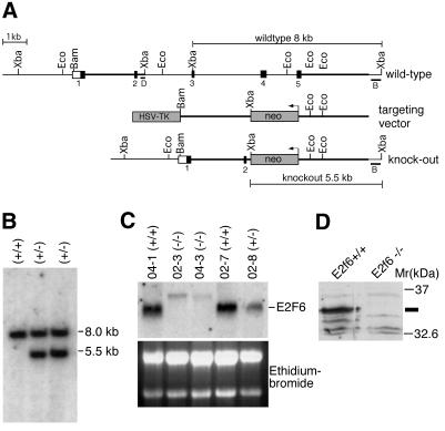
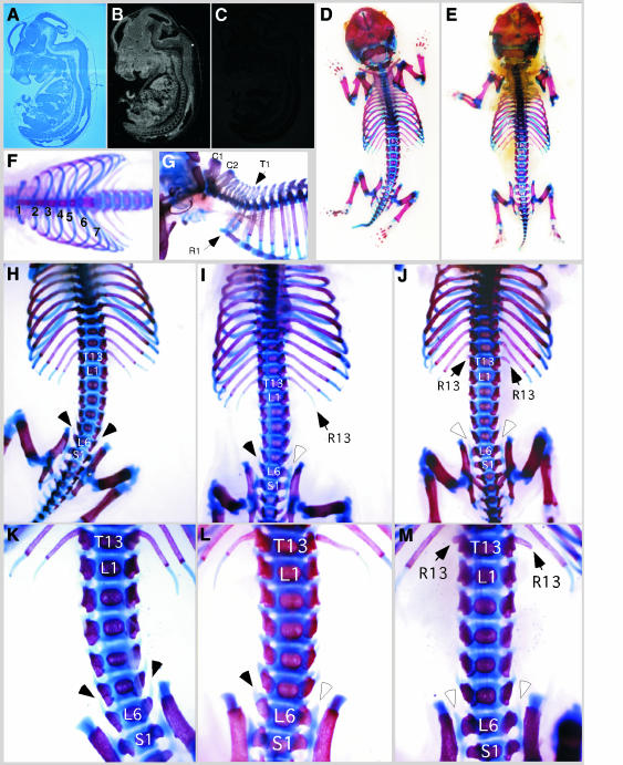
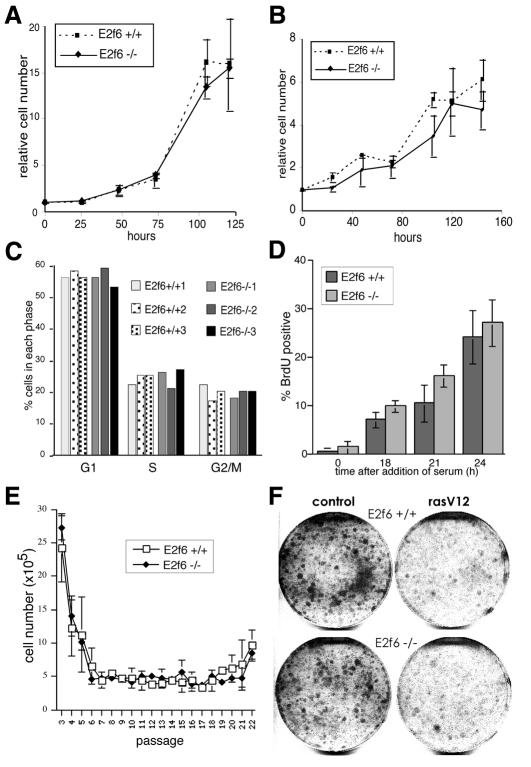
E2F transcription factors play an important role in regulating mammalian cell proliferation. E2F6, the most recently identified E2F family member, is a transcriptional repressor. In an effort to ascertain the in vivo biological function of E2F6, we have generated an E2f6 mutant mouse strain. Mice lacking E2F6 are viable and healthy. Surprisingly, E2f6-/- embryonic fibroblasts proliferate normally. However, E2f6-/- animals display overt homeotic transformations of the axial skeleton that are strikingly similar to the skeletal transformations observed in polycomb mutant mice. This observation is compatible with the recent finding that endogenous E2F6 and one or more mammalian polycomb proteins are components of the same multiprotein complex. The accumulated evidence suggests that, during development, E2F6 participates in the recruitment of polycomb proteins to specific target promoters.
Figures
Similar articles
-
E2f6 and Bmi1 cooperate in axial skeletal development.Dev Dyn. 2008 May;237(5):1232-42. doi: 10.1002/dvdy.21516. Dev Dyn. 2008. PMID: 18366140 Free PMC article.
-
A novel repressive E2F6 complex containing the polycomb group protein, EPC1, that interacts with EZH2 in a proliferation-specific manner.J Biol Chem. 2005 Jan 14;280(2):1199-208. doi: 10.1074/jbc.M412509200. Epub 2004 Nov 8. J Biol Chem. 2005. PMID: 15536069
-
The E2F6 transcription factor is a component of the mammalian Bmi1-containing polycomb complex.Proc Natl Acad Sci U S A. 2001 Feb 13;98(4):1519-24. doi: 10.1073/pnas.98.4.1519. Epub 2001 Jan 30. Proc Natl Acad Sci U S A. 2001. PMID: 11171983 Free PMC article.
-
The role of Bapx1 (Nkx3.2) in the development and evolution of the axial skeleton.J Anat. 2001 Jul-Aug;199(Pt 1-2):181-7. doi: 10.1046/j.1469-7580.2001.19910181.x. J Anat. 2001. PMID: 11523821 Free PMC article. Review.
-
MHox and vertebrate skeletogenesis: the long and the short of it.Bioessays. 1995 Sep;17(9):750-3. doi: 10.1002/bies.950170903. Bioessays. 1995. PMID: 8763826 Review.
Cited by
-
E2F6 associates with BRG1 in transcriptional regulation.PLoS One. 2012;7(10):e47967. doi: 10.1371/journal.pone.0047967. Epub 2012 Oct 17. PLoS One. 2012. PMID: 23082233 Free PMC article.
-
Characterization of E2F8, a novel E2F-like cell-cycle regulated repressor of E2F-activated transcription.Nucleic Acids Res. 2005 Sep 22;33(17):5458-70. doi: 10.1093/nar/gki855. Print 2005. Nucleic Acids Res. 2005. PMID: 16179649 Free PMC article.
-
Regulation of gene transcription by Polycomb proteins.Sci Adv. 2015 Dec 4;1(11):e1500737. doi: 10.1126/sciadv.1500737. eCollection 2015 Dec. Sci Adv. 2015. PMID: 26665172 Free PMC article.
-
Genome-wide occupancy reveals the localization of H1T2 (H1fnt) to repeat regions and a subset of transcriptionally active chromatin domains in rat spermatids.Epigenetics Chromatin. 2021 Jan 6;14(1):3. doi: 10.1186/s13072-020-00376-2. Epigenetics Chromatin. 2021. PMID: 33407810 Free PMC article.
-
L3MBTL2 protein acts in concert with PcG protein-mediated monoubiquitination of H2A to establish a repressive chromatin structure.Mol Cell. 2011 May 20;42(4):438-50. doi: 10.1016/j.molcel.2011.04.004. Mol Cell. 2011. PMID: 21596310 Free PMC article.
References
-
- Akasaka T., Kanno, M., Balling, R., Mieza, M.A., Taniguchi, M. and Koseki, H. (1996) A role for mel-18, a Polycomb group-related vertebrate gene, during the anteroposterior specification of the axial skeleton. Development, 122, 1513–1522. - PubMed
-
- Bates S., Phillips, A.C., Clark, P.A., Stott, F., Peters, G., Ludwig, R.L. and Vousden, K.H. (1998) p14ARF links the tumour suppressors RB and p53. Nature, 395, 124–125. - PubMed
-
- Cartwright P., Muller, H., Wagener, C., Holm, K. and Helin, K. (1998) E2F-6: a novel member of the E2F family is an inhibitor of E2F-dependent transcription. Oncogene, 17, 611–623. - PubMed
-
- Cillo C., Cantile, M., Faiella, A. and Boncinelli, E. (2001) Homeobox genes in normal and malignant cells. J. Cell Physiol., 188, 161–169. - PubMed
-
- Core N., Bel, S., Gaunt, S.J., Aurrand-Lions, M., Pearce, J., Fisher, A. and Djabali, M. (1997) Altered cellular proliferation and mesoderm patterning in Polycomb-M33-deficient mice. Development, 124, 721–729. - PubMed
Publication types
MeSH terms
Substances
LinkOut - more resources
Full Text Sources
Molecular Biology Databases
