. 2002 Jul 1;16(13):1640-58.
doi: 10.1101/gad.972202.

Nercc1, a mammalian NIMA-family kinase, binds the Ran GTPase and regulates mitotic progression

Affiliations

Nercc1, a mammalian NIMA-family kinase, binds the Ran GTPase and regulates mitotic progression

Joan Roig et al. Genes Dev. .

Abstract

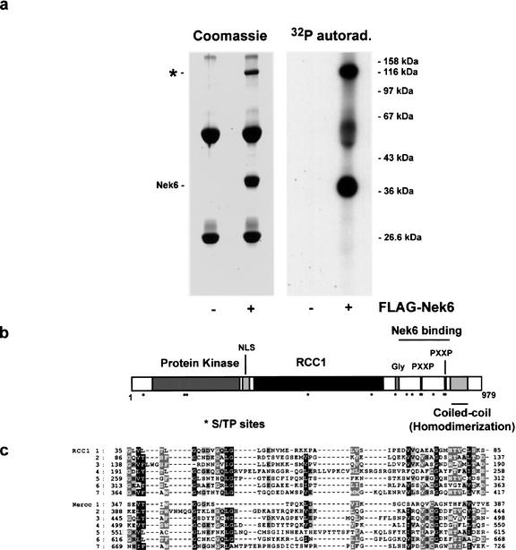
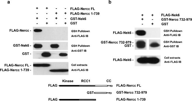
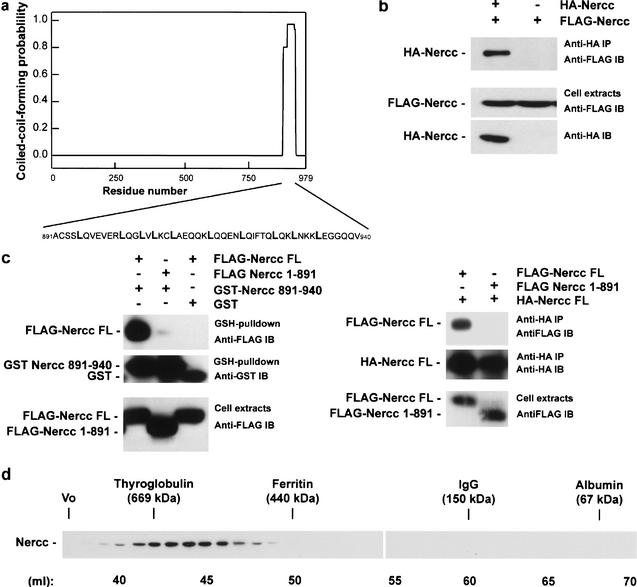
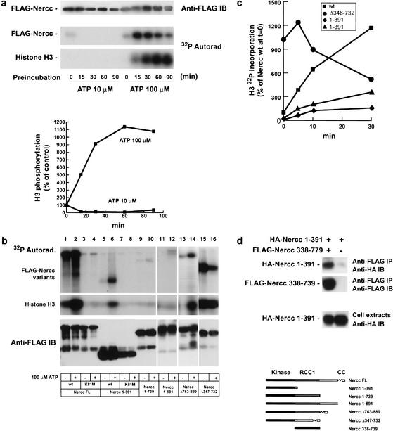
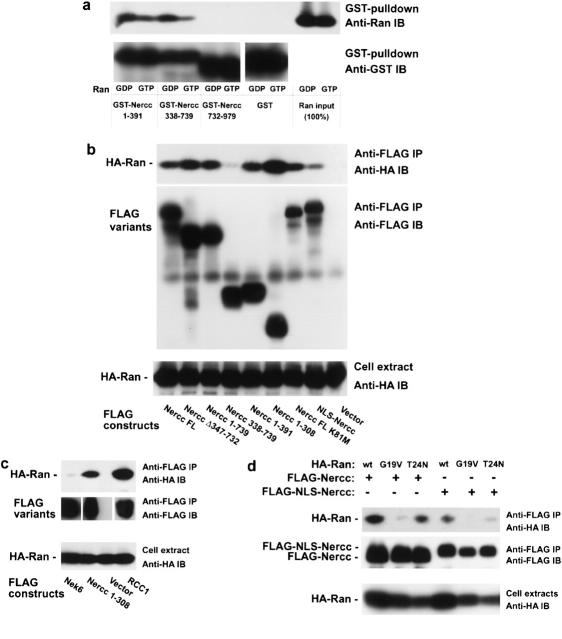
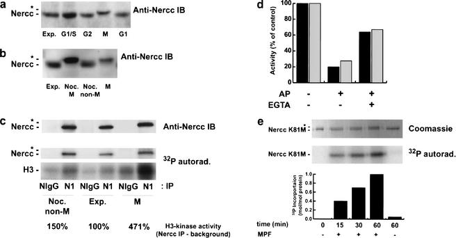
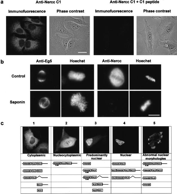
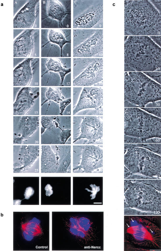
The protein kinase NIMA is an indispensable pleiotropic regulator of mitotic progression in Aspergillus. Although several mammalian NIMA-like kinases (Neks) are known, none appears to have the broad importance for mitotic regulation attributed to NIMA. Nercc1 is a new NIMA-like kinase that regulates chromosome alignment and segregation in mitosis. Its NIMA-like catalytic domain is followed by a noncatalytic tail containing seven repeats homologous to those of the Ran GEF, RCC1, a Ser/Thr/Pro-rich segment, and a coiled-coil domain. Nercc1 binds to another NIMA-like kinase, Nek6, and also binds specifically to the Ran GTPase through both its catalytic and its RCC1-like domains, preferring RanGDP in vivo. Nercc1 exists as a homooligomer and can autoactivate in vitro by autophosphorylation. Nercc1 is a cytoplasmic protein that is activated during mitosis and is avidly phosphorylated by active p34(Cdc2). Microinjection of anti-Nercc1 antibodies in prophase results in spindle abnormalities and/or chromosomal misalignment. In Ptk2 cells the outcome is prometaphase arrest or aberrant chromosome segregation and aneuploidy, whereas in CFPAC-1 cells prolonged arrest in prometaphase is the usual response. Nercc1 and its partner Nek6 represent a new signaling pathway that regulates mitotic progression.


Figures

Figure 1
Figure 1
Nek 6 coimmunoprecipitates with a 120-kD protein. Structure of Nercc1 polypeptide. (a) HEK293 cells were transfected with either empty vector (−) or pCM5 Flag–Nek6 (+). Cell lysates were prepared 48 h later, and Nek6 was immunoprecipitated using an anti-Flag antibody. The washed immunoprecipitates were incubated with Mg2+ [γ-32P]ATP. Coomassie stain (left panel) and 32P autoradiography (right panel) of the gel are shown. (b) Cartoon of Nercc1 polypeptide domain structure. (NLS) Nuclear localization signal; (RCC1) RCC1 homology domain; (Gly) polyglycine stretch; (PXXP) proline-rich motifs; (S/TP sites) Ser/ThrPro motifs. (c) Alignment of the RCC1 repeats of the human regulator of chromosome condensation (RCC1) with Nercc1 RCC1 domain repeats. Amino acid numbers are shown. Of the 10 residues known to be important for the exchange activity of RCC1 toward Ran (i.e., affecting the Km or Kcat; Ji et al. 1999), Nercc1 lacks a conserved residue corresponding to RCC1 D44, R206, D182, and H270, although a residue of similar charge exists at the position −1 and +1 for the latter two; D128 E157 and H304 are conserved, whereas H78, R101 and H410 are substituted by similarly charged residues. Notably, mutation of RCC1 D182 (an asparagine in Nercc1) results in a protein with no measurable GEF activity.
Figure 2
Figure 2
Nercc1 binding to Nek6. (a) HEK293 cells were cotransfected with full-length (FL) Flag–Nercc1 or Flag–Nercc1(1–739), and either GST–Nek6 or GST alone. GST fusion proteins were isolated from cell lysates using GSH-agarose beads. Western blots of the GSH-agarose isolate and the cell extract are shown. (b) HEK293 cells were cotransfected with Flag–Nek6 and either GST–Nercc1(732–979) or GST alone. Western blots of the GSH-agarose isolates and the cell extract are shown; a cartoon of the Nercc1 Flag-fusion proteins used is shown below.
Figure 3
Figure 3
Nercc1 oligomerizes through its C-terminal coiled-coil domain. (a) Nercc1 coiled-coil prediction carried out with the Coils 2.1. software (window 28). The propensity of a sequence to form coiled coils on a scale from 0 to 1 is plotted against the linear sequence of amino acids. The sequence of the predicted coiled coil is shown; leucine residues are shown in bold. (b) HEK293 cells were transfected with HA–Nercc1 and Flag–Nercc1. The anti-HA immunoprecipitate was blotted with anti-Flag antibody (upper panels); expression of the constructs is shown below. (c, left panels) HEK293 cells were transfected with Flag–Nercc1 full-length (FL) or Flag–Nercc1(1–891) and either GST or a GST fusion to the Nercc1 coiled-coil GST–Nercc1(891–940). GST-agarose isolates were blotted for Flag (upper panel) or GST (middle panel); Flag–Nercc1 expression in cell lysates is shown in the lower panel. (Right panels) Flag–Nercc1 (FL) or Flag–Nercc1(1–891) were cotransfected with HA–Nercc1 (FL). The HA immunoprecipitates were blotted for Flag (upper panel) or HA (middle panel); expression of Flag–Nercc1 in cell lysates is shown in the lower panel. (d) Gel filtration of Nercc1 endogenous to HEK293 cells. The median elution position of standard proteins (thyroglobulin, ferritin, IgG, and BSA) is indicated.
Figure 4
Figure 4
Nercc1 autoactivation in vitro. (a) Flag–Nercc1 was immunoprecipitated from HEK293 cells, washed, and incubated at 25°C in phosphorylation buffer for the indicated times with 10 μM or 100 μM ATP. Incubations were terminated by washing, followed by the addition of 10 μM [32P]ATP and histone H3 (1 μg/50 μL). After 10 min at 30°C, 32P incorporation was stopped by addition of SDS sample buffer, followed by SDS-PAGE and blot transfer. The anti-Flag immunoblot (upper panel), 32P autoradiography (middle panel), and the relative quantity of 32P incorporated into histone H3 (bottom panel) are shown. (b) Immobilized Flag-tagged Nercc1 variants, isolated after transient expression in HEK293 cells, were washed and incubated in phosphorylation buffer at 25°C for 30 min with Mg2+ and with or without 100 μM ATP. After an additional wash, samples were incubated at 30°C with Mg2+ plus 10 μM [32P]ATP and histone H3 (1 μg/50 μL). After 10 min the reaction was stopped by addition of SDS sample buffer followed by SDS-PAGE and blot transfer. The 32P autoradiogram (upper panel) and anti-Flag immunoblot (middle panel) are shown. (c) Time course of activation of the H3 kinase activity of wild-type and mutant Nercc1. (▪) Flag–Nercc1 (wild-type); (●) Flag–Nercc1(Δ346–732); (♦) Flag-Nercc1(1–391); and (▴) Flag–Nercc1(1–891), were expressed in HEK293 cells, immobilized on anti-Flag-agarose, washed and incubated at 25°C with Mg2+ plus 100 μM ATP. At the times indicated, samples were washed, followed by addition of Mg2+ plus 10 μM [32P]ATP and histone H3 (1 μg/50 μL). After 10 min at 30°C, SDS sample buffer was added, and 32P incorporation into H3 was measured (using a PhosphorImager) after SDS-PAGE and blot transfer. 32P incorporation is expressed as a percentage of Nercc1 wild-type value at t = 0, that is, no preincubation with 100 μM ATP. (d) The Nercc1 protein kinase domain and RCC1 domain interact in vivo. HEK293 cells were transfected with the HA–Nercc1 protein kinase domain, HA–Nercc1(1–391), and either Flag–Nercc1 RCC1 domain, Flag–Nercc1(338–778), or empty plasmid. Anti-Flag immunoprecipitates were immunoblotted with anti-HA (upper panel) or anti-Flag (middle panel). The expression of HA-Nercc1(1–391) is shown in the lower panel. A cartoon of the Nercc1 variant used in Figure 6 is below.
Figure 5
Figure 5
Nercc1 binding to Ran. (a) The binding of recombinant Ran to GST–Nercc1 variants in vitro. GST and GST–Nercc1 variants were purified and immobilized on GSH-agarose. Ran was produced in bacteria as a GST fusion, purified, cleaved from the immobilized GST fusion, and loaded with GDPβS (GDP) or GTPγS (GTP). Immobilized GST or GST–Nercc1 fusion proteins were incubated with Ran in the Ran binding buffer containing the indicated nucleotides (100 μM). After extensive washing, the proteins retained on the GSH-agarose were eluted into SDS sample buffer, and analyzed by immunoblot for Flag (upper panel) and GST (lower panel). The Ran input is shown. (b,c) HEK293 cells were cotransfected with HA–Ran and Flag–Nercc1 variants or Flag vector. Cells were extracted into Ran lysis buffer. Anti-Flag immunoprecipitates and aliquots of the extracts were subjected to immunoblot with anti-HA (upper panel) and anti-Flag (middle panel) antibodies. Expression of HA–Ran is shown in the lower panel. (d) HEK293 cells were transfected with Flag–Nercc1 (left) or Flag–NLS-Nercc1 (right) together with either HA–Ran wild type, HA–Ran G19V (constitutively GTP-bound), or HA–Ran T24N (constitutively GDP-bound or nucleotide-free). Cells were extracted into Ran lysis buffer. Anti-Flag immunoprecipitates were immunoblotted with anti-HA (upper panel) and anti-Flag (middle panel). The expression of the HA–Ran variants is shown in the lower panel.
Figure 6
Figure 6
Nercc1 kinase is activated during mitosis and can be phosphorylated in vitro by p34Cdc2. (a) HeLa cells were isolated in different phases of the cell cycle. (G1/S) Cells arrested with aphidicolin (2 μg/mL overnight); (G2/M) cells arrested with aphidicolin and released for 6 h; (M) mitotic cells isolated by shake-off from a culture treated with nocodazole (500 ng/mL overnight). (G1) Mitotic cells, isolated as above, were washed repeatedly, replated, and harvested 6 h later. (Exp.) Exponentially growing cells. Each cell cycle stage designation was confirmed by FACS. An immunoblot of endogenous Nercc1 (C1 antibody) at the different cell cycle stages is shown. (b) Slowing of Nercc1 on SDS-PAGE occurs during normal progression through mitosis. HeLa cells were partially synchronized using thymidine (2 mM thymidine overnight plus release). The resulting mitotic cells were collected by shake-off 9 h later and compared with exponentially growing cells (Exp.); mitotic, nocodazole-arrested cells detached after mitotic shake-off (Noc. M); and nocodazole-treated cells that remain attached after shake-off (non-mitotic cells; Noc. Non-M). Extracts of each cell type were subjected to immunoblot using anti-Nercc1 C1 antibody. (c) Nercc1 kinase is activated in mitosis. Immunoprecipitations were carried out using extracts from nocodazole-treated cells that remain attached after shake-off (non-mitotic cells; Noc. Non-M), exponentially growing cells (Exp.), or mitotic nocodazole-arrested cells (Noc. M), with both preimmune rabbit IgG (NIgG) and affinity-purified anti-Nercc1 antibody (N1). Immunoprecipitates were washed sequentially with lysis buffer and phosphorylation buffer, and incubated at 30°C for 10 min with Mg2+ plus [γ-32P]ATP (10 μM) and histone H3 (2 μg/50 μL). The reaction was stopped by addition of SDS sample buffer. Shown are an anti-Nercc1 (N1) immunoblot of the immunoprecipitates (upper panel) and the 32P incorporation into Nercc1 (middle panel) and histone H3 (lower panel). 32P incorporation into NIgG immunoprecipitates (background) was quantified by PhosphorImager and subtracted from H3 32P incorporation in anti-Nercc1 immunoprecipitates. The resulting Nercc1 activity was expressed as the percentage of activity in exponentially growing cells. (d) Flag–Nercc1 preactivated by incubation with 100 μM ATP (black bars) and endogenous Nercc1 immunoprecipitated from cells arrested in mitosis by nocodazole (gray bars) were incubated in alkaline phosphatase buffer with no addition (columns 1 and 2) or with 40 U of calf intestine alkaline phosphatase (columns 3–6), without (columns 3, 4) or with (columns 5, 6) 4 mM EGTA. After washing, Nercc1 activity was assayed and expressed as a percentage of non-phosphatase-treated enzyme (columns 1 and 2). (e) Nercc1 is an in vitro substrate of p34Cdc2. Flag–Nercc1 K81M was produced in HEK293 cells, immunopurified with anti-Flag antibody, and eluted from the immunoprecipitates with Flag peptide. Soluble K81M was incubated at 30°C for the indicated times in phosphorylation buffer containing 100 μM [γ-32P]ATP with and without purified active p34Cdc2/cyclin B from Xenopus MPF (maturation promoting factor). Coomassie staining and 32P autoradiography of Nercc1 K81M are shown. Quantitation of incorporated 32P into Nercc1 K81M was carried out by PhosphorImager.
Figure 7
Figure 7
Nercc1 cellular localization. (a) Nercc1 immunolocalization in interphase. Specificity of the anti-Nercc1 C1 antibody. Cells were immunostained as described, using anti-Nercc1 C1 antibody (left panels) or the C1 antibody preincubated with the immunizing peptide (right panels). Bar, 10 μm. (b) Immunocytochemical identification of Eg5, Nercc1, and DNA in mitotic HeLa cells, before and after a light (1-min) saponin treatment. Bar, 10 μm. (c) Flag–Nercc1 variants were transfected into HeLa cells, and their localization was visualized using anti-Flag antibody. The subcellular distribution of Flag–Nercc1 was assessed and assigned to four subgroups: cytoplasmic, nucleocytoplasmic, predominantly nuclear, and nuclear. Variants that induced abnormal nuclear morphologies are shown in panel 5; an example of the lobed nuclear morphology observed with several variants is shown.
Figure 8
Figure 8
The effect of microinjection of anti-Nercc1 IgG into PtK2 and CF-PAC1 cells. (a) PtK2 cells were microinjected with affinity-purified anti-Nercc1 (C1) IgG (2.5 mg/mL; typically, the volume of microinjected material comprised ∼10% of cell volume) in prophase. Representative phase contrast images from time-lapse recordings are shown. Recorded cells were fixed 3 min after the last image in the sequence shown and stained with Hoechst 33342 DNA stain (lowest image in each panel). Time in minutes is shown in the lower right-hand corner of the images, with acquisition of the last frame before the onset of anaphase serving as time 0 (A,B). The first image in panel C was taken 2 min after the nuclear envelope breakdown. Bar, 10 μm. (A) Anaphase A starts and proceeds normally, but the poles do not separate. Chromosomes remain trapped in the cytokinetic furrow and a bridge of DNA remains between the daughter cells. (B) An example of an extreme case of the absence of anaphase B phenotype. After moving the chromosomes apart in anaphase A, the substantial further separation typical of anaphase B does not occur, and a cytokinetic furrow separates the daughter cells into one containing all chromosomes and a cytoplast. Hoechst staining confirms the absence of DNA in the right cell. (C) After the nuclear envelope breakdown, the cell fails to form a mitotic spindle, or the spindle collapses soon after formation. Mitotic progression stops in prometaphase. See b. (b) Ptk2 cells were microinjected with normal IgG (Control) or anti-Nercc1 (C1) IgG (anti-Nercc) in prophase. Cells were fixed and stained with Hoechst 33342 DNA (blue), and anti-tubulin antibody (red). Control cells were fixed at metaphase; anti-Nercc1 injected cells failed to enter a normal metaphase, and were fixed at t = 120 min after microinjection. (c) CF-PAC1 cells were microinjected with affinity-purified anti-Nercc1 (C1) IgG in prophase. Representative phase contrast images from a time-lapse recording are shown. The recorded cell was fixed 3 min after the last image in the sequence shown and stained with Hoechst 33342 DNA stain and anti-tubulin (lowest image). Time after antibody microinjection in minutes is shown in the lower right-hand corner of the images (t = 0 taken 2 min after nuclear envelope breakdown). Arrows show monooriented chromosomes. Bar, 10 μm.

Similar articles

Cited by

References

    1. Azuma Y, Renault L, Garcia-Ranea JA, Valencia A, Nishimoto T, Wittinghofer A. Model of the ran–RCC1 interaction using biochemical and docking experiments. J Mol Biol. 1999;289:1119–1130. - PubMed
    1. Belham C, Comb MJ, Avruch J. Identification of the NIMA family kinases NEK6/7 as regulators of the p70 ribosomal S6 kinase. Curr Biol. 2001;11:1155–1167. - PubMed
    1. Bischoff JR, Plowman GD. The Aurora/Ipl1p kinase family: Regulators of chromosome segregation and cytokinesis. Trends Cell Biol. 1999;9:454–459. - PubMed
    1. Borodovsky M, McIninch J. GeneMark: Parallel gene recognition for both DNA strands. Comput Chem. 1993;17:123–133.
    1. Brunet S, Vernos I. Chromosome motors on the move: From motion to spindle checkpoint activity. EMBO Rep. 2001;2:669–673. - PMC - PubMed

Publication types

LinkOut - more resources