. 2002 Jul 1;16(13):1659-71.
doi: 10.1101/gad.975802.

Spo13 protects meiotic cohesin at centromeres in meiosis I

Affiliations

Spo13 protects meiotic cohesin at centromeres in meiosis I

Marion A Shonn et al. Genes Dev. .

Abstract

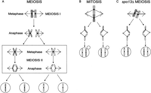
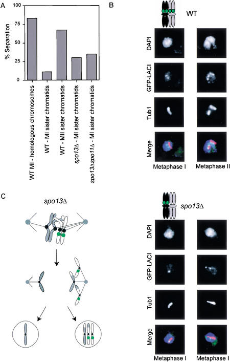
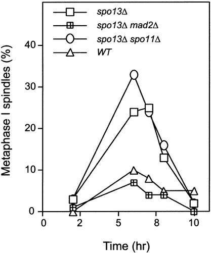
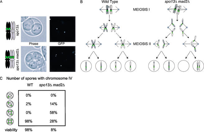
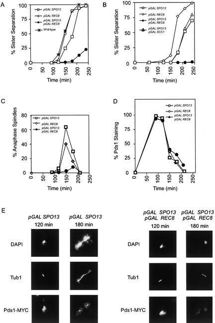
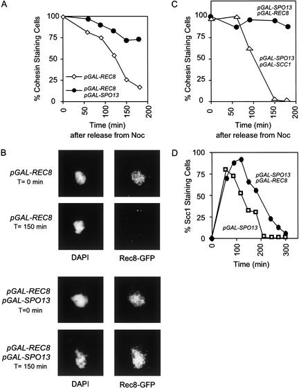
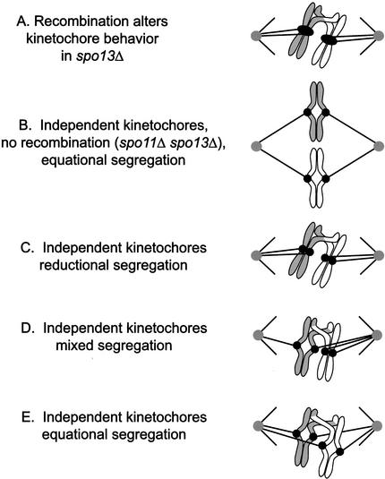
In the absence of Spo13, budding yeast cells complete a single meiotic division during which sister chromatids often separate. We investigated the function of Spo13 by following chromosomes tagged with green fluorescent protein. The occurrence of a single division in spo13Delta homozygous diploids depends on the spindle checkpoint. Eliminating the checkpoint accelerates meiosis I in spo13Delta cells and allows them to undergo two divisions in which sister chromatids often separate in meiosis I and segregate randomly in meiosis II. Overexpression of Spo13 and the meiosis-specific cohesin Rec8 in mitotic cells prevents separation of sister chromatids despite destruction of Pds1 and activation of Esp1. This phenotype depends on the combined overexpression of both proteins and mimics one aspect of meiosis I chromosome behavior. Overexpressing the mitotic cohesin, Scc1/Mcd1, does not substitute for Rec8, suggesting that the combined actions of Spo13 and Rec8 are important for preventing sister centromere separation in meiosis I.


Figures

Figure 1
Figure 1
Diagram of wild-type meiosis and mitosis, and meiosis in spo13Δ. (A) In meiosis I, homologous chromosomes are held together by chiasmata and segregate away from one another when cohesion is released from sister chromatid arms. In meiosis II, sister chromatids segregate away from one another when cohesion is released from sister centromeres. (B) Mitosis is a single division in which sister chromatids segregate from each other and cohesion is lost simultaneously at the arms and centromeres. (C) In spo13Δ mutants, a single meiotic division occurs in which sister chromatids segregate from each other as cohesion is lost simultaneously from arms and centromeres.
Figure 2
Figure 2
spo13Δ and spo13Δ spo11Δ mutants separate sister centromeres during meiosis. (A) Wild-type (MAS 651), spo13Δ (MAS 875), and spo13Δ spo11Δ (MAS 676) homozygous diploids were sporulated and fixed for indirect immunofluorescence against α tubulin and GFP–LacI binding to a Lac operator array located 2.1 kb from CEN VIII. The percentage of cells with separated homologous centromeres was quantified for wild-type cells in metaphase I. The percentage of cells with separated sister centromeres was quantified for wild-type cells in metaphase of meiosis II. The percentage cells with separated sister centromeres of at least 100 cells in meiosis I was quantified for spo13Δ and spo13Δ spo11Δ. Centromeres were considered separated if two LacI signals could be resolved. (B) Pictures of separated wild-type homologous centromeres in metaphase of meiosis I (left panel, top) and separated sister centromeres in metaphase of meiosis II (right panel, top). Pictures of spo13Δ metaphase I sister centromeres that fail to separate (left panel, bottom) and those that separate (right panel, bottom). (C) Cartoon of spo13Δ mutants illustrating bi-orientation of homologs and sister chromatid separation near the centromere.
Figure 3
Figure 3
The spindle checkpoint delays spo13Δ mutants in metaphase of meiosis I. (▵) Wild type (MAS118 × MAS119); (□) spo13Δ (MAS278 × MAS279); (○) spo13Δ spo11Δ (MAS 659 × MAS 660); and (│) spo13Δ mad2Δ (MAS442 × MAS443) homozygous diploids were sporulated and fixed for indirect immunofluorescence against α tubulin. The percentage of cells with metaphase spindles was quantified by fluorescence microscopy and graphed against time during sporulation. At least 100 cells were counted for each time point and the experiment was repeated three times with similar results. The spindle checkpoint delays spo13Δ and spo13Δ spo11Δ during metaphase I.
Figure 4
Figure 4
Two meiotic divisions occur in spo13Δ mad2Δ homozygous diploids. (A) GFP-tagged chromosomes were observed in spores by fluorescence microscopy. (Top panel) Pictures of sister chromatid separation in a dyad formed by spo13Δ homozygous diploids (MAS 314). One copy of chromosome IV is marked with a LacO array located 12 kb from CENIV. (Bottom panel) Pictures of random chromosome segregation in a tetrad formed by spo13Δ mad2Δ homozygous diploid (MAS 356). Both copies of chromosome IV are marked with GFP. (B) Cartoon of chromosome segregation in wild-type meiosis (left) and one of several possible patterns of chromosome segregation in spo13Δ mad2Δ homozygous diploids. Sister chromatids are shown separating to opposite poles at anaphase of meiosis I and then segregating at random in the second meiotic division. (C) Chromosome segregation and viability of wild-type and spo13Δ mad2Δ homozygous diploids. Cells were treated as in A. Viability was determined by picking and germinating spores. Chromosome segregation is close to random in spo13Δ mad2Δ homozygous diploids and 92% of spores are inviable.
Figure 5
Figure 5
Sister chromatids do not separate, but Pds1 is destroyed in strains overexpressing Spo13 and a meiotic cohesin. (A) GFP-tagged chromosomes were observed throughout the cell cycle by fluorescence microscopy in cells overexpressing various combinations of Spo13, a meiotic cohesin subunit (Rec8), and its mitotic counterpart (Scc1/Mcd1), as indicated. Sister chromatid separation was quantified and the percentage of cells with separated sister chromatids is graphed against time after release from a G1 arrest. For each timecourse, at least 100 cells were counted at every timepoint and the experiments were repeated at least twice. Strains: (□) pGAL–SPO13 (MAS 774); (⋄) pGAL–REC8 (MAS 786); (●) pGAL–REC8 and pGAL–SPO13 (MAS 775); or (✠) wild type (SBY 214). (B) Sister chromatid separation assessed as in A. Strains: (□) pGAL–SPO13 (MAS 774); (⋄) pGAL–REC8 (MAS 786); (●) pGAL–REC8 and pGAL–SPO13 (MAS 775); (▵) pGAL–SCC1 pGAL–SPO13 (MAS 874). (C) Indirect immunofluorescence microscopy was performed against α tubulin and percentage of cells with anaphase spindles were quantified and graphed against time after release from a G1 arrest into galactose-containing medium. Strains: (□) pGAL–SPO13 (MAS 774); (⋄) pGAL–REC8 (MAS 786); (●) pGAL–REC8 and pGAL–SPO13 (MAS 775). (D) Pds1 destruction was quantified and graphed against time after release from a G1 arrest into galactose-containing medium. PDS1–MYC was detected by indirect immunofluorescence throughout the cell cycle in the indicated strains: (□) pGAL–SPO13 (MAS 774); (⋄) pGALREC8 (MAS785); (●) pGAL–SPO13 and pGAL–REC8 (MAS 775). Punctate staining is GFP–LacI. (E) Images from the experiment quantified in D, T = 120 min (left panels), T = 180 min (right panels). DAPI staining shown at top panels, anti-α tubulin staining (middle panels), and anti-MYC staining (bottom panels). Sister chromatids failed to separate and the spindle did not elongate in cells expressing pGAL–SPO13 and pGAL–REC8 despite the destruction of Pds1.
Figure 6
Figure 6
Rec8 is protected on chromosomes in the presence of Spo13. (A) A Rec8–GFP fusion was detected by indirect immunofluorescence at the indicated times after release from nocodazole arrest in strains expressing either (⋄) pGAL–REC8–GFP (MAS 850) or (●) pGAL–REC8–GFP pGAL–SPO13 (MAS 866). Rec8–GFP destruction was quantified by fluorescence microscopy and graphed against time after release from nocodazole arrest. (B) Images from the experiment quantified in A, at T = 0 min (top panels), T = 150 (bottom panels). DAPI staining is shown in left panels, anti-GFP staining is shown in right panels. Rec8–GFP is protected on chromosomes in cells expressing Spo13. (C) Cohesin staining was detected by indirect immunofluorescence against SCC1–HA3 (Uhlmann and Nasmyth 1998) after release from nocodazole arrest into glucose-containing medium. Strains expressed either (●) pGAL–REC8–GFP pGAL–SPO13 (MAS 866) or (▵) pGAL–SCC1–HA3 (Uhlmann and Nasmyth 1998), pGAL–SPO13 (MAS 874). (D) Indirect immunofluorescence was performed on Scc1/Mcd1–3XHA expressed from its own promoter in strains expressing either (□) pGAL–SPO13 (MAS 823) or (●) pGAL–SPO13 pGAL–REC8 (MAS 821). Staining of endogenous Scc1/Mcd1–3XHA is graphed against time after release from a G1 arrest. Scc1/Mcd1–3XHA is removed from chromosomes in the presence of Spo13 and Rec8.
Figure 7
Figure 7
Centromere behavior in spo13Δ mutants. Model for centromere behavior in spo13Δ mutants. (A) The act of recombination in a spo13Δ cell induces the two sister kinetochores to act as a single functional unit, as they do in wild-type cells in meiosis I. (B) In spo11Δ spo13Δ cells, no recombination occurs. Kinetochores can only come under tension if they attach to opposite poles, explaining why sister centromeres always segregate from each other at meiosis I in these cells. (C–E) Recombination has no direct effect on kinetochore behavior, but connects together four independent kinetochores. (C) Homologs segregate to opposite poles (reductional division) when one set of sister kinetochores attaches to one pole, and the other pair attaches to the opposite pole. (D) A mixed division occurs when one pair of sister kinetochores attaches to opposite poles and the other pair attaches to the same pole. (E) Sister centromeres separate from each other at anaphase (equational segregation) when they attach to opposite poles of the spindle.

Similar articles

Cited by

References

    1. Bernard P, Maure JF, Javerzat JP. Fission yeast Bub1 is essential in setting up the meiotic pattern of chromosome segregation. Nat Cell Biol. 2001;3:522–526. - PubMed
    1. Bhatt AM, Lister C, Page T, Fransz P, Findlay K, Jones GH, Dickinson HG, Dean C. The DIF1 gene of Arabidopsis is required for meiotic chromosome segregation and belongs to the REC8/RAD21 cohesin gene family. Plant J. 1999;19:463–472. - PubMed
    1. Biggins S, Severin FF, Bhalla N, Sassoon I, Hyman AA, Murray AW. The conserved protein kinase Ipl1 regulates microtubule binding to kinetochores in budding yeast. Genes & Dev. 1999;13:532–544. - PMC - PubMed
    1. Buonomo SB, Clyne RK, Fuchs J, Loidl J, Uhlmann F, Nasmyth K. Disjunction of homologous chromosomes in meiosis I depends on proteolytic cleavage of the meiotic cohesin Rec8 by separin. Cell. 2000;103:387–398. - PubMed
    1. Ciosk R, Zachariae W, Michaelis C, Shevchenko A, Mann M, Nasmyth K. An ESP1/PDS1 complex regulates loss of sister chromatid cohesion at the metaphase to anaphase transition in yeast. Cell. 1998;93:1067–1076. - PubMed

Publication types