. 2002 Jul 1;16(13):1696-706.
doi: 10.1101/gad.231702.

Spot 42 RNA mediates discoordinate expression of the E. coli galactose operon

Affiliations

Spot 42 RNA mediates discoordinate expression of the E. coli galactose operon

Thorleif Møller et al. Genes Dev. .

Abstract

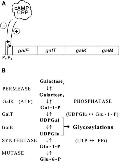
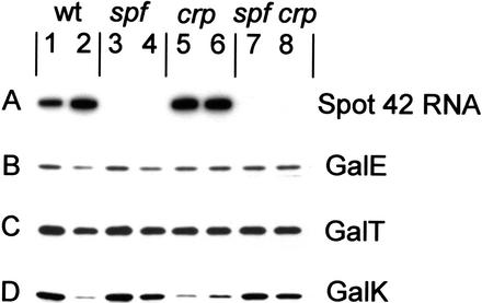
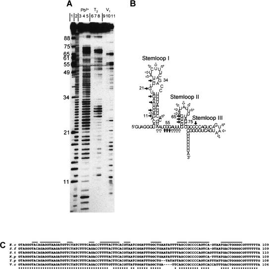
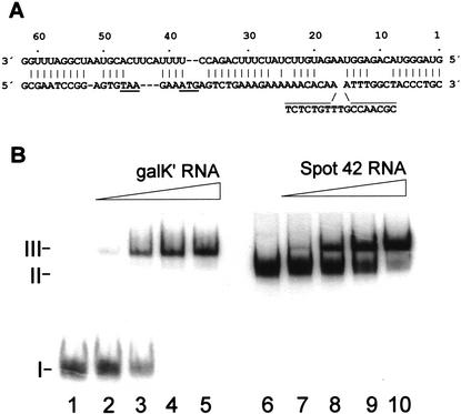
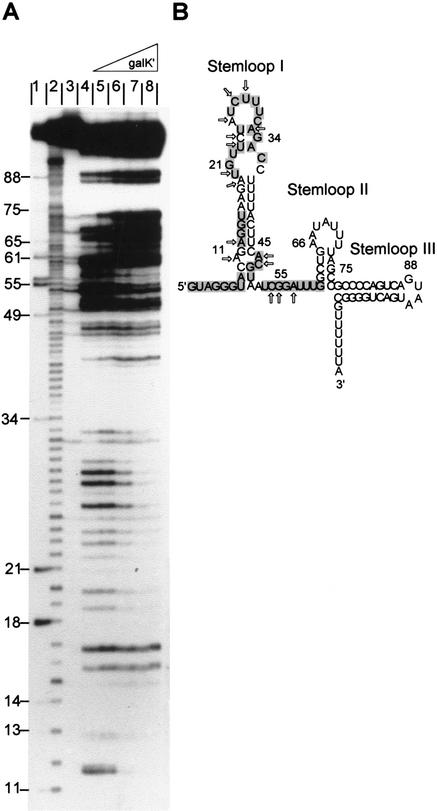
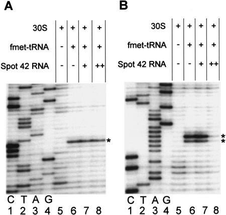
The physiological role of Escherichia coli Spot 42 RNA has remained obscure, even though the 109-nucleotide RNA was discovered almost three decades ago. Structural features of Spot 42 RNA and previous work suggested to us that the RNA might be a regulator of discoordinate gene expression of the galactose operon, a control that is only understood at the phenomenological level. The effects of controlled expression of Spot 42 RNA or deleting the gene (spf) encoding the RNA supported this hypothesis. Down-regulation of galK expression, the third gene in the gal operon, was only observed in the presence of Spot 42 RNA and required growth conditions that caused derepression of the spf gene. Subsequent biochemical studies showed that Spot 42 RNA specifically bound at the galK Shine-Dalgarno region of the galETKM mRNA, thereby blocking ribosome binding. We conclude that Spot 42 RNA is an antisense RNA that acts to differentially regulate genes that are expressed from the same transcription unit. Our results reveal an interesting mechanism by which the expression of a promoter distal gene in an operon can be modulated and underline the importance of antisense control in bacterial gene regulation.


Figures

Figure 1
Figure 1
The E. coli galactose operon. (A) Schematic diagram of the E. coli galactose operon. Relative positions of the four genes are shown. The genes encode: galE, epimerase (GalE); galT, transferase (GalT); galK, kinase (GalK); and galM, mutarotase. The two promoters (P1 and P2) are modulated negatively by Gal repressor, and the cAMP–CRP complex abolishes transcription from P2 and is required for P1 transcription. (B) The pathway for galactose metabolism. Note that the GalE, GalT, and GalK enzymes are part of an amphibolic pathway that produces substrates for biosynthetic glycosylations (UDP-glucose and UDP-galactose). Moreover, the pathway generates galactose from UDP-galactose when cells are growing in other carbon sources. Subscripts: (e) extracellular; (i) intracellular.
Figure 2
Figure 2
Effects of controlled spf expression on the Gal enzyme pattern. The figure shows the relevant part of two-dimensional gels of SØ928 Δspf cells harboring pGal4 (a derivative of pBR322 carrying the galETKM genes) and pSpf33 (a derivative of pBAD33 carrying the spf gene under the control of the inducible araBAD promoter). Uninduced and induced cultures (induced with 1 mM arabinose for 20 min) were labeled with [35S]methionine for 1 min and concentrated. Following this, their proteins were analyzed by two-dimensional gel electrophoresis and autoradiography. (A) Uninduced cells. (B) Induced cells. Arrows mark the positions of the four Gal enzymes. After quantification of the Gal protein spots from uninduced cells (A) and induced cells (B), the following ratios were obtained: GalEB/GalEA = 1.09, GalTB/GalTA = 0.92, GalKB/GalKA = 0.47, and GalMB/GalMA = 0.83 (all normalized to standard spots that were not affected by Spot 42 RNA).
Figure 3
Figure 3
Effects of Spot 42 RNA on the cellular levels of the Gal enzymes. (A) Northern blot analysis of Spot 42 RNA levels and (BD) immunoblots of GalE, GalT, and GalK levels in isogenic gal constitutive spf+ and Δspf strains. The RNA and the proteins from equal amounts of cells growing to early-exponential phase were electrophoretically separated on a urea–8% polyacrylamide gel and on an SDS–10% polyacrylamide gel, respectively, and probed as described in Materials and Methods. (Lanes 1,3,5,7) Glycerol-grown cells; (lanes 2,4,6,8) glucose-grown cells. In all experiments casamino acids (0.05%) were included in the growth medium. After quantification of the Gal proteins from glycerol- and glucose-grown ΔgalR and ΔgalR, Δspf cells, the following ratios were obtained for GalE, GalT, and GalK, respectively: ΔgalR strain (lane 1/lane 2) 1.5, 1.4, and 4.9; ΔgalR, Δspf strain (lane 3/lane 4) 1.4, 1.4, and 1.4. The results are the average of three independent experiments.
Figure 4
Figure 4
Secondary structure of Spot 42 RNA. (A) Nuclease and lead (II) acetate probing of 5′-end-labeled Spot 42 RNA. (Lane 1) RNase T1 cleavage of Spot 42 RNA under denaturing condition (G-specific cleavage); (lane 2) alkaline hydrolysis ladder; (lanes 3,6,9) control (untreated Spot 42 RNA); (lanes 4,5), partial cleavage by lead (II) acetate; (lanes 7,8) partial cleavage by RNase T2; (lanes 10,11) partial cleavage by RNase V1. (B) Structure predictions of Spot 42 RNA based on thermodynamic criteria and summary of the structural probing. Single-stranded-specific and double-stranded-specific nuclease susceptibility are indicated by open and filled arrows, respectively. (C) Phylogenetic structure analysis of Spot 42 RNA. The DNA sequences of Spot 42 RNA from E. coli, Shigella flexneri, Salmonella typhimurium, Klebsiella pneumoniae, Yersinia pestis and Vibrio cholerae were aligned. A star indicates identity. The inverted repeats predicted to fold into stem structures are overlined. Sequences for Shigella, Salmonella, Klebsiella, and Yersinia were obtained from a BLAST search of the E. coli sequence with the unfinished microbial genome database at the Institute for Genomic Research Web site at http://www.tigr.org. Shigella and Yersinia sequencing data were from the University of Wisconsin–Madison Genome Project. The Washington University Consortium sequencing project obtained the data for the Salmonella and Klebsiella sequences.
Figure 5
Figure 5
Spot 42 RNA–galK mRNA interaction. (A) The complementarity between Spot 42 RNA (top sequence) and the galK translation initiation region (bottom sequence). The stop codon of galT and the start codon of galK are underlined. (B) Gel mobility shift assays of Spot 42 RNA binding to galK‘ RNA. (Lanes 15) 5′-End-labeled transcript of Spot 42 RNA (0.05 pmole) and 500-fold molar excess tRNA were incubated with increasing amounts of unlabeled galK‘ RNA (0, 1, 5, 10, and 20 pmole) to allow complex formation and then resolved on a native polyacrylamide gel. (Lanes 610) 5′-End-labeled galK‘ RNA (0.05 pmole) and 500-fold molar excess of tRNA were incubated with increasing amounts of unlabeled Spot 42 RNA (0, 1, 5, 10, and 20 pmole) to allow complex formation and then resolved on a native polyacrylamide gel.
Figure 6
Figure 6
Nuclease probing of Spot 42 RNA–galK‘ RNA complexes. (A) In vitro synthesized 5′-end-labeled Spot 42 RNA (0.05 pmole) was mixed with increasing concentrations of unlabeled galK‘ mRNA (1, 5, 10, and 20 pmole), and the incubations were treated with RNase T2. (Lane 1) RNase T1 cleavage of Spot 42 RNA under denaturing condition (G-specific cleavage); (lane 2) alkaline hydrolysis ladder; (lane 3) control (untreated Spot 42 RNA); (lanes 48) RNase T2 footprinting reactions in absence (lane 4) and in presence (lanes 58) of galK‘ RNA. (B) Summary of changes in the cleavage pattern induced by galK‘ RNA. The galK complementary regions are shown on a gray background. Arrows indicate reduced cleavage.
Figure 7
Figure 7
Toeprinting analysis of 30S ribosomal subunit binding to galT‘ and galK‘ RNA. (A) Ternary complex formation on galT‘ RNA. The toeprint signal is at position +15 relative to A of the start codon (marked by a star). (B) Ternary complex formation on galK‘ RNA. The toeprint signal is at positions +16 and +17 relative to A of the start codon (marked by stars). In all reactions the concentration of mRNA and 30S subunits was 0.04 μM. fMet-tRNA was in molar excess over 30S subunits. When present, Spot 42 RNA was added prior to the addition of 30S subunits and fMet-tRNA. Spot 42 RNA was added at the molar ratios to mRNA, 1:2 (lane 7) and 5:1 (lane 8). (Lanes 14) The DNA sequence reactions (G A T C) were carried out with the same end-labeled oligonucleotide used in the toeprinting assays.

References

    1. Adhya S. The galactose operon. In: In: Neidhardt FC, et al., editors. Escherichia coli and Salmonella thyphimurium: Cellular and molecular biology. Washington, DC.: American Society for Microbiology; 1987. pp. 1503–1512.
    1. Adhya S, Echols H. Glucose effect and the galactose enzymes of Escherichia coli: Correlation between glucose inhibition of induction and inducer transport. J Bacteriol. 1966;92:601–608. - PMC - PubMed
    1. Adoutte A. Evolutionary biology. Small but mighty timekeepers. Nature. 2000;408:37–38. - PubMed
    1. Altuvia S, Wagner EG. Switching on and off with RNA. Proc Natl Acad Sci. 2000;97:9824–9826. - PMC - PubMed
    1. Altuvia S, Zhang A, Argaman L, Tiwari A, Storz G. The Escherichia coli OxyS regulatory RNA represses fhlA translation by blocking ribosome binding. EMBO J. 1998;17:6069–6075. - PMC - PubMed

Publication types

MeSH terms