

Detection of the CD56+/CD45- immunophenotype by flow cytometry in neuroendocrine malignancies
- PMID: 12101203
- PMCID: PMC1769705
- DOI: 10.1136/jcp.55.7.535
Detection of the CD56+/CD45- immunophenotype by flow cytometry in neuroendocrine malignancies
Abstract
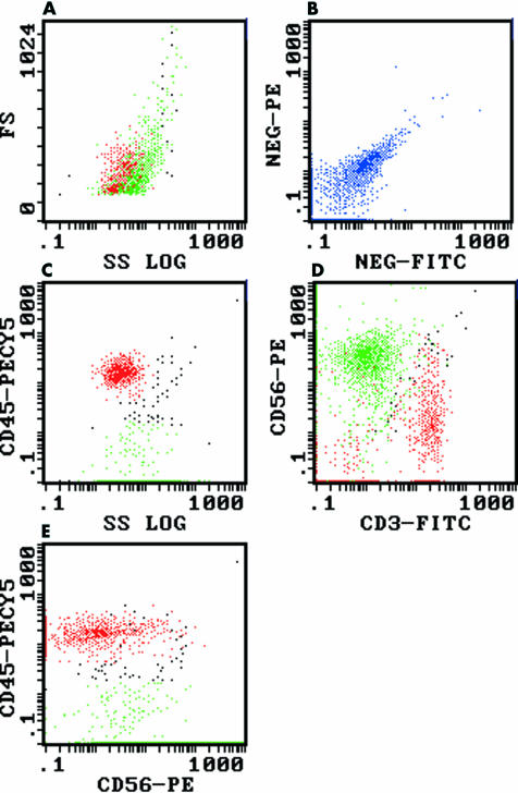
Aims: Antibodies against CD56 are primarily used in flow cytometric studies to detect natural killer cells. However, they may be useful in the identification of neuroendocrine malignancies, especially if the cells do not express CD45, indicating a non-leucocyte origin.
Methods: A retrospective review was conducted on all solid tissue flow cytometric studies performed between January 1997 and September 2001, to identify all cases with a CD56+/CD45- immunophenotype.
Results: Twelve neuroendocrine malignancies (five metastatic small cell carcinomas, three Merkel cell carcinomas, two metastatic undifferentiated neuroendocrine carcinomas, one metastatic pancreatic neuroendocrine carcinoma, and one neuroblastoma) were identified.
Conclusions: CD56+/CD45- neuroendocrine malignancies are only rarely detected in the flow cytometric analysis of solid tissue samples. However, the recognition of this immunophenotype is important to avoid their misclassification as natural killer cell malignancies. Furthermore, flow cytometry assists in the rapid identification of such cases, so that appropriate immunohistochemical studies can be performed to facilitate their correct diagnosis.
Figures
Similar articles
-
CD56 expression of neuroendocrine neoplasms on immunophenotyping by flow cytometry: a novel diagnostic approach to fine-needle aspiration biopsy.Cancer. 2003 Aug 25;99(4):240-6. doi: 10.1002/cncr.11458. Cancer. 2003. PMID: 12925986
-
Detection of Nonhematologic Neoplasms by Routine Flow Cytometry Analysis.Am J Clin Pathol. 2020 Jan 1;153(1):99-104. doi: 10.1093/ajcp/aqz138. Am J Clin Pathol. 2020. PMID: 31587038
-
Interference of bone marrow CD56+ mesenchymal stromal cells in minimal residual disease investigation of neuroblastoma and other CD45- /CD56+ pediatric malignancies using flow cytometry.Pediatr Blood Cancer. 2019 Aug;66(8):e27799. doi: 10.1002/pbc.27799. Epub 2019 May 7. Pediatr Blood Cancer. 2019. PMID: 31066205
-
Collision tumor of primary merkel cell carcinoma and chronic lymphocytic leukemia/small lymphocytic lymphoma, diagnosed on ultrasound-guided fine-needle aspiration biopsy: a unique case report and review of literature.Diagn Cytopathol. 2015 Jan;43(1):66-71. doi: 10.1002/dc.23127. Epub 2014 Mar 8. Diagn Cytopathol. 2015. PMID: 24610800 Review.
-
Comprehensive phenotyping of endothelial cells using flow cytometry 2: Human.Cytometry A. 2021 Mar;99(3):257-264. doi: 10.1002/cyto.a.24293. Epub 2020 Dec 23. Cytometry A. 2021. PMID: 33369145 Free PMC article. Review.
Cited by
-
CD56, HLA-DR, and CD45 recognize a subtype of childhood AML harboring CBFA2T3-GLIS2 fusion transcript.Cytometry A. 2021 Aug;99(8):844-850. doi: 10.1002/cyto.a.24339. Epub 2021 Apr 2. Cytometry A. 2021. PMID: 33811445 Free PMC article.
-
Dynamics of Minimal Residual Disease in Neuroblastoma Patients.Front Oncol. 2019 Jun 4;9:455. doi: 10.3389/fonc.2019.00455. eCollection 2019. Front Oncol. 2019. PMID: 31214500 Free PMC article. Review.
-
Aggressive high-grade Ewing's sarcoma of maxilla: A rare case report.J Oral Maxillofac Pathol. 2018 Jan;22(Suppl 1):S48-S53. doi: 10.4103/jomfp.JOMFP_89_17. J Oral Maxillofac Pathol. 2018. PMID: 29491605 Free PMC article.
-
Comparison of Bone Marrow Biopsy and Flow Cytometry in Demonstrating Bone Marrow Metastasis of Neuroblastoma.Diagnostics (Basel). 2024 Dec 11;14(24):2776. doi: 10.3390/diagnostics14242776. Diagnostics (Basel). 2024. PMID: 39767137 Free PMC article.
-
Acute Myeloid Leukemia with RAM Immunophenotype: A Pediatric Case with Unusual Morphologic Features.Hematol Rep. 2017 Jun 14;9(2):7057. doi: 10.4081/hr.2017.7057. eCollection 2017 Jun 1. Hematol Rep. 2017. PMID: 28670435 Free PMC article.
References
-
- Knapp W, Dorken B, Rieber P, et al. CD antigens 1989. Blood 1989;74:1448–50. - PubMed
-
- Brackenbury R. Expression of neural cell adhesion molecules in normal and pathologic tissues. Ann N Y Acad Sci 1988;540:39–46. - PubMed
-
- Fischler DF, Bauer TW, Tubbs RR. Tissue reactivity of anti-Leu19. Histopathology 1992;21:563–7. - PubMed
-
- Wick MR. Immunohistology of neuroendocrine and neuroectodermal tumors. Semin Diagn Pathol 2000;17:194–203. - PubMed
MeSH terms
Substances
LinkOut - more resources
Full Text Sources
Other Literature Sources
Research Materials
Miscellaneous