. 2002 Aug;22(15):5281-95.
doi: 10.1128/MCB.22.15.5281-5295.2002.

PIN1 is an E2F target gene essential for Neu/Ras-induced transformation of mammary epithelial cells

Affiliations

PIN1 is an E2F target gene essential for Neu/Ras-induced transformation of mammary epithelial cells

Akihide Ryo et al. Mol Cell Biol. 2002 Aug.

Abstract

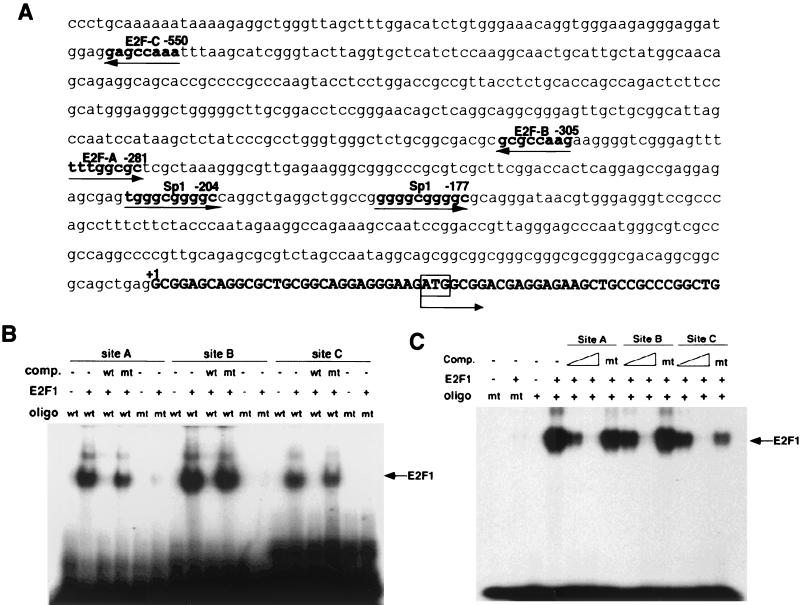
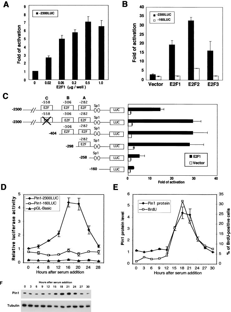
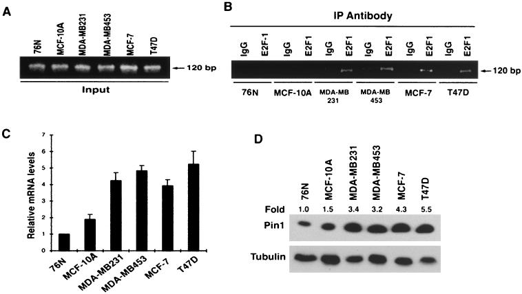
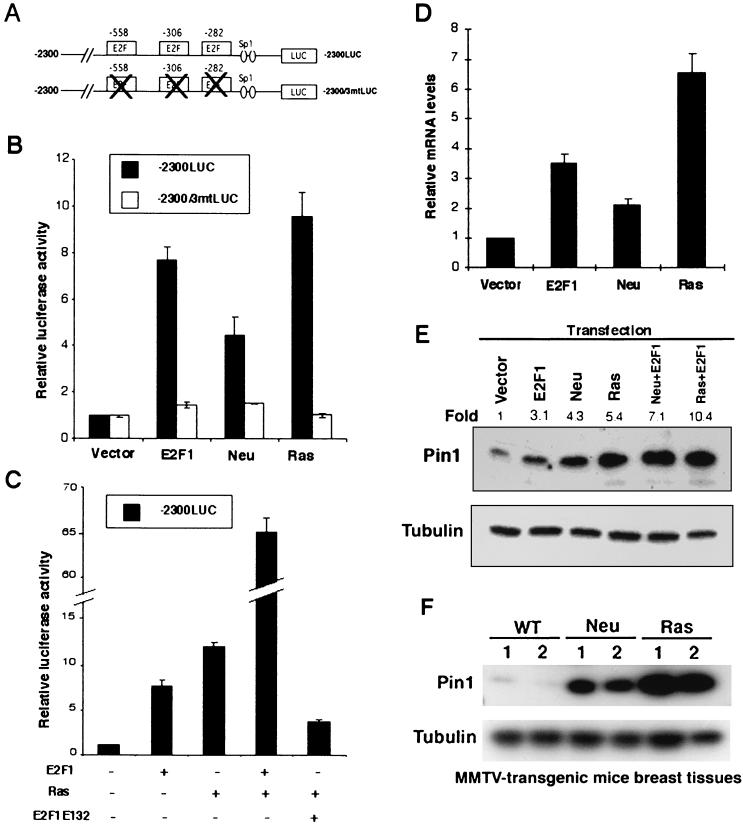
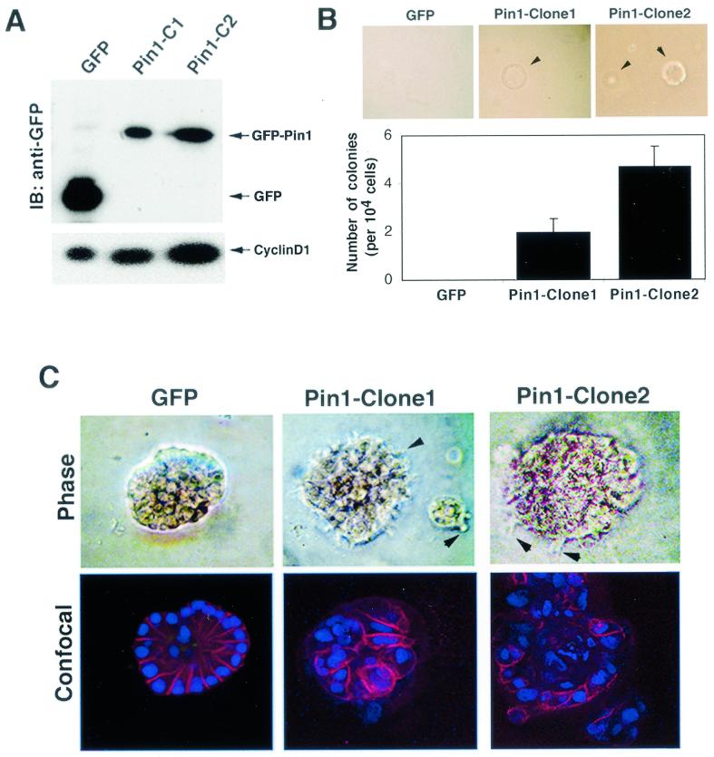
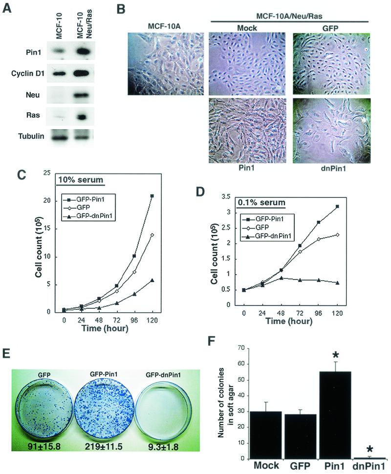
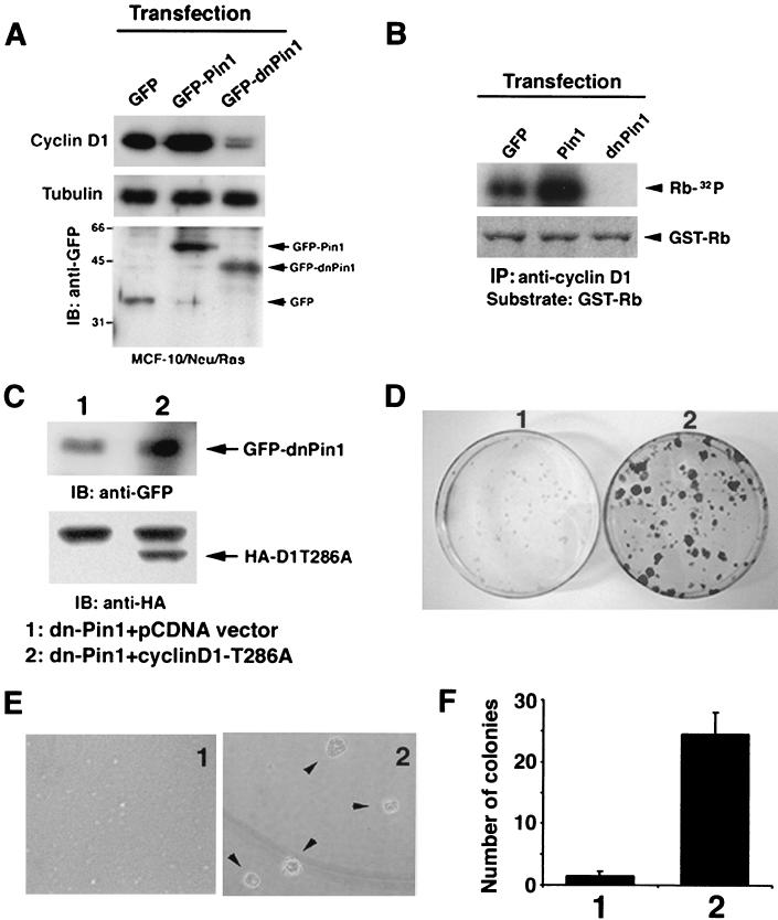
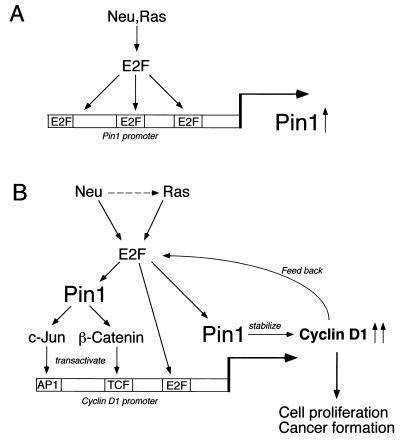
Oncogenes Neu/HER2/ErbB2 and Ras can induce mammary tumorigenesis via upregulation of cyclin D1. One major regulatory mechanism in these oncogenic signaling pathways is phosphorylation of serines or threonines preceding proline (pSer/Thr-Pro). Interestingly, the pSer/Thr-Pro motifs in proteins exist in two completely distinct cis and trans conformations, whose conversion is catalyzed specifically by the essential prolyl isomerase Pin1. By isomerizing pSer/Thr-Pro bonds, Pin1 can regulate the conformation and function of certain phosphorylated proteins. We have previously shown that Pin1 is overexpressed in breast tumors and positively regulates cyclin D1 by transcriptional activation and posttranslational stabilization. Moreover, in Pin1 knockout mice, mammary epithelial cells fail to undergo massive proliferation during pregnancy, as is the case in cyclin D1 null mice. These results indicate that Pin1 is upregulated in breast cancer and may be involved in mammary tumors. However, the mechanism of Pin1 overexpression in cancer and its significance in cell transformation remain largely unknown. Here we demonstrate that PIN1 expression is mediated by the transcription factor E2F and enhanced by c-Neu and Ha-Ras via E2F. Furthermore, overexpression of Pin1 not only confers transforming properties on mammary epithelial cells but also enhances the transformed phenotypes of Neu/Ras-transformed mammary epithelial cells. In contrast, inhibition of Pin1 suppresses Neu- and Ras-induced transformed phenotypes, which can be fully rescued by overexpression of a constitutively active cyclin D1 mutant that is refractory to the Pin1 inhibition. Thus, Pin1 is an E2F target gene that is essential for the Neu/Ras-induced transformation of mammary epithelial cells through activation of cyclin D1.


Figures

FIG. 1.
FIG. 1.
E2F binds the PIN1 promoter. (A) Human PIN1 promoter sequence. The nucleotide sequence of the human PIN1 gene that includes the 5′-flanking region and first exon is listed. Putative binding sites for transcription factors are underlined. The ATG translation initiation codon is in the first exon typed in boldface. (B) Electrophoretic mobility shift assays were performed with recombinant E2F1 protein and end-labeled double-stranded oligonucleotides (oligo) corresponding to the PIN1 promoter sequence containing either wild-type (wt) or mutant (mt) E2F binding sites. A consensus E2F site from the adenovirus E2 promoter was used as a competitor (comp.) in a 100× molar excess of labeled probe. (C) Competitive activity of PIN1 promoter sequences for E2F binding. Labeled oligonucleotides corresponding to E2F binding sites from the adenovirus E2 promoter were incubated with recombinant E2F1 protein in the presence or absence of unlabeled PIN1 promoter sequences. Three different oligonucleotides corresponding to E2F binding sites in the PIN1 promoter (sites A to C) were used as competitors. Wild-type oligonucleotides were mixed at a 10- or 100-fold molar excess and mutants were mixed at a 100-fold molar excess of labeled probe.
FIG. 2.
FIG. 2.
Activation of the PIN1 promoter by E2F. (A) E2F1 activates PIN1 promoter activity in a dose-dependent manner. MCF-7 cells were transfected with the PIN1 promoter-luciferase construct (−2300LUC) and E2F-1 expression vector. Cells were harvested at 36 h after transfection and subjected to a gene reporter assay. (B) E2F family proteins enhance PIN1 promoter activity. Cells were cotransfected with vectors expressing E2F1, E2F2, or E2F3 together with the −2300LUC or −160LUC reporter construct. (C) Mapping of the PIN1 promoter region responsible for transcriptional activation by E2F. A series of 5′ deletion and site-directed mutants were transfected into HeLa cells together with the E2F1 expression vector or a control vector. (D) Cell growth-dependent regulation of PIN1 gene expression in normal fibroblasts. MEFs were transfectedwith the indicated luciferase reporter constructs and induced to enter quiescence by serum starvation (0.05% serum) for 48 h. The medium was then supplemented with serum (15%), allowing cells to reenter the cell cycle as a synchronous population. Cells were harvested at various time points and subjected to gene reporter assays. (E and F) MEFs were synchronized by serum starvation as for panel D. Prior to harvesting, cells were treated with BrdU for 30 min. Cells were collected at indicated time points and subjected to immunoblotting analysis with anti-Pin1 antibody or flow cytometory analysis with anti-BrdU antibody. Band intensities in Pin1 protein levels (F) were quantified by using NIH-Image and graphed with the results from the BrdU study (E).
FIG. 3.
FIG. 3.
E2F binding to the PIN1 promoter sequence in vivo correlates with Pin1 expression level in breast cancer cell lines. (A and B) Levels of E2F binding to the PIN1 promoter in different breast cell lines. Cross-linked chromatin from exponentially growing breast cancer cell lines was incubated with either antibodies against E2F1 or control IgG. Immunoprecipitates from each sample were analyzed by PCR with primers specific for the PIN1 promoter sequence (B). As an input control, total input chromatin was analyzed by PCR with the same primer set (A). (C and D) Levels of PIN1 mRNA and protein in different breast cell lines. mRNAs were isolated from the cell types indicated, and PIN1 mRNA was quantified by real-time RT-PCR analysis and normalized to GAPDH mRNA (C). PIN1 levels were determined by subjecting cell lysates to immunoblotting analysis with a monoclonal anti-Pin1 antibody (D), Numbers above the gel image indicate the induction (fold) of Pin1 protein level normalized to α-tubulin.
FIG. 4.
FIG. 4.
Ras and Neu stimulate the PIN1 promoter through E2F activation. (A) Schematic representation of wild-type and mutant PIN1 promoter reporter constructs. (B) MCF-7 cells were cotransfected with a reporter construct (0.1 μg) and E2F-1, Ha-Ras, or Neu (0.5 μg), followed by gene reporter assays. (C) Dominant-negative E2F1 inhibits activation of the PIN1 promoter by Ras. Gene reporter assays were performed in MCF-7 cells as shown in panel B. Wild-type E2F-1 or its dominant-negative mutant E2F1E132 was cotransfected with the Ras and −2300LUC reporter constructs. (D) Neu and Ras upregulate PIN1 mRNA levels in MCF-10A cells. MCF-10A cells were transiently transfected with plasmids encoding E2F1, Ha-Ras, or Neu. For each transfection, a plasmid encoding a puromycin resistance gene (pIRES-puro) was cotransfected as a selection marker. Puromycin (1.3 μg/ml) was added to the medium 24 h after transfection. At 36 h following addition of puromycin, puromycin-resistant cells were reseeded and cultured for an additional 24 h. Total RNA was collected and subjected to real-time RT-PCR as described in Materials and Methods. (E) MCF-7 cells were transfected with the indicated expression vectors for 48 h, and cell lysates were subjected to immunoblotting analysis with anti-Pin1 and antitubulin antibodies. Numbers above the gel image indicate the fold induction of the Pin1 protein level normalized to α-tubulin. (F) PIN1 is overexpressed in breast tissues from MMTV-c-Neu and MMTV-Ha-Ras mice. Mammary tissues from two wild-type (WT), two MMTV-Neu (Neu), and two MMTV-Ras (Ras) mice were lysed and subjected to immunoblotting analysis with anti-Pin1 and antitubulin antibodies.
FIG. 5.
FIG. 5.
PIN1 overexpression confers a transformed phenotype on MCF-10A cells. (A) Establishment of MCF-10A cells stably expressing GFP or GFP-Pin1. Immunoblotting (IB) analysis was performed with anti-GFP and anti-cyclin D1 antibodies. (B) To measure anchorage-independent cell growth and survival, GFP- or GFP-Pin1-transfected MCF-10A cells were suspended in 0.3% soft agar for 14 days. (C) Cell lines stably expressing GFP and GFP-Pin1 were plated on Matrigel for 15 days. Phase images of an acinus at higher magnification are shown in the upper panels. The acini were stained with anti-E-cadherin antibodies and the DNA dye TOPRO-3, and confocal images though the middle of an acinus are shown in the lower panels. Arrows indicate cell surface spikes protruding into the Matrigel.
FIG. 6.
FIG. 6.
PIN1 is essential for Neu/Ras-induced cell transformation. (A) Establishment of stable MCF-10A cell lines expressing both Neu and Ras. Cells were cotransfected with Neu and Ras expression vectors and selected with G418. A selected clone was checked for expression of Ras, Neu, PIN1, and cyclin D1 by immunoblotting analysis. (B) Morphological changes in MCF-10A cells stably expressing Neu and Ras (MCF-10/Neu/Ras) and additional PIN1 or dn-PIN1. Cells were seeded in 60-mm dishes and photographed with a phase-contrast microscope before reaching confluence. (C and D) Manipulation of Pin1 levels alters proliferation in MCF-10/Neu/Ras cells. Transfected cells were selected with puromycin for 48 h and reseeded in 35-mm dishes. Cells were grown in high-serum (10%) (C) or low-serum (0.1%) (D) medium and trypsinized at various time points. Viable cells were counted by the trypan blue dye exclusion method. (E) PIN1 is necessary for Neu/Ras-induced focus formation. The same number of cells (104) transfected with either the GFP, GFP-Pin1, or GFP-dnPin1 expression vector were seeded in 10-cm plastic dishes after selection with puromycin. After 14 days, cells were fixed and stained with crystal violet. Numbers below plates indicate colony numbers (mean ± SD) in three independent experiments. (F) Cells were plated in 0.3% soft agar and cultured for 2 weeks. After 14 days, colony formation was scored microscopically. MCF-10/Neu/Ras cells expressing GFP-Pin1 demonstrated significant increases in anchorage-independent growth, whereas dn-Pin1 overexpession significantly blocked the growth of MCF-10/Neu/Ras cells. ∗, P < 0.01, t test.
FIG. 7.
FIG. 7.
Pin1 inhibition is complemented by overexpression of a constitutively active cyclin D1. (A) Pin1 is essential to maintain cyclin D1 level and activity in Neu/Ras-transformed MCF-10 cells. MCF-10/Neu/Ras cells transfected with either GFP, GFP-Pin1, or GFP-dnPin1 were lysed and immunoblotted with anti-cyclin D1, -tubulin, and -GFP antibodies. (B) The same cell lysates as in panel A were immunoprecipitated with anti-cyclin D1 antibodies, followed by the in vitro kinase assay with GST-pRB as a substrate. Phosphorylation of the GST-pRB substrate is shown in the upper panel. The lower panel shows input GST-pRB stained with Coomassie blue. (C) MCF-10/Neu/Ras cells were transfected with either dnPin1 and pCDNA vector or dnPin1 and the cyclin D1T286A mutant (1:10 ratio) and selected with puromycin for 48 h. Cells were subjected to immunoblotting analysis with anti-HA and anti-GFP antibodies. (D) Cells were transfected as described for C and seeded on plastic plates for 3 weeks. Cells were fixed and stained with crystal violet. (E and F) Cells were transfected as described for panel C and cultured in 0.3% soft agar for 3 weeks. The number of colonies formed was scored. Representative phase pictures are shown in panel E. Colony numbers are the mean ± SD of three independent experiments (F).
FIG. 8.
FIG. 8.
Schematic models for Pin1 transcriptional regulation and its role in regulation of cyclin D1 by Neu/Ras signaling. (A) Oncogenic Neu-Ras signaling transactivates the PIN1 promoter through E2F activity. (B) PIN1 is a downstream target of oncogenic Neu/Ras signaling and is essential for Neu/Ras-induced cyclin D1 activation and cell transformation. PIN1 upregulated by Neu/Ras signaling enhances β-catenin and c-Jun signaling to transactivate the cyclin D1 gene. Furthermore, Pin1 binds directly to cyclin D1 and stabilizes it via a posttranslational mechanism. It is possible that cyclin D1 also regulates Pin1 expression via E2F in a positive feedback loop.

Similar articles

Cited by

References

    1. Albanese, C., J. Johnson, G. Watanabe, N. Eklund, D. Vu, A. Arnold, and R. G. Pestell. 1995. Transforming p21ras mutants and c-Ets-2 activate the cyclin D1 promoter through distinguishable regions. J. Biol. Chem. 270:23589-23597. - PubMed
    1. Alt, J. R., J. L. Cleveland, M. Hannink, and J. A. Diehl. 2000. Phosphorylation-dependent regulation of cyclin D1 nuclear export and cyclin D1-dependent cellular transformation. Genes Dev. 14:3102-3114. - PMC - PubMed
    1. Andrechek, E. R., and W. J. Muller. 2000. Tyrosine kinase signalling in breast cancer: tyrosine kinase-mediated signal transduction in transgenic mouse models of human breast cancer. Breast Cancer Res. 2:211-216. - PMC - PubMed
    1. Arber, N., Y. Doki, E. K. Han, A. Sgambato, P. Zhou, N. H. Kim, T. Delohery, M. G. Klein, P. R. Holt, and I. B. Weinstein. 1997. Antisense to cyclin D1 inhibits the growth and tumorigenicity of human colon cancer cells. Cancer Res. 57:1569-1574. - PubMed
    1. Bartkova, J., J. Lukas, H. Muller, D. Lutzhoft, M. Strauss, and J. Bartek. 1994. Cyclin D1 protein expression and function in human breast cancer. Int. J. Cancer 57:353-361. - PubMed

Publication types

MeSH terms

LinkOut - more resources