

E2F1 induces phosphorylation of p53 that is coincident with p53 accumulation and apoptosis
- PMID: 12101227
- PMCID: PMC133953
- DOI: 10.1128/MCB.22.15.5308-5318.2002
E2F1 induces phosphorylation of p53 that is coincident with p53 accumulation and apoptosis
Abstract
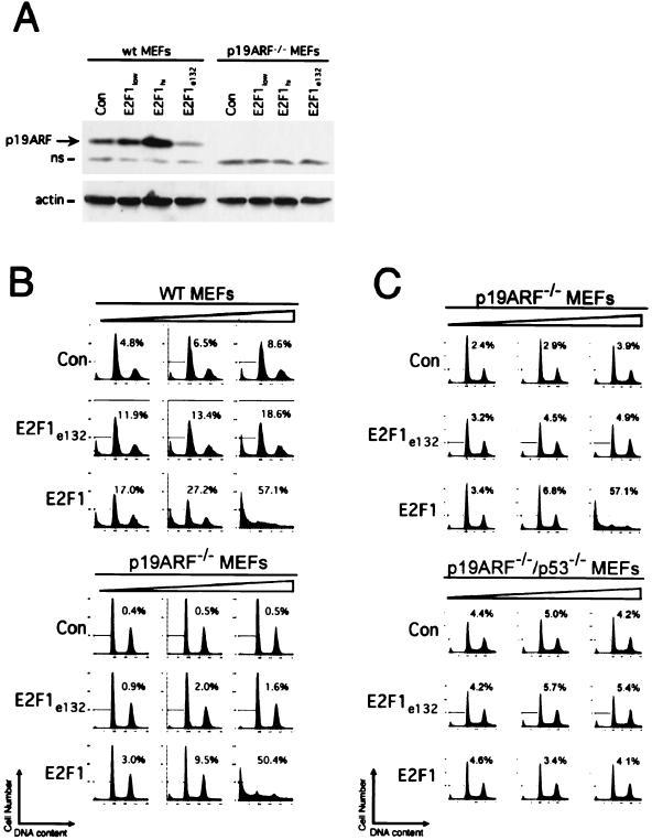
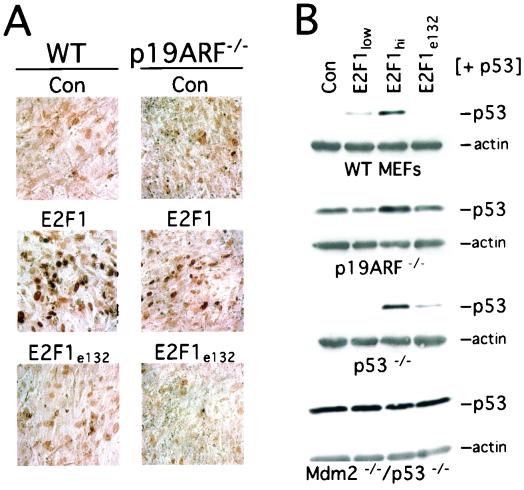
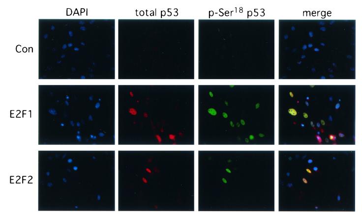
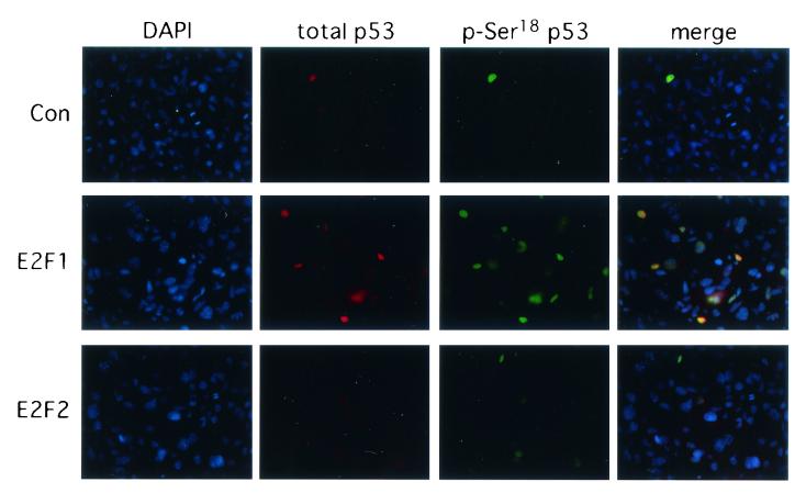
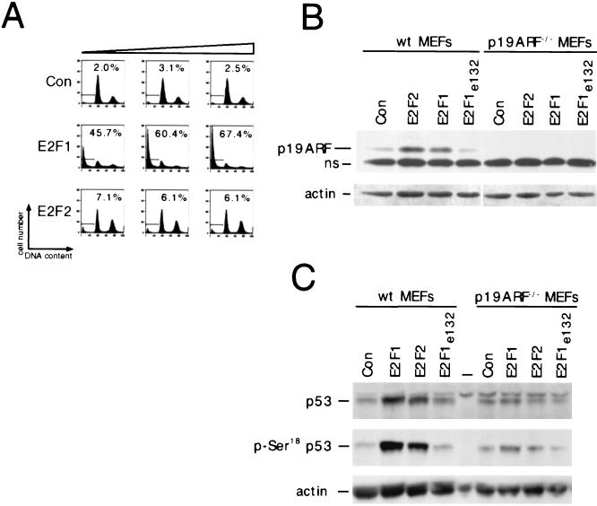
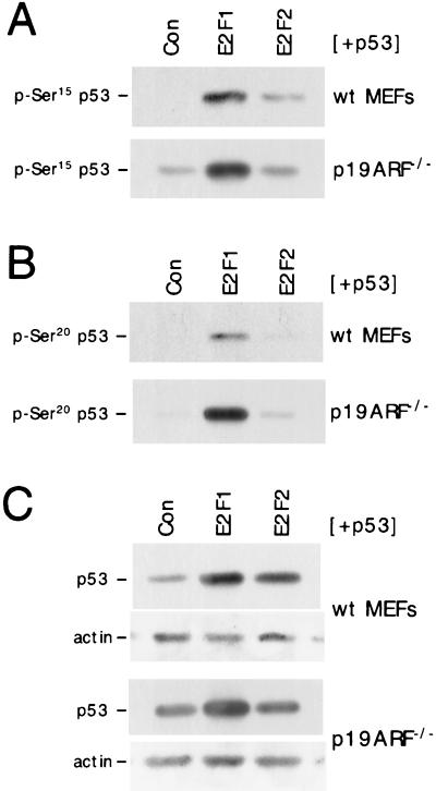
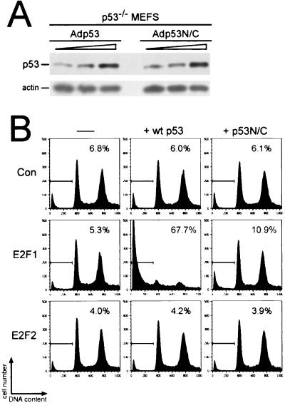
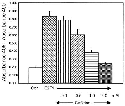
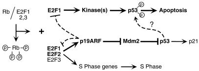
It has been proposed that the E2F1 transcription factor serves as a link between the Rb/E2F proliferation pathway and the p53 apoptosis pathway by inducing the expression of p19ARF, a protein that regulates p53 stability. We find that although p19ARF contributes to p53 accumulation in response to E2F expression, p19ARF is not required for E2F1-mediated apoptosis. E2F1 can signal p53 phosphorylation in the absence of p19ARF, similar to the observed modifications to p53 in response to DNA damage. These modifications are not observed in the absence of p19ARF following expression of E2F2, an E2F family member that does not induce apoptosis in mouse embryo fibroblasts but can induce p19ARF and p53 protein expression. p53 modification is found to be crucial for E2F1-mediated apoptosis, and this apoptosis is compromised when E2F1 is coexpressed with a p53 mutant lacking many N- and C-terminal phosphorylation sites. Additionally, E2F1-mediated apoptosis is abolished in the presence of caffeine, an inhibitor of phosphatidylinositol 3-kinase-related kinases that phosphorylate p53. These findings suggest that p53 phosphorylation is a key step in E2F1-mediated apoptosis and that this modification can occur in the absence of p19ARF.
Figures
Similar articles
-
Mdm2 inhibition of p53 induces E2F1 transactivation via p21.Oncogene. 2002 Jun 27;21(28):4414-21. doi: 10.1038/sj.onc.1205541. Oncogene. 2002. PMID: 12080472
-
Myc and E2F1 induce p53 through p14ARF-independent mechanisms in human fibroblasts.Oncogene. 2003 Aug 7;22(32):4993-5005. doi: 10.1038/sj.onc.1206659. Oncogene. 2003. PMID: 12902982
-
p19(ARF) is dispensable for oncogenic stress-induced p53-mediated apoptosis and tumor suppression in vivo.Mol Cell Biol. 2002 Jan;22(1):370-7. doi: 10.1128/MCB.22.1.370-377.2002. Mol Cell Biol. 2002. PMID: 11739748 Free PMC article.
-
The role of the transcription factor DP in apoptosis.Apoptosis. 2003 Oct;8(5):461-8. doi: 10.1023/a:1025586207239. Apoptosis. 2003. PMID: 12975577 Review.
-
E2F1 and c-Myc in cell growth and death.Cell Cycle. 2003 Jul-Aug;2(4):333-8. Cell Cycle. 2003. PMID: 12851485 Review.
Cited by
-
Human cytomegalovirus IE1-72 activates ataxia telangiectasia mutated kinase and a p53/p21-mediated growth arrest response.J Virol. 2005 Sep;79(17):11467-75. doi: 10.1128/JVI.79.17.11467-11475.2005. J Virol. 2005. PMID: 16103197 Free PMC article.
-
p53 and E2f: partners in life and death.Nat Rev Cancer. 2009 Oct;9(10):738-48. doi: 10.1038/nrc2718. Nat Rev Cancer. 2009. PMID: 19776743 Review.
-
Apoptosis inducing effects of Kuding tea polyphenols in human buccal squamous cell carcinoma cell line BcaCD885.Nutrients. 2014 Aug 5;6(8):3084-100. doi: 10.3390/nu6083084. Nutrients. 2014. PMID: 25100434 Free PMC article.
-
Balancing the decision of cell proliferation and cell fate.Cell Cycle. 2009 Feb 15;8(4):532-5. doi: 10.4161/cc.8.4.7609. Epub 2009 Feb 11. Cell Cycle. 2009. PMID: 19182518 Free PMC article. Review.
-
Novel role for RbAp48 in tissue-specific, estrogen deficiency-dependent apoptosis in the exocrine glands.Mol Cell Biol. 2006 Apr;26(8):2924-35. doi: 10.1128/MCB.26.8.2924-2935.2006. Mol Cell Biol. 2006. PMID: 16581768 Free PMC article.
References
-
- Appella, E., and C. W. Anderson. 2001. Post-translational modifications and activation of p53 by genotoxic stresses. Eur. J. Biochem. 268:2764-2772. - PubMed
-
- Banin, S., L. Moyal, S. Shieh, Y. Taya, C. W. Anderson, L. Chessa, N. I. Smorodinsky, C. Prives, Y. Reiss, Y. Shiloh, and Y. Ziv. 1998. Enhanced phosphorylation of p53 by ATM in response to DNA damage. Science 281:1674-1677. - PubMed
-
- Bates, S., A. C. Phillips, P. A. Clark, F. Stott, G. Peters, R. L. Ludwig, and K. H. Vousden. 1998. p14ARF links the tumour suppressors RB and p53. Nature 395:124-125. - PubMed
-
- Blasina, A., B. D. Price, G. A. Turenne, and C. H. McGowan. 1999. Caffeine inhibits the checkpoint kinase ATM. Curr. Biol. 9:1135-1138. - PubMed
Publication types
MeSH terms
Substances
Grants and funding
LinkOut - more resources
Full Text Sources
Research Materials
Miscellaneous