. 2002 Aug;22(15):5319-36.
doi: 10.1128/MCB.22.15.5319-5336.2002.

Oxygen-dependent ubiquitination and degradation of hypoxia-inducible factor requires nuclear-cytoplasmic trafficking of the von Hippel-Lindau tumor suppressor protein

Affiliations

Oxygen-dependent ubiquitination and degradation of hypoxia-inducible factor requires nuclear-cytoplasmic trafficking of the von Hippel-Lindau tumor suppressor protein

Isabelle Groulx et al. Mol Cell Biol. 2002 Aug.

Abstract

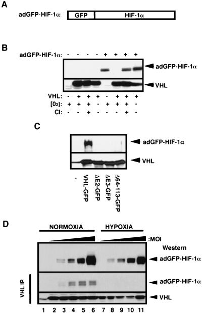
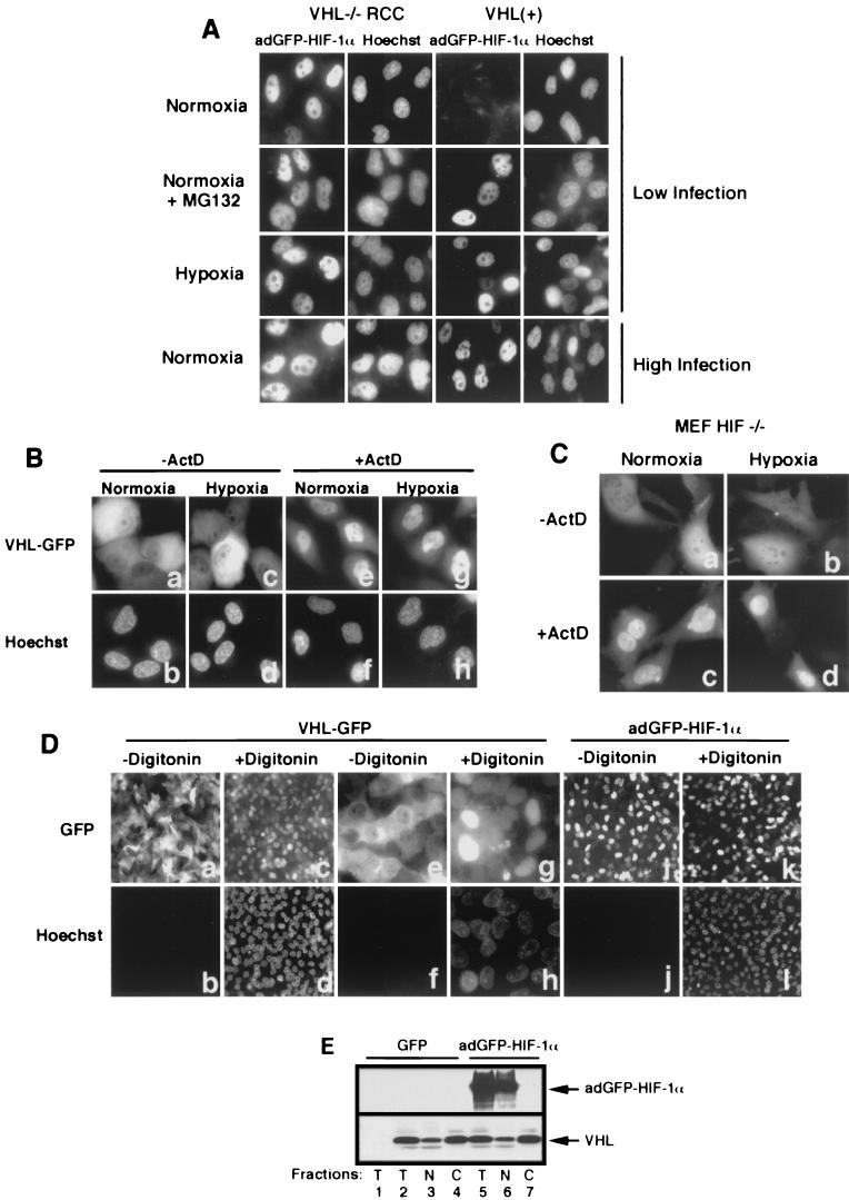
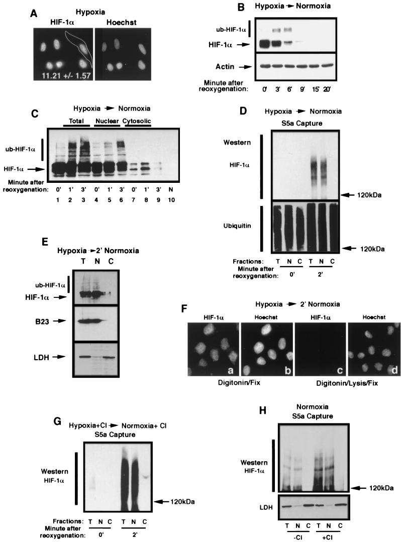
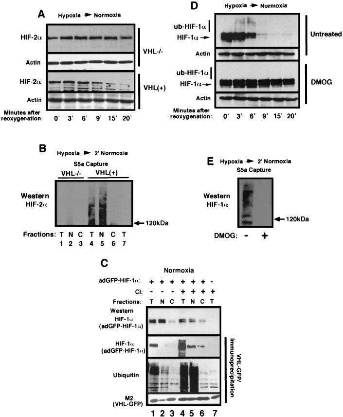
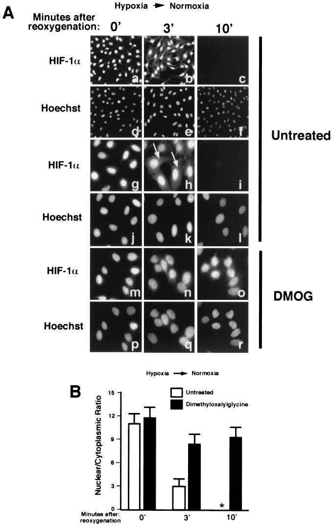
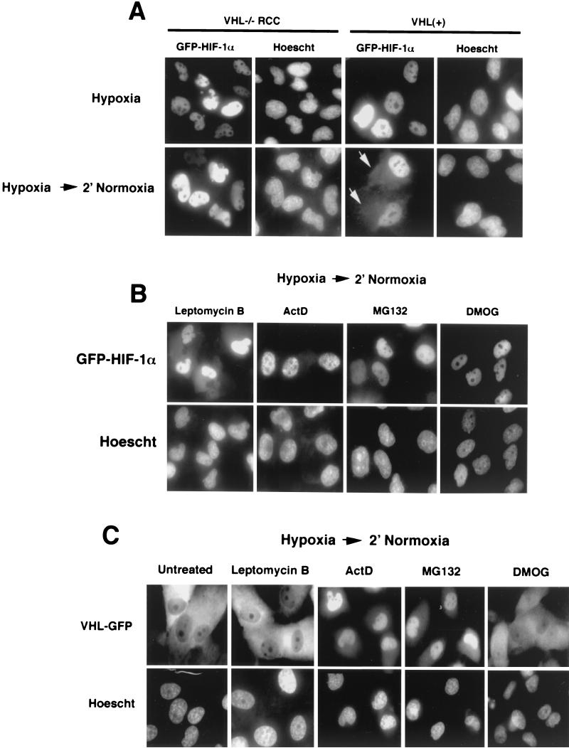
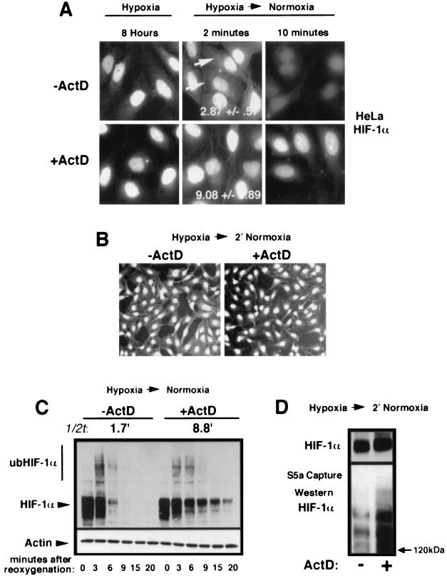
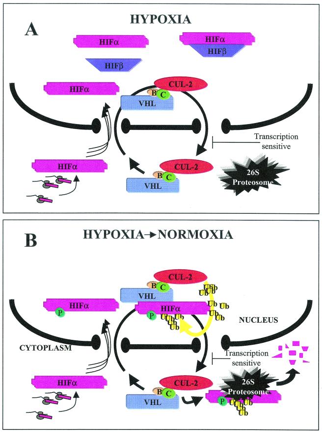
It is becoming increasingly evident that the degradation of nuclear proteins requires nuclear-cytoplasmic trafficking of both the substrate proteins, as well as the E3 ubiquitin-ligases. Here, we show that nuclear-cytoplasmic trafficking of the von Hippel-Lindau tumor suppressor protein (VHL) is required for oxygen-dependent ubiquitination and degradation of the alpha subunits of hypoxia-inducible factor (HIF-alpha). VHL engages in a constitutive transcription-sensitive nuclear-cytoplasmic shuttle unaffected by oxygen tension or levels of nuclear substrate HIF-alpha. Ubiquitinated forms of HIF-alpha, as well as VHL/ubiquitinated HIF-alpha complexes, are found solely in the nuclear compartment of normoxic or reoxygenated VHL-competent cells. HIF-alpha localizes exclusively in the nucleus of hypoxic cells but is exported to the cytoplasm upon reoxygenation. Oxygen-dependent nuclear ubiquitination and nuclear export of HIF-alpha can be prevented by treatment with an HIF-specific prolyl hydroxylase inhibitor. Treatment with inhibitors of RNA polymerase II activity, which interfere with the ability of VHL to engage in nuclear export, also prevents cytoplasmic accumulation of HIF-alpha in reoxygenated cells. This caused a marked increase in the HIF-alpha half-life without affecting its nuclear ubiquitination. We present a model by which VHL-mediated ubiquitination of HIF-alpha and its subsequent degradation are dependent upon dynamic nuclear-cytoplasmic trafficking of both the E3 ubiquitin-ligase and the nuclear substrate protein.


Figures

FIG. 1.
FIG. 1.
Characterization of the adGFP-HIF-1α fusion protein. (A) Schematic representation of HIF-1α fused to GFP. The cDNA encoding for the alpha subunit of the HIF-1α was fused to the C terminus of the cDNA of the GFP, resulting in GFP-HIF-1α fusion protein. (B) Proteolysis of adGFP-HIF-1α is proteasome mediated and VHL and oxygen dependent. VHL−/− RCC cells (cell line 786-0) or the VHL(+) WT-7 stable cell line (expressing HA-tagged VHL) were either not infected (−) or infected (+) with low titers (MOI) of the adGFP-HIF-1α. Cells were incubated in normal oxygen tension (O2+) or in hypoxic conditions (1% oxygen; O2−) for 4 h. Normoxic cells were also incubated in the presence (CI+) or the absence (CI−) of calpain inhibitor I for 2 h. Cell lysates were subjected to an SDS-8% PAGE analysis, and a Western blot was detected with anti-HIF-1α or anti-HA antibody. (C) The fusion protein adGFP-HIF-1α binds to VHL-GFP in vitro. Lysates of adGFP-HIF-1α infected VHL−/− RCC cells were incubated in reticulocyte lysates programmed with pcDNA vector alone (−), expressing FLAG tagged-VHL-GFP or mutants of the α or β domain (ΔE2-GFP, ΔE3-GFP, and Δ64-113-GFP). The resulting mixture was immunoprecipitated with M2 monoclonal antibody and immunoblotted with anti-HIF-1α or anti-M2 antibody. (D) adGFP-HIF-1α binds to VHL in normoxia but not in hypoxia. VHL-GFP stable cell lines were infected with increasing amounts of adGFP-HIF-1α virus and incubated in normoxia or hypoxia for 4 h. Immunoprecipitation was carried out in normoxic or hypoxic conditions with M2 antibody. Immunodetection of input (top panel) and of the immunoprecipitation was done with an anti-HIF-1α or anti-M2 antibody. Lane 1 represents M2 beads alone.
FIG.2.
FIG.2.
Subcellular localization of VHL and adGFP-HIF-1α is independent from each other. (A) adGFP-HIF-1α localize to the nucleus. Fluorescence analysis of VHL−/− RCC cells and the VHL(+) WT-7 stable cell line expressing adGFP-HIF-1α in normoxia at low levels of infection (first row) and of normoxic cells in the presence of proteasome inhibitor MG132 (second row) or in hypoxia (third row). Normoxic VHL−/− RCC and VHL(+) cells infected with high titers of adGFP-HIF-1α are shown in the last row. (B) Transcription-dependent shuttling of VHL is unaffected by hypoxic treatment. Fluorescence analysis of VHL−/− RCC cells expressing VHL-GFP in normoxia or hypoxia (4 h) in the absence or the presence of ActD (10 μg/ml) for 2 h. (C) Transcription-dependent shuttling of VHL occurs in the absence of substrate HIF-α. Fluorescence analysis of HIF−/− mouse embryonic fibroblasts expressing VHL-GFP in normoxia or hypoxia (4 h) in the absence or the presence of ActD (10 μg/ml) for 2 h. (D) Digitonin-permeabilization system to achieve nuclear-cytoplasmic fractionation of cells. VHL-GFP-expressing VHL−/− RCC cells were either not treated (a and b and, at a higher magnification, e and f) or treated for 5 min with 50 μg of digitonin/ml (c and d and, at a higher magnification, g and h) and observed for GFP fluorescence (a, c, e, and g) or for cell-impermeable Hoechst stain 33258. Notice the complete loss of cytoplasmic VHL-GFP signal but the retention of nuclear signal in digitonin-treated cells and that essentially all of the nuclei of permeabilized cells have been stained with Hoechst. Panels j to l show that nuclear adGFP-HIF-1α remains in the nuclear compartment after digitonin treatment. (E) VHL steady-state localization is independent of levels of adGFP-HIF-1α in normoxic cells. VHL(+) cells were either infected with GFP adenovirus or with high levels of adGFP-HIF-1α virus. Biochemical subcellular fractionation was performed with the digitonin system to obtain total (T), nuclear (N), and cytosolic (C) fractions, which were analyzed by Western blot with anti-M2 and anti-HIF-1α antibody. A negative control is present in the first lane and represents VHL−/− RCC infected with GFP virus.
FIG.3.
FIG.3.
Ubiquitination of HIF-1α occurs in the nuclear compartment. (A) HIF-1α localizes to the nuclear compartment of hypoxic HeLa cells. HeLa cells grown on coverslips were incubated for 8 h in hypoxia prior to fixation with 1% formaldehyde and staining with an anti-HIF-1α antibody and a secondary Texas red-labeled anti-mouse antibody. A CCD-captured image was analyzed as described in Materials and Methods for the nuclear/cytoplasmic ratio of the distribution of the fluorescence signal. The mean ± the standard deviation of 32 cells of three independent experiments is shown. (B) HIF-1α is efficiently degraded upon the return of hypoxic cells to normoxia. HeLa cells were incubated for 8 h in hypoxia prior to a return to a normoxic incubator for the indicated time. Cells were lysed in 4% SDS-PBS and analyzed by SDS-8% PAGE, followed by Western blotting with an anti-HIF-1α antibody. Notice the appearance of higher-molecular-weight bands at 3 and 6 min, which is most likely ub-HIF-1α. (C) ub-HIF-1α appears promptly in the nucleus after reoxygenation of hypoxic cells. HeLa cells were incubated in hypoxia for 8 h (0′) prior to being transferred to an oxygenated environment at 37°C for 1 and 3 min. Samples from each time point were submitted to subcellular fractionation, and the resulting cell fractions (total, nuclear, and cytosolic) were analyzed by SDS-8% PAGE, followed by Western blotting with an anti-HIF-1α antibody. Normoxic (N) HeLa cell lysate is shown in lane 10. (D) ub-HIF-α is found strictly in the nuclear compartment upon reoxygenation. Capture of polyubiquitinated proteins with agarose-GST-S5a beads was carried out in lysates of total (T), nuclear (N), or cytosolic (C) fractions of hypoxic or reoxygenated (2 min) HeLa cells. The captured polyubiquitinated proteins were analyzed by SDS-6% PAGE, followed by Western blot with an anti-HIF-1α or anti-ubiquitin antibody. (E) Reoxygenated HeLa cells were treated with digitonin, and nuclear or cytosolic fractions were blotted with an anti-HIF-1α antibody, an anti-LDH antibody, or an anti-B23 antibody. (F) HIF-1α can be detected solely in the nucleus of digitonin-treated cells after a return of hypoxic cells to normoxia. Hypoxic HeLa cells grown on coverslips were transferred to an oxygenated incubator for 2 min prior to permeabilization with digitonin for 5 min at 4°C. Permeabilized cells were either fixed with 1% formaldehyde (a and b) or treated with lysis buffer prior to fixation (c and d). Fixed and permeabilized cells were staining with an anti-HIF-1α antibody and a secondary Texas red-labeled anti-mouse antibody (a and c) or with Hoechst stain (b and d). Note that the specific HIF-1α signal can be detected only in the nuclear compartment in panel a and is completely extractable with the lysis buffer (see panel c). (G) ub-HIF-1α can only be detected in the nuclear compartment in proteasome-treated and reoxygenated HeLa cells. HeLa cells were treated for 1 h with proteasome inhibitors prior to reoxygenation and cells were treated as described for panel D to detect ub-HIF-1α. (H) ub-HIF-1α are found solely in the nucleus of normoxic cells. Normoxic HeLa cells treated (+) or not (−) with calpain inhibitor (CI) were fractionated into total (T), nuclear (N), and cytosolic (C) lysates, which were then subjected to agarose-GST-S5a immunoprecipitation and anti-HIF-1α Western blot analysis. The bottom panel shows the fractions blotted with anti-LDH.
FIG. 4.
FIG. 4.
Nuclear ubiquitination of HIF-α requires VHL and prolyl hydroxylase activity. (A) Degradation of HIF-2α requires VHL upon a return of hypoxic cells to an oxygenated environment. VHL−/− RCC and VHL(+) cells were incubated for 8 h in hypoxia and returned to an oxygenated incubator for the indicated time. Cells were lysed in 4% SDS-PBS and analyzed by SDS-8% PAGE, followed by Western blotting with an anti-HIF-2α antibody (Novus). (B) Nuclear ubiquitination of HIF-2α requires VHL. VHL−/− RCC (lanes 1 to 3) and VHL(+) (lanes 4 to 6) cells were incubated in hypoxia for 8 h prior to reoxygenation for 2 min. Cells were fractionated into total (T), nuclear (N), and cytosolic (C) compartments, followed by immunoprecipitation with agarose-GST-S5a beads. Captured polyubiquitinated proteins were analyzed by SDS-6% PAGE for ub-HIF-2α content with an anti-HIF-2α antibody. Lane 7 consists of a total normoxic fraction of VHL(+) cells in which we failed to detect ub-HIF-2α, most likely due to low abundance. (C) VHL/ub-adGFP-HIF-1α complexes can be found in the nuclear compartment of normoxic cells. Normoxic VHL-GFP-expressing stable cells were either infected (lanes 1 to 6) or not infected (lane 7) with adGFP-HIF-1α virus, and the cells were either not treated (lanes 1 to 3) or were treated (lanes 4 to 7) with a proteasome inhibitor (calpain inhibitor [CI]) for 2 h. Cells were fractionated into total (T), nuclear (N), and cytoplasmic (C) lysates, which were immunoprecipitated with agarose M2-beads to capture FLAG-tagged VHL-GFP. The top panel shows adGFP-HIF-1α input by straight Western analysis. The lower three panels consists of agarose M2-bead immunoprecipitations of VHL-GFP, followed by Western blotting with an anti-HIF-1α antibody to detect adGFP-HIF-1α (second panel from the top), an antiubiquitin antibody (third panel from the top), or with a M2 anti-FLAG antibody to detect VHL-GFP (bottom panel). Lane 7 is an immunoprecipitation of VHL-GFP from lysates of uninfected VHL-GFP cells. Note the lack of ubiquitin signal in this lane, demonstrating that the ubiquitin signals in lanes 1 to 6 are from ub-adGFP-HIF-1α. (D) The degradation of HIF-1α can be abolished by treatment with HIF-1α-specific prolyl hydroxylase inhibitor DMOG. HeLa cells were incubated for 8 h in hypoxia prior to a return to a normoxic incubator for the indicated time. Cells were either treated with dimethyl sulfoxide alone (untreated) or treated with DMOG (1 mM) for 8 h. Cells were lysed in 4% SDS-PBS and analyzed by SDS-8% PAGE, followed by Western blotting with an anti-HIF-1α antibody. Note the absence of higher-molecular-weight bands at 3 and 6 min in the DMOG-treated cells. (E) DMOG treatment abolishes ubiquitination of HIF-1α in reoxygenated cells. Hypoxic HeLa cells were transferred to an oxygenated incubator for 2 min prior to lysis and incubation with agarose S5a beads. The captured polyubiquitinated proteins were analyzed by SDS-6% PAGE, followed by Western blotting with an anti-HIF-1α antibody.
FIG. 5.
FIG. 5.
Nuclear export of HIF-1α occurs in reoxygenated HeLa cells through a pathway that requires nuclear prolyl hydroxylase activity. (A) Hypoxic HeLa cells (panels a to l) were either fixed with 1% formaldehyde or transferred to an oxygenated incubator for 3 or 10 min prior to fixation. Cells were also treated with DMOG (1 mM) for 8 h (panels m to r). Cells were stained with an anti-HIF-1α antibody or with Hoechst. Fields a to c are the same exposure. Fields g, h, i, m, n, and o are also equivalent exposures. (B) Nuclear/cytoplasmic ratios were calculated for ca. 30 cells for each condition obtained from at least three independent experiments. Mean averages with the standard deviations are shown. ✽, data not available since there is no signal.
FIG. 6.
FIG. 6.
Cytoplasmic accumulation of adGFP-HIF-1α upon reoxygenation requires VHL and occurs through an ActD-dependent, but leptomycin B-independent pathway. (A) Nuclear export of adGFP-HIF-1α requires VHL. VHL−/− RCC and VHL(+) cells were infected with adGFP-HIF-1α, incubated for 8 h in hypoxia, and transferred for 2 to 15 min in normoxia. The arrows point to the cytoplasmic signal. The same exposure time was used for all of the images. (B) Effect of drugs on cytoplasmic accumulation of adGFP-HIF-1α upon reoxygenation of hypoxic cells. VHL(+) cells were infected with adGFP-HIF-1α and incubated in hypoxia for 8 h in the presence of the indicated drug for last 2 h, except for DMOG, which was incubated for 8 h. Cells were transferred to an oxygenated incubator for 2 to 15 min. The same exposure time was used for all of the images. (C) Nuclear accumulation of VHL-GFP occurs upon treatment with ActD and MG132 but not with leptomycin B or DMOG. VHL−/− RCC cells were infected with adVHL-GFP and incubated for 8 h in hypoxia with the indicated drugs for the last 2 h except for DMOG, which was incubated for 8 h. Cells were transferred to an oxygenated incubator for 2 to 15 min. The same exposure time was used for all of the images.
FIG. 7.
FIG. 7.
ActD treatment increases the half-life of HIF-α without a decrease in its nuclear ubiquitination. (A) Cytoplasmic accumulation of endogenous HIF-1α in HeLa cells upon a return to normoxia is inhibited by ActD treatment. HeLa cells were incubated for 8 h in hypoxia in the absence or presence of ActD (10 μg/ml) for the last 2 h. Hypoxic cells were fixed with 1% formaldehyde or fixed 2 or 10 min after reoxygenation and then stained with an anti-HIF-1α antibody. (B) Lower magnification of HIF-1α immunostaining of reoxygenated HeLa cells. Experiments were performed as in panel A. (C) Efficient degradation of endogenous HIF-1α is dependent upon ongoing transcription. HeLa cells were incubated for 8 h in hypoxia in the absence or presence of ActD (10 μg/ml) for the last 2 h. Cells were returned to an oxygenated environment for the indicated times and Western blotting was performed with an anti-HIF-1α. The half-life (1/2t) of endogenous HIF-1α is indicated. (D) Nuclear ubiquitination of HIF-1α is unaffected by ActD treatment. Hypoxic HeLa cells were either not treated or treated with ActD (10 μg/ml) for 2 h and then returned to an oxygenated environment for 2 min. Lysates were incubated with agarose S5a beads, and captured polyubiquitinated proteins were analyzed by SDS-6% PAGE, followed by Western blotting with an anti-HIF-1α antibody. The top panel shows a straight Western blot for HIF-1α content. The experiment was conducted to obtain similar HIF-1α levels in untreated and in ActD-treated cells.
FIG. 8.
FIG. 8.
Model linking VBC/Cul-2 nuclear-cytoplasmic trafficking and oxygen-dependent ubiquitination and degradation of HIF-α. (A) In hypoxic conditions, HIF-α (pink) is stabilized and is immediately imported into the nucleus, where it binds to its partner HIF-β (purple) to form the HIF transcription factor that transactivates hypoxia-inducible genes such as vascular endothelial growth factor and glucose transporter-1. VBC/Cul-2 engages in a constitutive nuclear-cytoplasmic shuttle unaffected by oxygen tension or levels of substrate HIF-α. (B) Upon a return to normoxia, HIF-α incurs a posttranslational modification in the nuclear compartment, which is most likely the hydroxylation of proline of the ODD domains since ubiquitination is preventable by treatment with DMOG. Nuclear HIF-α binds to VHL and undergoes Cullin-2-mediated ubiquitination (yellow) prior to being exported to the cytoplasm for 26S proteasomal degradation. Nuclear HIF-α embarks on the VBC/Cul-2 “merry-go-round” depending upon its ability to assemble with nuclear VHL. The proposed model is applicable for hypoxic cells that are returned to an oxygenated environment. As for cells maintained in normoxia, we cannot totally exclude cytoplasmic ubiquitination and degradation of HIF-α. However, we prefer a model by which HIF-α in normoxia is rapidly imported for nuclear ubiquitination and reexported to the cytoplasm in a manner similar to that observed for reoxygenated cells.

Similar articles

Cited by

References

    1. Berra, E., D. E. Richard, E. Gothie, and J. Pouyssegur. 2001. HIF-1-dependent transcriptional activity is required for oxygen-mediated HIF-1α degradation. FEBS Lett. 491:85-90. - PubMed
    1. Berra, E., D. Roux, D. E. Richard, and J. Pouyssegur. 2001. Hypoxia-inducible factor-1 alpha (HIF-1α) escapes O2-driven proteasomal degradation irrespective of its subcellular localization: nucleus or cytoplasm. EMBO Rep. 2:615-620. - PMC - PubMed
    1. Bird, C. H., E. J. Blink, C. E. Hirst, M. S. Buzza, P. M. Steele, J. Sun, D. A. Jans, and P. I. Bird. 2001. Nucleocytoplasmic distribution of the ovalbumin serpin PI-9 requires a nonconventional nuclear import pathway and the export factor Crm1. Mol. Cell. Biol. 21:5396-5407. - PMC - PubMed
    1. Blankenship, C., J. G. Naglich, J. M. Whaley, B. Seizinger, and N. Kley. 1999. Alternate choice of initiation codon produces a biologically active product of the von Hippel-Lindau gene with tumor suppressor activity. Oncogene 18:1529-1535. - PubMed
    1. Blondel, M., J. M. Galan, Y. Chi, C. Lafourcade, Longaretti, C., R. J. Deshaies, and M. Peter. 2000. Nuclear-specific degradation of Far1 is controlled by the localization of the F-box protein Cdc4. EMBO J. 19:6085-6097. - PMC - PubMed

Publication types

MeSH terms

LinkOut - more resources