. 2002 Aug;22(15):5380-94.
doi: 10.1128/MCB.22.15.5380-5394.2002.

The UV (Ribotoxic) stress response of human keratinocytes involves the unexpected uncoupling of the Ras-extracellular signal-regulated kinase signaling cascade from the activated epidermal growth factor receptor

Affiliations

The UV (Ribotoxic) stress response of human keratinocytes involves the unexpected uncoupling of the Ras-extracellular signal-regulated kinase signaling cascade from the activated epidermal growth factor receptor

Mihail S Iordanov et al. Mol Cell Biol. 2002 Aug.

Abstract

In mammals, UVB radiation is of biological relevance primarily for the cells of the epidermis. We report here the existence of a UVB response that is specific for proliferating human epidermal keratinocytes. Unlike other cell types that also display a UVB response, keratinocytes respond to UVB irradiation with a transient but potent downregulation of the Ras-extracellular signal-regulated kinase (ERK) signaling cascade. The downregulation of ERK precedes a profound decrease in the steady-state levels of cyclin D1, a mediator of the proliferative action of ERK. Keratinocytes exhibit high constitutive activity of the Ras-ERK signaling cascade even in culture medium lacking supplemental growth factors. The increased activity of Ras and phosphorylation of ERK in these cells are maintained by the autocrine production of secreted molecules that activate the epidermal growth factor receptor (EGFR). Irradiation of keratinocytes increases the phosphorylation of EGFR on tyrosine residues Y845, Y992, Y1045, Y1068, Y1086, Y1148, and Y1173 above the basal levels and leads to the increased recruitment of the adaptor proteins Grb2 and ShcA and of a p55 form of the regulatory subunit of the phosphatidylinositide 3-kinase to the UVB-activated EGFR. Paradoxically, however, UVB causes, at the same time, the inactivation of Ras and a subsequent dephosphorylation of ERK. By contrast, the signaling pathway leading from the activated EGFR to the phosphorylation of PKB/Akt1 is potentiated by UVB. The UVB response of keratinocytes appeared to be a manifestation of the more general ribotoxic stress response inasmuch as the transduction of the UVB-generated inhibitory signal to Ras and ERK required the presence of active ribosomes at the time of irradiation.


Figures
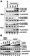
FIG. 1.
FIG. 1.
Dose-dependent regulation of the phosphorylation of MAP kinases by UVB. HEKn and HeLa cells (A) and HEKn-E6/E7 and HaCaT cells (B) (grown in BKM+exoGF) were treated with the indicated doses of UVB and were harvested 30 min later. Shown are the results of immunoblot analyses using phosphorylation-specific antibodies against the indicated proteins.
FIG. 2.
FIG. 2.
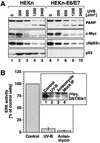
(A) Effects of UVB irradiation on cell cycle and apoptosis markers in HEKn and HEKn-E6/E7. Cells grown in BKM+exoGF were irradiated with the indicated doses of UVB, and the expression of the indicated proteins was determined in immunoblot analyses 21 h postirradiation. PARP, poly(ADP)ribose polymerase. (B) Inhibition of ERK activity by UVB and anisomycin. HEKn (grown in BKM+exoGF) were treated with UVB (1,200 J/m2) or anisomycin (10 μg/ml). ERK activity was assessed 30 min later in immunocomplex kinase assays (see Materials and Methods). Lane 4 of the boxed insert shows the control immunoprecipitation (IP) with an irrelevant antibody. Error bars represent the standard deviations from experimental points in triplicates.
FIG. 3.
FIG. 3.
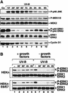
Time-dependent inhibition of ERK phosphorylation by UVB. Shown are the results of immunoblot analyses using phosphorylation-specific antibodies against the indicated proteins. (A) HEKn were treated with UVB (1,200 J/m2) UVB and later harvested at the indicated times. Co, control. (B) HEKn and HEKn-E6/E7, placed 20 h earlier in either BKM+exoGF or BKM-exoGF, were irradiated with UVB (1,200 J/m2) and harvested at the indicated times after the irradiation. -, control cells. Note that a prior hybridization of a membrane with antibodies against the phosphorylated forms of ERK or EGFR interferes with a subsequent hybridization of the same membrane with the antibodies against nonphosphorylated ERK or EGFR at any location in the membrane where the phospho-specific signal was strong. This explains the apparent weaker signal of the nonphosphorylated p42 ERK2 seen in the bottom panel of lane 6. The same phenomenon can also be seen in Fig. 4A, lanes 3 and 10 (ERK2); Fig. 5A, lane 2 (EGFR and ERK2); Fig. 6A, lanes 1 to 3 and 7 (ERK2); Fig. 7A, lanes 5 to 7 (ERK2); Fig 9, panel l, lanes 7 and 12 (ERK2); and Fig. 12A, lanes 2 to 4 (ERK2).
FIG. 4.
FIG. 4.
Production of a soluble ligand(s) of EGFR by HEKn-E6/E7 but not by HaCaT cells. Shown are the results of immunoblot analyses using antibodies specific for the phosphorylated states of EGFR (Y1173) and ERK. (A) The phosphorylation state of EGFR (Y1173) and ERK in HeLa (lanes 1 to 8) or HaCaT (lanes 9 to 12) cells that have been subjected to unconditioned, HEKn-E6/E7-conditioned, or HaCaT-conditioned BKM-exoGF according to the method described in the figure body. The cells were pretreated for 30 min with 10 μM AG1478 (+) or the vehicle solvent DMSO(−). (B) HEKn-E6/E7, placed 20 h earlier in either BKM+exoGF (+ GF) or BKM-exoGF (- GF), were treated with 10 μM AG1478 or the vehicle solvent DMSO (lanes 1 to 6) for the indicated times.
FIG. 5.
FIG. 5.
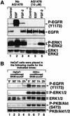
Human epidermal keratinocytes maintain high steady-state levels of ERK activity via the autocrine production of EGFR ligand(s). (A) Results of immunoblot analyses showing that AG1478 inhibits specifically the EGF-induced signaling but not the FGF-2-induced signaling to ERK. HEKn-E6/E7 were pretreated for 30 min with 10 μM AG1478 (lanes 4 to 6) or the vehicle solvent DMSO (lanes 1 to 3) and were then treated for 15 min with either EGF (100 ng/ml) or FGF-2 (20 ng/ml). (B) Immunoblot analyses of the phosphorylation state of EGFR (Y1173), ERK, and PKB/Akt1 (Ser473) in HaCaT cells that have been subjected to either HaCaT-conditioned or HEKn-E6/E7-conditioned BKM-exoGF according to the method described in the figure body.
FIG. 6.
FIG. 6.
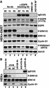
Expression of cyclin D1 in keratinocytes is dependent on the activities of EGFR and MEK. (A) HEKn-E6/E7, placed 20 h earlier in BKM-exoGF, were left untreated or were treated with the EGFR-neutralizing antibody (Ab) LA1 (5 μg/ml), with AG1478 (10 μM), or with DMSO (vehicle control for AG1478). The cells were harvested at the indicated times, and the expression and/or phosphorylation status of the indicated proteins was assayed in immunoblot analyses. IgG1(H), the heavy immunoglobulin chain of the neutralizing anti-EGFR antibody used. (B) HEKn, placed 20 h earlier in BKM-exoGF, were left untreated or were treated with UVB (1,200 J/m2), AG1478 (10 μM), UO126 (10 μM), or the EGFR-neutralizing antibody LA1 (5 μg/ml). The cells were harvested at the indicated times, and the expression and/or phosphorylation status of the indicated proteins was assayed in immunoblot analyses.
FIG. 7.
FIG. 7.
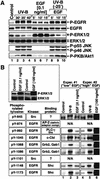
UVB activates EGFR but triggers the functional uncoupling of ERK (but not of PKB/Akt1) from EGFR. (A) Immunoblot analyses with HEKn-E6/E7 placed in BKM-exoGF and treated 2 h later with UVB (1,200 J/m2), with a dose of EGF that is submaximal with respect to EGFR phosphorylation (0.1 ng/ml), or with EGF after a pretreatment with UVB. The phosphorylation statuses of EGFR (Y1173), ERK, JNK, and PKB/Akt1 (Ser473) were assessed at the indicated times after the treatments. (B) Immunoblot analyses with HEKn-E6/E7 handled as described for panel A, except that two doses of EGF (0.1 ng/ml for experiment 1 and 100 ng/ml for experiment 2) were applied. The cells were harvested 10 min after the EGF treatments and 30 min after the UVB (1,200 J/m2) treatments (identical UVB treatments were done in experiments 1 and 2). Lanes 1 to 6, each panel represents the analysis of the EGFR phosphorylation with phosphorylation-specific antibodies against the tyrosine residues indicated at left. N/A, antibody not available. The boxed insert shows the EGF-induced phosphorylation of ERK in experiment 1 or 2.
FIG. 8.
FIG. 8.
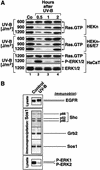
(A) Inhibition of Ras activity by UVB in HEKn and HEKn-E6/E7 but not in HaCaT cells. Cells (plated and maintained in BKM+exoGF) were irradiated with the indicated doses of UVB, and the levels of GTP loading of Ras (and ERK phosphorylation in the case of HaCaT cells) were determined by immunoblot analysis at the indicated times after the irradiation as described in the text. (B) Existence of a multiprotein complex containing Sos1, Grb2, and ShcA in untreated or UVB-treated HEKn. HEKn (grown in BKM+exoGF) were treated with UVB (1,200 J/m2). Sos1 was immunoprecipitated 30 min later, and the presence of Sos1, Grb2, and the three forms of ShcA (p46, p52, and p66) in the immunoprecipitate was detected in immunoblot analyses.
FIG. 9.
FIG. 9.
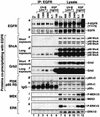
UVB-induced association of the activated EGFR with ShcA, Grb2, and a p55 PI3K. HEKn were maintained in BKM+exoGF and were treated for 30 min with the indicated doses of UVB or for 10 min with the indicated doses of EGF. Lanes 1 to 6, coimmunoprecipitation of the indicated proteins with EGFR; lanes 7 to 12, cell lysates before the immunoprecipitation. Shown are the results of immunoblot analyses using antibodies as indicated in each panel. For the detection of ShcA (panels c and d) and Grb2 (panels e and f), the presentation of short and long film exposures was necessary. IgG, the heavy immunoglobulin chain of the anti-EGFR antibody used for precipitation.
FIG. 10.
FIG. 10.
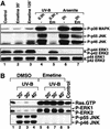
The ribotoxic stress response of human keratinocytes. (A) Immunoblot analyses with HEKn (plated and maintained in BKM+exoGF) treated with anisomycin (10 μg/ml) (lanes 1 to 9) or with UVB (1,200 J/m2) (lanes 11 and 12) in the absence or in the presence of a pretreatment (30 min) with actinomycin D (20 μM). The phosphorylation statuses of JNK and ERK were determined at the indicated times after the treatments. (B) Immunoblot analyses with HEKn treated with UVB (1,200 J/m2) in the absence or in the presence of a pretreatment (5 min) with either emetine (Em; 100 μg/ml) or pactamycin (Pa; 0.2 μg/ml). The phosphorylation statuses of JNK and ERK were determined 30 min after the irradiation.
FIG. 11.
FIG. 11.
The ribotoxic stress response of human keratinocytes. (A) Immunoblot analyses with HEKn (plated and maintained in BKM+exoGF) treated with UVB (1,200 J/m2) or sodium arsenite (200 μg/ml) in the absence or in the presence of a pretreatment (5 min) with emetine (Em, 100 μg/ml). The phosphorylation statuses of p38α MAP kinase, JNK, and ERK were determined at the indicated times after the treatments. -, cells pretreated with the vehicle solvent DMSO. (B) Immunoblot analyses with HEKn-E6/E7 (plated and maintained in BKM+exoGF) treated with UVB (1,200 J/m2) in the absence or in the presence of a pretreatment (5 min) with emetine (100 μg/ml). RasGTP levels were determined at the indicated times after the treatments as shown in Fig. 8A. The phosphorylation statuses of JNK and ERK were assessed from the same cell lysates before the precipitation of Ras with GST/Raf-RBD.
FIG. 12.
FIG. 12.
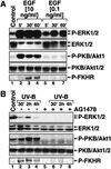
Activation of PKB/Akt1 in response to EGF and UVB in keratinocytes. (A) Immunoblot analyses with HEKn-E6/E7 placed in BKM-exoGF overnight and treated with low (0.1 ng/ml) or high (10 ng/ml) doses of EGF. The phosphorylation statuses of ERK, PKB/Akt1 (Ser473), and FKHR (Ser256) were assessed at the indicated times after the treatments. (B) Immunoblot analyses with HEKn-E6/E7 placed in BKM-exoGF and treated 2 h later with UVB (1,200 J/M2) in the absence (-) or presence (+) of a 30-min pretreatment with AG1478 (10 μM). The phosphorylation statuses of ERK, PKB/Akt1 (Ser473), and FKHR (Ser256) were assessed at the indicated times after the treatments.
FIG. 13.
FIG. 13.
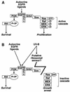
A model for the selective interference of UVB radiation with the EGFR-dependent signal transduction in keratinocytes. (A) In the absence of UVB, the autocrine stimulation of EGFR maintains an active Ras-ERK cascade that is involved in cell proliferation (see Discussion). (B) EGFR also generates an antiapoptotic signal that is mediated via the PI3K-PKB/Akt1 cascade (reference and references therein). UVB irradiation causes the selective inactivation of the Ras-ERK cascade, whereas the signal transduction pathway leading to the activation of PKB/Akt1 remains intact (or is even stimulated [Fig. 12]).

Similar articles

Cited by

References

    1. Albanell, J., J. Codony-Servat, F. Rojo, J. M. Del Campo, S. Sauleda, J. Anido, G. Raspall, J. Giralt, J. Rosello, R. I. Nicholson, J. Mendelsohn, and J. Baselga. 2001. Activated extracellular signal-regulated kinases: association with epidermal growth factor receptor/transforming growth factor alpha expression in head and neck squamous carcinoma and inhibition by anti-epidermal growth factor receptor treatments. Cancer Res. 61:6500-6510. - PubMed
    1. Assoian, R. K., and M. A. Schwartz. 2001. Coordinate signaling by integrins and receptor tyrosine kinases in the regulation of G1 phase cell-cycle progression. Curr. Opin. Genet. Dev. 11:48-53. - PubMed
    1. Balmanno, K., and S. J. Cook. 1999. Sustained MAP kinase activation is required for the expression of cyclin D1, p21Cip1 and a subset of AP-1 proteins in CCL39 cells. Oncogene 18:3085-3097. - PubMed
    1. Batzer, A. G., P. Blaikie, K. Nelson, J. Schlessinger, and B. Margolis. 1995. The phosphotyrosine interaction domain of Shc binds an LXNPXY motif on the epidermal growth factor receptor. Mol. Cell. Biol. 15:4403-4409. - PMC - PubMed
    1. Batzer, A. G., D. Rotin, J. M. Urena, E. Y. Skolnik, and J. Schlessinger. 1994. Hierarchy of binding sites for Grb2 and Shc on the epidermal growth factor receptor. Mol. Cell. Biol. 14:5192-5201. - PMC - PubMed

Publication types

MeSH terms