. 2002 Aug;22(15):5451-66.
doi: 10.1128/MCB.22.15.5451-5466.2002.

Jun dimerization protein 2 functions as a progesterone receptor N-terminal domain coactivator

Affiliations

Jun dimerization protein 2 functions as a progesterone receptor N-terminal domain coactivator

Suzanne E Wardell et al. Mol Cell Biol. 2002 Aug.

Abstract

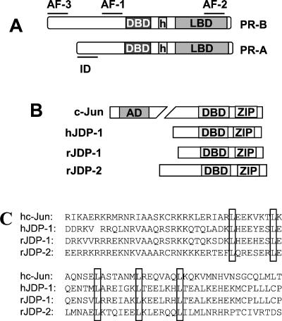
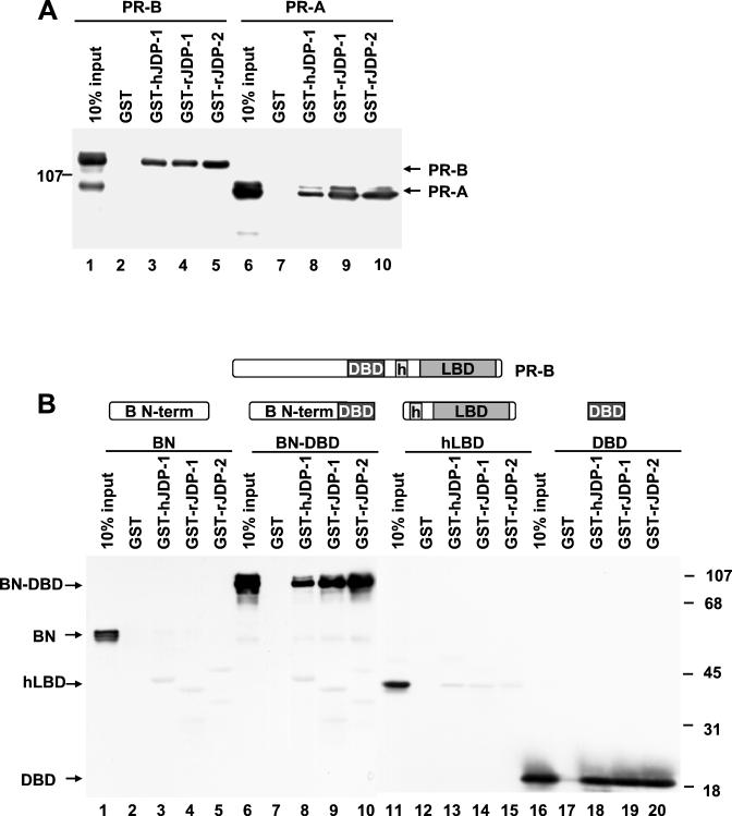
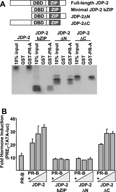
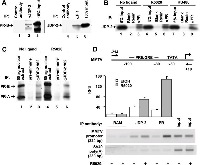
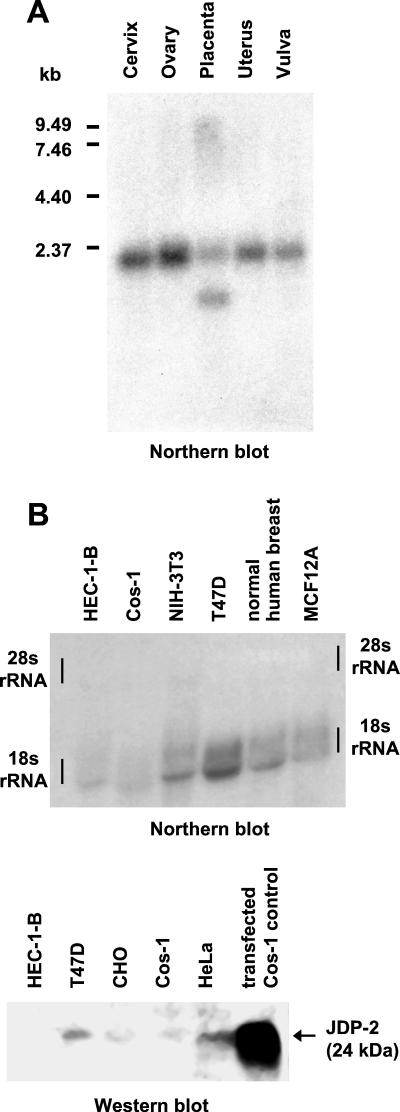
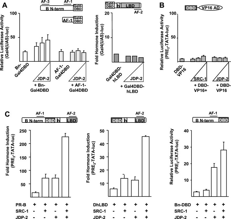
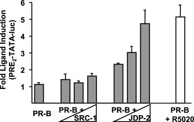
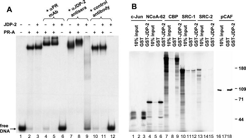
The progesterone receptor (PR) contains two transcription activation function (AF) domains, constitutive AF-1 in the N terminus and AF-2 in the C terminus. AF-2 activity is mediated by a hormone-dependent interaction with a family of steroid receptor coactivators (SRCs). SRC-1 can also stimulate AF-1 activity through a secondary domain that interacts simultaneously with the primary AF-2 interaction site. Other protein interactions and mechanisms that mediate AF-1 activity are not well defined. By interaction cloning, we identified an AP-1 family member, Jun dimerization protein 2 (JDP-2), as a novel PR-interacting protein. JDP-2 was first defined as a c-Jun interacting protein that functions as an AP-1 repressor. PR and JDP-2 interact directly in vitro through the DNA binding domain (DBD) of PR and the basic leucine zipper (bZIP) region of JDP-2. The two proteins also physically associate in mammalian cells, as detected by coimmunoprecipitation, and are recruited in vivo to a progesterone-inducible target gene promoter, as detected by a chromatin immunoprecipitation (ChIP) assay. In cell transfection assays, JDP-2 substantially increased hormone-dependent PR-mediated transactivation and worked primarily by stimulating AF-1 activity. JDP-2 is a substantially stronger coactivator of AF-1 than SRC-1 and stimulates AF-1 independent of SRC-1 pathways. The PR DBD is necessary but not sufficient for JDP-2 stimulation of PR activity; the DBD and AF-1 are required together. JDP-2 lacks an intrinsic activation domain and makes direct protein interactions with other coactivators, including CBP and p300 CBP-associated factor (pCAF), but not with SRCs. These results indicate that JDP-2 stimulates AF-1 activity by the novel mechanism of docking to the DBD and recruiting or stabilizing N-terminal PR interactions with other general coactivators. JDP-2 has preferential activity on PR among the nuclear receptors tested and is expressed in progesterone target cells and tissues, suggesting that it has a physiological role in PR function.


Figures

FIG. 1.
FIG. 1.
(A) Domain structures of PR-A and PR-B isoforms. (B) Diagrammatic alignment with c-Jun of human and rat JDP-1 and rat JDP-2. (C) Sequence alignment of human c-Jun with human and rat JDP-1 and JDP-2 in the conserved basic and leucine zipper regions. Conserved leucines required for zipper-mediated dimerization are boxed.
FIG. 2.
FIG. 2.
JDP-1 and JDP-2 interact in vitro with PR through binding the DBD. (A) WCEs of Sf9 cells expressing polyhistidine-tagged PR-A or PR-B bound to R5020 were incubated with free GST, GST-JDP-1, or GST-JDP-2 fusion proteins immobilized to glutathione Sepharose resin. Resins were washed in a buffer containing 100 mM NaCl, and interacting proteins were eluted and analyzed by Western blotting with a MAb to the polyhistidine tag. Lanes 1 and 6 show the results for 10% input of PR-B and PR-A, respectively. Lanes 2 to 5 show the results for PR-B interaction with GST, GST-hJDP-1, GST-rJDP-1, and GST-rJDP-2 as indicated. Lanes 7 to 10 show the results for PR-A interaction with the same GST constructs in the same order as described for lanes 2 to 5. (B) Sf9 WCEs expressing polyhistidine-tagged domains of PR were incubated with immobilized GST or GST-JDPs as described above, and eluted proteins were detected by Western blotting. PR domain constructs are schematically represented above appropriate lanes. BN, input of PR-B N terminus (1); BN-DBD, input of B N terminus linked to the DBD (6). Inputs of hLBD (11) and DBD (16) are labeled. Lanes 2 to 5, 7 to 10, 12 to 15, and 17 to 20 represent results for interaction of the B N terminus, B N terminus plus DBD, hLBD, and DBD, respectively, with GST or GST-JDP constructs. Results are representative of at least three independent experiments.
FIG. 3.
FIG. 3.
PR interacts with the bZIP region of JDP-2. (A) Cos-1 cells were transfected with plasmids expressing JDP-2 or JDP-2 domain constructs, including bZIP, JDP-2ΔN, and JDP-2ΔC (2 μg of each plasmid). Lysates were analyzed by Western blotting with JDP-2 antibody to normalize pull-down inputs for equal expression of JDP-2 constructs. Lysates were incubated with GST or R5020-bound GST-PR-A immobilized to glutathione-Sepharose. Resins were washed as described in the Fig. 2 legend, and interacting proteins were eluted and detected by Western blotting with an antibody to JDP-2. (B) Cos-1 cells were transfected with phPR-B (0.35 ng) together with PRE2-TATA-luc (200 ng) in the presence or absence of pCDNA.1-JDP-2 or JDP-2 domains in pCI-neo (25 to 200 ng). Cells were treated with vehicle or 10 nM R5020 for 24 h prior to harvest. Severalfold hormone induction was calculated as the ratio of relative luciferase activity of hormone-treated samples divided by the relative luciferase activity of corresponding vehicle-treated samples. Values are averages ± standard errors of the means (SEM) of three experiments.
FIG. 4.
FIG. 4.
JDP-2 and PR physically associate in mammalian cells and are recruited to a progesterone-inducible promoter in vivo. (A) Cos-1 cells were transfected with 1.0 μg of pCDNA1-PR-B and 1.5 μg of pCDNA3-hisJDP-2 and then treated with 100 nM R5020 for 1 h, followed by lysis in a high-salt buffer. Cleared WCEs were incubated with RAM control antibody or PR- or JDP-2-specific antibodies, using protein A-Sepharose as an adsorbent. Resins were washed in a buffer containing 100 mM NaCl, and bound proteins were detected by Western blotting with antibodies to PR or JDP-2. Lanes 1 and 4 show the results of PR and JDP-2 immunoprecipitation with a control unrelated antibody, respectively. Lane 2 shows the results of PR immunoprecipitation with JDP-2 antisera. Lane 5 shows the results of JDP-2 immunoprecipitation with MAb 1294 to PR. Lanes 3 and 6 show the results with 10% input of PR-B and JDP-2, respectively. (B) Cos-1 cells were transfected with 1.5 μg of pCDNA1-PR-B or 0.75 μg of pCR3.1-JDP-2, treated for 2 h with vehicle or 100 nM R5020 or RU486, and then lysed in high-salt buffer. Cleared WCEs were incubated with PR-specific MAb 1294 using protein G-Sepharose as an adsorbent or blank protein G-Sepharose resin as a control, and bound proteins were detected as described above. Lanes 1, 4, and 7 show the results of 10% input of PR-B in the presence of vehicle (ethanol), R5020, and RU486, respectively. Lanes 2, 5, and 8 show the results for nonspecific JDP-2 binding to blank resins alone. Lanes 3, 6, and 9 show the results for JDP-2 immunoprecipitation with the PR-specific MAb 1294 without ligand and in the presence R5020 and RU486, respectively. (C) T47D cells were hormone treated as described for panel A and lysed in buffer without salt. Nuclei were isolated by centrifugation and lysed in high-salt buffer. Cleared nuclear extracts were incubated with preimmune or JDP-2 specific antiserum, using protein A-Sepharose as an adsorbent. Resins were washed and eluted as described above, and bound PR was detected by Western blotting with an antibody to PR. (D) A6 cells were treated for 1 h with vehicle or 10 nM R5020, cross-linked with formaldehyde, and lysed in buffer containing SDS. Lysates were sonicated to shear chromatin and cleared by centrifugation. Clear lysates were incubated with a control unrelated antibody (FSG-RAM) or antibodies to PR or JDP-2, using protein A Sepharose as an adsorbent. Resins were washed in multiple buffers containing various salts and detergents, followed by elution, reversal of cross-links, and isolation of DNA fragments. Immunoprecipitated DNA fragments were analyzed by PCR using primers specific to the MMTV promoter of the integrated reporter gene or to the SV40 poly(A) 2,700-bp fragment removed from the promoter. Amplified products were normalized to similarly amplified input DNA of the corresponding treatment groups (vehicle or R5020). Relative phosphorimage units (RPU) were calculated as the ratio of volume (pixels × intensity) × 100 of immunoprecipitated DNA to the volume of corresponding input DNA, and values are averages ± SEM of four independent experiments.
FIG. 5.
FIG. 5.
JDP-2 increases ligand-dependent PR-mediated transactivation. (A) Cos-1 cells were transfected with phPR-B (1.5 ng) and the progesterone-responsive reporter PRE2-TATA-Luc (200 ng) in the presence or absence of increasing amounts of pCR3.1-SRC-1 or pCDNA3-hisJDP-2 (25, 50, 100, and 200 ng). Cells were treated with the vehicle or 10 nM R5020 for 24 h prior to harvest. Relative luciferase activity was calculated by setting normalized luciferase activity of the reporter alone to 1.0 and all other treatment group values as severalfold relative to 1.0. Values are averages ± SEM of three independent experiments. (B) Cos-1 cells were cotransfected as described for panel A above with PR-B and JDP-2, except for the use of a different progesterone-responsive reporter gene, MMTV-Luc. (C) HEC-1-B cells were transfected with phPR-B (2.5 ng) and PRE2-TATA-luc (200 ng) in the presence or absence of pCR3.1-SRC-1 (50 ng) or pCR3.1-JDP-2 (34 ng) and were hormone treated as described above. Severalfold hormone induction was calculated as the ratio of the relative luciferase activity of hormone-treated samples divided by the relative luciferase activity of corresponding vehicle-treated samples. Values are averages ± SEM of three independent experiments. (D) Cos-1 cells were transfected with indicated nuclear receptors [phPR-B (1 ng), pRSV-GR (2 ng), SVMT-ER (0.1 ng), TRβ (0.5 ng), or RSV-VDR (1.5 ng)], together with 200 ng of their cognate hormone-responsive luciferase reporter genes [PRE2-TATA-luc (PR and GR), ERE3-TATA-luc (ER), TRE-luc (TR), or VDRE(1,2)-ΔMTV-luc (VDR)] in the presence or absence of pCR3.1-JDP-2 (110 ng). At 24 h prior to harvest, cells were treated with the appropriate ligands: 10 nM R5020 (PR), 100 nM dexamethasone (GR), 10 nM estradiol (ER), 100 nM T3 (TR), or 10 nM 1,25-vitamin D3. Severalfold hormone induction was calculated as described for panel C, and values are averages ± SEM of three independent experiments.
FIG. 6.
FIG. 6.
JDP-2 is expressed in progesterone target tissues and variably expressed in cell lines. (A) JDP-2 mRNA was detected in human tissues by Northern blotting of a human female reproductive system (MessageBlot; Stratagene) using 32P-labeled rat JDP-2 cDNA. (B) JDP-2 mRNA present in total RNA (25 μg) from indicated cell lines was analyzed by Northern blotting using 32P-labeled JDP-2 cDNA. (C) Nuclear extracts (300 μg) of indicated cell lines were prepared as described for Fig. 4C and analyzed for JDP-2 expression by Western blotting with antibody to JDP-2. Extracts (10 μg) of Cos-1 cells transfected with pCR3.1-JDP-2 served as a positive control.
FIG. 7.
FIG. 7.
JDP-2 enhances transactivation of the N-terminal AF-1 domain of PR. Cos-1 cells were transfected with the PR domain constructs indicated together with PRE2-TATA-luc reporter (200 ng) in the presence or absence of various amounts of pCR3.1-SRC-1 or pCR3.1-JDP-2. Equimolar doses of SRC-1 and JDP-2 expression vectors were used (50 to 200 ng or 34 to 137 ng of SRC-1 or JDP-2, respectively). Cells transfected with DhLBD were treated with vehicle or 10 nM R5020 24 h prior to harvest. Other constructs lacking the LBD did not receive hormone. Relative luciferase activity and severalfold hormone induction were calculated as for Fig. 5. Cos-1 cells were transfected with BnDBD (10 ng) or DhLBD (50 ng) (A), with BnDBD (1.5 ng) or AnDBD (10 ng) (B), or with AF-1-DBD (20 ng) (C). Values are averages ± SEM of at least three independent experiments.
FIG. 8.
FIG. 8.
PR-DBD is required, but not sufficient, for JDP-2 enhancement of PR transactivation domains, while JDP-2 and SRC-1 synergize to coactivate AF-2 but not the N terminus. (A) Cos-1 cells were cotransfected with the Gal4DBD-PR chimeric constructs indicated, including BnGal4DBD (1 ng), AF-1-Gal4DBD (1 ng), or GalDBD-LBD (50 ng), together with the 5×-GalUAS-luc reporter (500 ng), in the presence or absence of various amounts of pCR3.1-JDP-2 (34 to 137 ng). Cells transfected with GalDBD-LBD were treated with vehicle or 10 nM R5020 24 h prior to harvest. Constructs lacking the LBD did not receive hormone. (B) Cos-1 cells were transfected with pfPR-DBD-VP16 (50 ng) together with PRE2-TATA-luc (200 ng) in the presence or absence of pCR3.1-SRC-1 (50 to 200 ng) or pCR3.1-JDP-2 (34 to 137 ng). (C) Cos-1 cells were transfected with phPR-B (1.5 ng) (leftmost panel), DhLBD (50 ng) (middle panel), or BnDBD (1.5 ng) (rightmost panel), together with PRE2-TATA-luc (200 ng), in the presence or absence of pCR3.1-SRC-1 (200 ng) or pCR3.1-JDP-2 (137 ng). Cells transfected with PR-B or DhLBD were treated with vehicle or 10 nM R5020 for 24 h. Relative luciferase activity and severalfold hormone induction values were calculated as for Fig. 5. Values are averages ± SEM of at least three independent experiments.
FIG. 9.
FIG. 9.
JDP-2 potentiates the partial agonist activity of RU486. Cos-1 cells were transfected with phPR-B (1.5 ng) and PRE2-TATA-luc (200 ng) in the presence or absence of equimolar doses of pCR3.1-SRC-1 (50 to 200 ng) or pCR3.1-JDP-2 (34 to 137 ng). Cells were treated with vehicle, 10 nM RU486 (dark bars), or 10 nM R5020 (open bar) for the last 24 h of transfection. Severalfold hormone induction values were calculated as for Fig. 5, and values are averages ± SEM of three independent experiments.
FIG. 10.
FIG. 10.
JDP-2 forms a ternary complex with PR on DNA and interacts with general coactivators but not with SRCs (p160s). (A) Purified recombinant PR-A (30 nM) bound to R5020 and purified JDP-2 (200 ng) were incubated alone or together with 0.3 ng of a 32-bp synthetic [32P]PRE oligonucleotide probe for 1 h on ice. To supershift PR-DNA-JDP-2 complexes, 2 μg of the PR MAb, 4 μg of JDP-2 antiserum, or an unrelated control antibody (as indicated above appropriate lanes) were added for the final 30 min of incubation with DNA. Samples were separated on 5% nondenaturing polyacrylamide gels, followed by drying of the gels and detection of protein-DNA complexes by autoradiography. (B) c-Jun, NCoA-62, CBP, and SRC-1 and SRC-2 (GRIP-1) were transcribed and translated in vitro using rabbit reticulocyte lysate supplemented with [35S]methionine. pCAF with an N-terminal FLAG tag was expressed in Sf9 cells. Labeled coactivators or FLAG-pCAF were incubated with GST or GST-JDP-2 immobilized to glutathione-Sepharose. Resins were washed in a buffer containing 100 mM NaCl, and bound proteins were eluted and detected either by autoradiography or by Western blotting with an antibody to the FLAG tag (pCAF). Lanes contain 10% input or coactivator interaction with GST or GST-JDP-2 as indicated.

Similar articles

Cited by

References

    1. Alen, P., F. Claessens, G. Verhoeven, W. Rombauts, and B. Peeters. 1999. The androgen receptor amino-terminal domain plays a key role in p160 coactivator-stimulated gene transcription. Mol. Cell. Biol. 19:6085-6097. - PMC - PubMed
    1. Allan, G., S. Leng, S. Tsai, N. Weigel, D. Edwards, M.-J. Tsai, and B. O'Malley. 1992. Hormone and antihormone induce distinct conformational changes which are central to steroid receptor activation. J. Biol. Chem. 267:19513-19520. - PubMed
    1. Aronheim, A., E. Zandi, H. Hennemann, S. Elledge, and M. Karin. 1997. Isolation of an AP-1 repressor by a novel method for detecting protein-protein interactions. Mol. Cell. Biol. 17:3094-3102. - PMC - PubMed
    1. Bain, D., M. Franden, J. McManaman, G. Takimoto, and K. Horwitz. 2000. The N-terminal region of the human progesterone A-receptor. J. Biol. Chem. 275:7313-7320. - PubMed
    1. Baudino, T., D. Kraichely, S. Jefcoat, Jr., S. Winchester, N. Partridge, and P. MacDonald. 1998. Isolation and characterization of a novel coactivator protein, NCoA-62, involved in vitamin D-mediated transcription. J. Biol. Chem. 273:16434-16441. - PubMed

Publication types

MeSH terms

LinkOut - more resources