A novel colonic repressor element regulates intestinal gene expression by interacting with Cux/CDP
- PMID: 12101240
- PMCID: PMC133930
- DOI: 10.1128/MCB.22.15.5467-5478.2002
A novel colonic repressor element regulates intestinal gene expression by interacting with Cux/CDP
Abstract
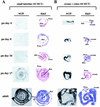
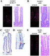
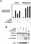
Intestinal gene regulation involves mechanisms that direct temporal expression along the vertical and horizontal axes of the alimentary tract. Sucrase-isomaltase (SI), the product of an enterocyte-specific gene, exhibits a complex pattern of expression. Generation of transgenic mice with a mutated SI transgene showed involvement of an overlapping CDP (CCAAT displacement protein)-GATA element in colonic repression of SI throughout postnatal intestinal development. We define this element as CRESIP (colon-repressive element of the SI promoter). Cux/CDP interacts with SI and represses SI promoter activity in a CRESIP-dependent manner. Cux/CDP homozygous mutant mice displayed increased expression of SI mRNA during early postnatal development. Our results demonstrate that an intestinal gene can be repressed in the distal gut and identify Cux/CDP as a regulator of this repression during development.
Figures


Similar articles
-
Sucrase-isomaltase gene transcription requires the hepatocyte nuclear factor-1 (HNF-1) regulatory element and is regulated by the ratio of HNF-1 alpha to HNF-1 beta.J Biol Chem. 2001 Aug 24;276(34):32122-8. doi: 10.1074/jbc.M102002200. Epub 2001 Jun 6. J Biol Chem. 2001. PMID: 11395488
-
Hepatocyte nuclear factor-1 alpha, GATA-4, and caudal related homeodomain protein Cdx2 interact functionally to modulate intestinal gene transcription. Implication for the developmental regulation of the sucrase-isomaltase gene.J Biol Chem. 2002 Aug 30;277(35):31909-17. doi: 10.1074/jbc.M204622200. Epub 2002 Jun 11. J Biol Chem. 2002. PMID: 12060663
-
Spatio-temporal patterns of intestine-specific transcription factor expression during postnatal mouse gut development.Gene Expr Patterns. 2006 Apr;6(4):426-32. doi: 10.1016/j.modgep.2005.09.003. Epub 2005 Dec 27. Gene Expr Patterns. 2006. PMID: 16377257
-
Role of the multifunctional CDP/Cut/Cux homeodomain transcription factor in regulating differentiation, cell growth and development.Gene. 2001 May 30;270(1-2):1-15. doi: 10.1016/s0378-1119(01)00485-1. Gene. 2001. PMID: 11403998 Review.
-
Contribution of CDP/Cux, a transcription factor, to cell cycle progression.Acta Biochim Biophys Sin (Shanghai). 2007 Dec;39(12):923-30. doi: 10.1111/j.1745-7270.2007.00366.x. Acta Biochim Biophys Sin (Shanghai). 2007. PMID: 18064384 Review.
Cited by
-
Nuclear receptor co-repressor is required to maintain proliferation of normal intestinal epithelial cells in culture and down-modulates the expression of pigment epithelium-derived factor.J Biol Chem. 2009 Sep 11;284(37):25220-9. doi: 10.1074/jbc.M109.022632. Epub 2009 Jul 16. J Biol Chem. 2009. PMID: 19608741 Free PMC article.
-
Regulation of cell proliferation and differentiation in the kidney.Front Biosci (Landmark Ed). 2009 Jun 1;14(13):4978-91. doi: 10.2741/3582. Front Biosci (Landmark Ed). 2009. PMID: 19482600 Free PMC article. Review.
-
CCAAT displacement protein regulates nuclear factor-kappa beta-mediated chemokine transcription in melanoma cells.Melanoma Res. 2007 Apr;17(2):91-103. doi: 10.1097/CMR.0b013e3280a60888. Melanoma Res. 2007. PMID: 17496784 Free PMC article.
-
Enteroendocrine cells-sensory sentinels of the intestinal environment and orchestrators of mucosal immunity.Mucosal Immunol. 2018 Jan;11(1):3-20. doi: 10.1038/mi.2017.73. Epub 2017 Aug 30. Mucosal Immunol. 2018. PMID: 28853441 Review.
-
Polycomb repressive complex 2 impedes intestinal cell terminal differentiation.J Cell Sci. 2012 Jul 15;125(Pt 14):3454-63. doi: 10.1242/jcs.102061. Epub 2012 Mar 30. J Cell Sci. 2012. PMID: 22467857 Free PMC article.
References
-
- Antes, T. J., J. Chen, A. D. Cooper, and B. Levy-Wilson. 2000. The nuclear matrix protein CDP represses hepatic transcription of the human cholesterol-7alpha hydroxylase gene. J. Biol. Chem. 275:26649-26660. - PubMed
-
- Beaulieu, J. F., M. M. Weiser, L. Herrera, and A. Quaroni. 1990. Detection and characterization of sucrase-isomaltase in adult human colon and in colonic polyps. Gastroenterology 98:1467-1477. - PubMed
-
- Blochlinger, K., R. Bodmer, J. Jack, L. Y. Jan, and Y. N. Jan. 1988. Primary structure and expression of a product from cut, a locus involved in specifying sensory organ identity in Drosophila. Nature 333:629-635. - PubMed
Publication types
MeSH terms
Substances
Grants and funding
LinkOut - more resources
Full Text Sources
Molecular Biology Databases
