. 2002 Aug;22(15):5492-505.
doi: 10.1128/MCB.22.15.5492-5505.2002.

Nrf2 transcription factor, a novel target of keratinocyte growth factor action which regulates gene expression and inflammation in the healing skin wound

Affiliations

Nrf2 transcription factor, a novel target of keratinocyte growth factor action which regulates gene expression and inflammation in the healing skin wound

Susanne Braun et al. Mol Cell Biol. 2002 Aug.

Abstract

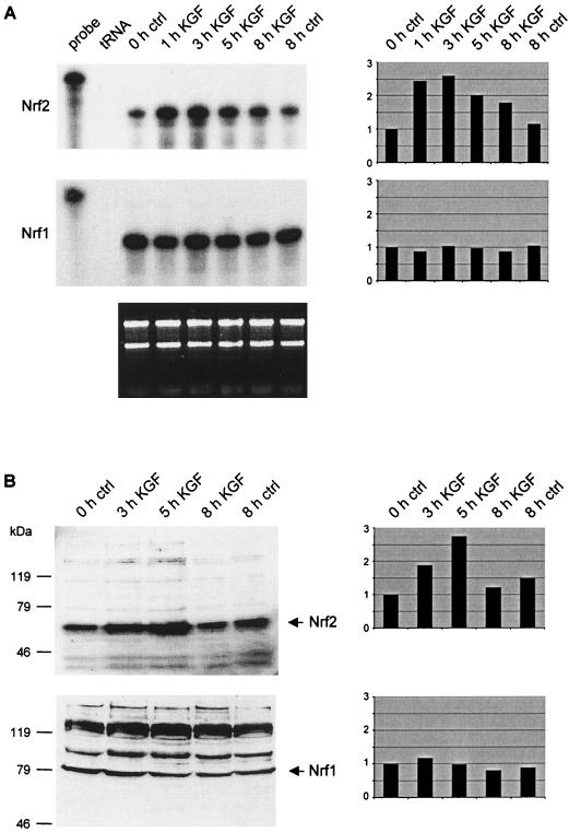
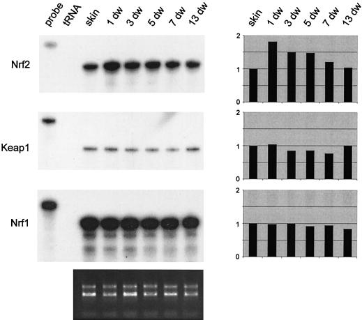
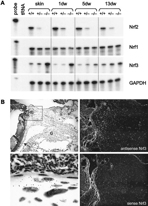
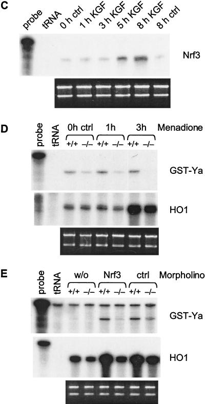
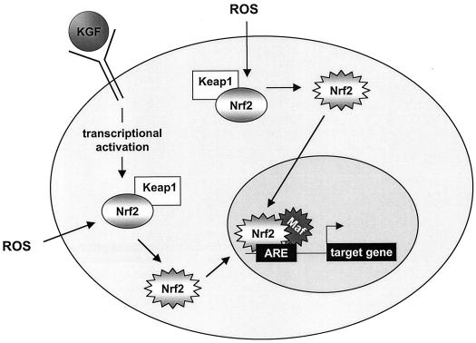
Keratinocyte growth factor (KGF) is a potent mitogen for epithelial cells, and it promotes survival of these cells under stress conditions. In a search for KGF-regulated genes in keratinocytes, we identified the gene encoding the transcription factor NF-E2-related factor 2 (Nrf2). Nrf2 is a key player in the cellular stress response. This might be of particular importance during wound healing, where large amounts of reactive oxygen species are produced as a defense against invading bacteria. Therefore, we studied the wound repair process in Nrf2 knockout mice. Interestingly, the expression of various key players involved in wound healing was significantly reduced in early wounds of the Nrf2 knockout animals, and the late phase of repair was characterized by prolonged inflammation. However, these differences in gene expression were not reflected by obvious histological abnormalities. The normal healing rate appears to be at least partially due to an up-regulation of the related transcription factor Nrf3, which was also identified as a target of KGF and which was coexpressed with Nrf2 in the healing skin wound. Taken together, our results reveal novel roles of the KGF-regulated transcription factors Nrf2 and possibly Nrf3 in the control of gene expression and inflammation during cutaneous wound repair.


Figures

FIG. 1.
FIG. 1.
KGF induces expression of Nrf2 in HaCaT keratinocytes. Cells were rendered quiescent by serum starvation and treated with KGF as described in Materials and Methods. (A) RNase protection assays were performed with total cellular RNA (10 μg) from quiescent and KGF-treated cells to analyze the Nrf2 (upper panel) and Nrf1 (middle panel) mRNA levels. tRNA (50 μg) was used as a negative control. The hybridization probes (1,000 cpm) were loaded in the lanes labeled “probe” and used as size markers. One microgram of each RNA sample was loaded on a 1% agarose gel and stained with ethidium bromide (bottom panel). (B) Western blot analyses of lysates (50 μg) from quiescent and KGF-stimulated HaCaT cells for the presence of Nrf2 (top panel) and Nrf1 (bottom panel). Densitometric quantification of each RNase protection assay and Western blot is shown on the right-hand side. The signal intensities of nontreated keratinocytes were arbitrarily set as 1.
FIG. 2.
FIG. 2.
Increased expression of Nrf2 after skin injury. Mice were wounded and sacrificed at different time points (e.g., 1-day wound ([1 dw]) after injury as described in Materials and Methods. Twenty micrograms of total cellular RNA from normal and wounded skin was analyzed by RNase protection assay for the expression of Nrf2, Keap1, and Nrf1. For an explanation of lanes labeled “probe” and “tRNA,” see the legend to Fig. 1. Ethidium bromide-stained 1% agarose gel loaded with each RNA sample (1 μg) served as a loading control (shown below the protection assays). Densitometric quantification of each RNase protection assay is shown on the right-hand side. The signal intensities of unwounded skin were arbitrarily set as 1.
FIG. 3.
FIG. 3.
Localization of Nrf2 (A) and Nrf1 (B) mRNAs in 5-day wounds by in situ hybridization. Paraformaldehyde-fixed frozen sections from the middle of 5-day full-thickness excisional wounds from BALB/c mice were hybridized with 35S-labeled sense (lower right panels; magnification, ×100) or antisense (upper right panels; ×100) riboprobes. The upper left panels show an overview over the right wound margin (×50), and the rectangles mark the area of the wounds, which are shown in the panels on the right hand side. Details of the hyperproliferative epithelium (marked with rectangles in the upper right panels) are shown in the lower left panels (×1,000). Signals appear as black dots in the bright field survey (lower left panels) and as white dots in the dark field survey (right panels). D, dermis; E, epidermis; ES, eschar; G, granulation tissue; HE, hyperproliferative epithelium. Sections were counterstained with H/E.
FIG. 4.
FIG. 4.
(A to D): Histology of 5- and 13-day wounds of wild-type and homozygous Nrf2 knockout mice. Full-thickness excisional wounds were made on the backs of control and Nrf2−/− mice (8 to 12 weeks old). Sections from the middle of 5-day and 13-day wounds were stained with H/E. (A) Five-day wound of a control mouse (+/+); (B) Five-day wound of an Nrf2−/− mouse (−/−); (C) Thirteen-day wound of a control mouse; (D) Thirteen-day wound of an Nrf2−/− mouse. For abbreviations, see the legend to Fig. 3. Magnification, ×50. (E and F) Analysis of cell proliferation in 5-day wounds. Mice were injected with BrdU and sacrificed 2 h after injection. Paraffin sections of wounds from control mice (E) and Nrf2 null mice (F) were incubated with a peroxidase-conjugated antibody against BrdU and stained with the diaminobenzidine-peroxidase staining kit.
FIG. 5.
FIG. 5.
(A) Altered expression of proinflammatory cytokines in wounded skin of Nrf2 knockout mice. mRNA levels of IL-1β, IL-6, and TNF-α were determined by RNase protection assay on 10-μg samples from normal and wounded skin of wild-type (+/+), heterozygous (+/-), and homozygous (−/−) Nrf2 knockout mice. As a loading control the RNA samples were also hybridized with an antisense probe to the housekeeping GAPDH gene. The same set of RNAs was used for the protection assays shown in Fig. 5, 6, and 8. For an explanation of lanes labeled “probe” and “tRNA,” see the legend to Fig. 1. The mRNA levels of the various cytokines during the wound repair process in control, heterozygous, and homozygous Nrf2 knockout mice were quantified by laser scanning densitometry of the autoradiograms (panels on the right-hand side). Results of a representative experiment are shown. The band intensities of the RNase protection assays are expressed as a percentage of the maximal mRNA level for each gene assayed. 1dw, 5dw, and 13dw, 1-, 5-, and 13-day wounds. (B) IL-1β protein content in wild-type (+/+) and homozygous (−/−) knockout mice during wound repair determined by ELISA.
FIG. 6.
FIG. 6.
Elevated number of macrophages in 13-day wounds of Nrf2 knockout mice. Paraffin sections from 13-day wounds of control mice (+/+) and Nrf2 knockout mice (−/−) were incubated with an antibody against F4/80 and a Cy3-coupled secondary antibody (left panels; magnification, ×200). Serial sections were stained with H/E (right panels; ×50), and the part of the wound that is shown in the respective immunofluorescence is marked with a rectangle.
FIG. 7.
FIG. 7.
Altered expression of extracellular matrix molecules, TGFβ1, and VEGF in wounded skin of Nrf2 knockout mice. mRNA levels of collagens α1 (I) and α1 (III), fibronectin, TGFβ1, and VEGF were determined by RNase protection assay on 10-μg RNA samples from nonwounded back skin and wounds. Hybridization with a GAPDH probe served as a loading control. For an explanation of lanes labeled “probe” and “tRNA,” see the legend to Fig. 1. 1dw, 5dw, and 13dw, 1-, 5-, and 13-day wounds
FIG. 8.
FIG. 8.
Altered expression of the Nrf2 target genes HO1 and GST-Ya in wounded skin of Nrf2 knockout mice. (A) The band intensities of RNase protection assays (not shown) were quantified by laser scanning densitometry of the autoradiograms and are expressed as a percentage of the maximal mRNA level for each gene assayed. Results from at least two RNase protection assays with RNAs from independent wound healing experiments were used for this quantitative analysis. (B) Western blot analysis of tissue lysates from normal and wounded skin of control (+/+), Nrf2+/−, and Nrf2−/− mice for the presence of HO1. 1dw, 5dw, and 13dw, 1-day, 5-day, and 13-day wounds.
FIG. 9.
FIG. 9.
A possible compensatory effect of Nrf3 in Nrf2 knockout mice. (A) Ten-microgram RNA samples from unwounded skin and wound tissue of control (+/+), Nrf2+/−, and Nrf2−/− mice were analyzed by RNase protection assay for the presence of Nrf1, Nrf2, and Nrf3 mRNAs. Hybridization with a GAPDH probe served as a loading control. 1dw, 5dw, and 13dw, 1-day, 5-day, and 13-day wounds. (B) Paraformaldehyde-fixed frozen sections from the middle of 5-day full-thickness excisional wounds from BALB/c mice were hybridized with a 35S-labeled sense (lower right panel; magnification, ×200) or antisense (upper right panel; ×200) Nrf3 riboprobes. An overview of half of the wound area is shown in the upper left panel (magnification, ×50). The area indicated by the rectangle is shown in the panels on the right-hand side. Details of the migrating epithelial tongue (marked with an arrow in the upper left panel) are shown in the lower left panel (magnification, ×1,000). Signals appear as black dots in the bright field survey (lower left panel) and as white dots in the dark field survey (right panels). For abbreviations, see the legend to Fig. 3. Sections were counterstained with H/E. (C) HaCaT cells were rendered quiescent by serum starvation and treated with KGF. Total cellular RNA (10 μg) isolated from quiescent and KGF-treated cells was analyzed by RNase protection assay for the expression of Nrf3. (D) Freshly isolated keratinocytes from 3-day-old Nrf2−/− mice and control (ctrl) littermates were rendered quiescent and subsequently treated with menadione. Cells were harvested before and after 1 or 3 h of menadione treatment. (E) Primary keratinocytes from the skin of 3-day-old Nrf2−/− mice and control littermates were transfected either with an antisense morpholino oligonucleotide directed against Nrf3 or with a control morpholino oligonucleotide and treated with menadione for 3 h. Nontransfected cells served as a control. w/o, without morpholino oligonucleotide. Total cellular RNA (10 μg [D] or 5 μg [E]) was analyzed by RNase protection assay for the expression of GST-Ya and HO1 as indicated. Note that the GST-Ya signal seen in menadione-treated, nontransfected cells is visible only in panel D and not in panel E due to the smaller amounts of RNA used in the protection assay shown in panel E. Ethidium bromide-stained agarose gels loaded with each RNA sample (1 μg) served as a loading control for the RNase protection assays shown in panels C, D, and E (shown below the RNase protection assays). For an explanation of lanes in panels C, D, and E labeled “probe” and “tRNA,” see the legend to Fig. 1.
FIG. 9.
FIG. 9.
A possible compensatory effect of Nrf3 in Nrf2 knockout mice. (A) Ten-microgram RNA samples from unwounded skin and wound tissue of control (+/+), Nrf2+/−, and Nrf2−/− mice were analyzed by RNase protection assay for the presence of Nrf1, Nrf2, and Nrf3 mRNAs. Hybridization with a GAPDH probe served as a loading control. 1dw, 5dw, and 13dw, 1-day, 5-day, and 13-day wounds. (B) Paraformaldehyde-fixed frozen sections from the middle of 5-day full-thickness excisional wounds from BALB/c mice were hybridized with a 35S-labeled sense (lower right panel; magnification, ×200) or antisense (upper right panel; ×200) Nrf3 riboprobes. An overview of half of the wound area is shown in the upper left panel (magnification, ×50). The area indicated by the rectangle is shown in the panels on the right-hand side. Details of the migrating epithelial tongue (marked with an arrow in the upper left panel) are shown in the lower left panel (magnification, ×1,000). Signals appear as black dots in the bright field survey (lower left panel) and as white dots in the dark field survey (right panels). For abbreviations, see the legend to Fig. 3. Sections were counterstained with H/E. (C) HaCaT cells were rendered quiescent by serum starvation and treated with KGF. Total cellular RNA (10 μg) isolated from quiescent and KGF-treated cells was analyzed by RNase protection assay for the expression of Nrf3. (D) Freshly isolated keratinocytes from 3-day-old Nrf2−/− mice and control (ctrl) littermates were rendered quiescent and subsequently treated with menadione. Cells were harvested before and after 1 or 3 h of menadione treatment. (E) Primary keratinocytes from the skin of 3-day-old Nrf2−/− mice and control littermates were transfected either with an antisense morpholino oligonucleotide directed against Nrf3 or with a control morpholino oligonucleotide and treated with menadione for 3 h. Nontransfected cells served as a control. w/o, without morpholino oligonucleotide. Total cellular RNA (10 μg [D] or 5 μg [E]) was analyzed by RNase protection assay for the expression of GST-Ya and HO1 as indicated. Note that the GST-Ya signal seen in menadione-treated, nontransfected cells is visible only in panel D and not in panel E due to the smaller amounts of RNA used in the protection assay shown in panel E. Ethidium bromide-stained agarose gels loaded with each RNA sample (1 μg) served as a loading control for the RNase protection assays shown in panels C, D, and E (shown below the RNase protection assays). For an explanation of lanes in panels C, D, and E labeled “probe” and “tRNA,” see the legend to Fig. 1.
FIG. 10.
FIG. 10.
Model of Nrf2 activation by KGF and ROS. Triggered by ROS, the inactive Nrf2 (gray shaded oval) is liberated from its cytosolic inhibitor, Keap1, and therefore activated. Subsequently, the active Nrf2 (jagged gray shaded oval) is translocated into the nucleus, where it dimerizes with small Maf proteins, binds to the ARE sequence, and initiates the transcription of target genes (26). As shown in this study, KGF stimulates the expression of Nrf2. In inflamed tissues, e.g., in wounded skin, the newly synthesized Nrf2 protein is likely to be activated by ROS.

Similar articles

Cited by

References

    1. Beer, H. D., C. Florence, J. Dammeier, L. McGuire, S. Werner, and D. R. Duan. 1997. Mouse fibroblast growth factor 10: cDNA cloning, protein characterization, and regulation of mRNA expression. Oncogene 15:2211-2218. - PubMed
    1. Bloch, W., K. Huggel, R. Sasaki, R. Grose, P. Bugnon, K. Addicks, R. Timpl, and S. Werner. 2000. The angiogenesis inhibitor endostatin impairs blood vessel maturation during wound healing. FASEB J. 14:2373-2376. - PubMed
    1. Boukamp, P., R. T. Petrussevska, D. Breitkreutz, J. Hornung, A. Markham, and N. E. Fusenig. 1988. Normal keratinization in a spontaneously immortalized aneuploid human keratinocyte cell line. J. Cell Biol. 106:761-771. - PMC - PubMed
    1. Caldelari, R., M. M. Suter, D. Baumann, A. De Bruin, and E. Müller. 2000. Long-term culture of murine epidermal keratinocytes. J. Investig. Dermatol. 114:1064-1065. - PubMed
    1. Chan, J. Y., X. L. Han, and Y. W. Kan. 1993. Isolation of cDNA encoding the human NF-E2 protein. Proc. Natl. Acad. Sci. USA 90:11366-11370. - PMC - PubMed

Publication types

MeSH terms

Substances

LinkOut - more resources