. 2002 Aug;22(15):5554-62.
doi: 10.1128/MCB.22.15.5554-5562.2002.

Testis-specific cytochrome c-null mice produce functional sperm but undergo early testicular atrophy

Affiliations

Testis-specific cytochrome c-null mice produce functional sperm but undergo early testicular atrophy

Sonoko Narisawa et al. Mol Cell Biol. 2002 Aug.

Abstract

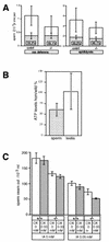
Differentiating male germ cells express a testis-specific form of cytochrome c (Cyt c(T)) that is distinct from the cytochrome c expressed in somatic cells (Cyt c(S)). To examine the role of Cyt c(T) in germ cells, we generated mice null for Cyt c(T). Homozygous Cyt c(T)(-/-) pups were statistically underrepresented (21%) but developed normally and were fertile. However, spermatozoa isolated from the cauda epididymis of Cyt c(T)-null animals were less effective in fertilizing oocytes in vitro and contain reduced levels of ATP compared to wild-type sperm. Sperm from Cyt c(T)-null mice contained a greater number of immotile spermatozoa than did samples from control mice, i.e., 53.1% +/- 13.7% versus 33.2% +/- 10.3% (P < 0.0001) for vas deferens sperm and 40.1% +/- 9.6% versus 33.2% +/- 7.5% (P = 0.0104) for epididymal sperm. Cyt c(T)-null mice often exhibit early atrophy of the testes after 4 months of age, losing germ cells as a result of increased apoptosis. However, no difference in the activation of caspase-3, -8, or -9 was detected between the Cyt c(T)(-/-) testes and controls. Our data indicate that the Cyt c(T)-null testes undergo early atrophy equivalent to that which occurs during aging as a consequence of a reduction in oxidative phosphorylation.


Figures

FIG. 1.
FIG. 1.
Disruption of the Cyt cT gene. (a) Amino acid sequences of Cyt cS and Cyt cT. Vertical lines indicate unmatched amino acids. Underlined amino acid sequences shown in this panel were used for the production of antibodies (anti-cS antibody, carboxyl-terminal 16 amino acids; anti-cT antibody, carboxyl-terminal 16 amino acids). (b) Genomic organization of the Cyt cT gene. White boxes are exons and shadowed regions are introns. Translation starts at ATG in exon III. Probes used for Southern blot analysis are shown beneath the genomic DNA. (c) Targeting vector. (d) PCR screening performed for ES cell clones obtained from the vector. PCR using the 5′ primer shown in panel c and the 3′ primer shown in panel b amplified a predicted 3-kb fragment (left panel). EcoRI digestion of this fragment showed the predicted 1.8- and 1.2-kb fragments (right panel). D3, parental ES cell. 6 and 64 are ES clones used for generation of mouse colonies. (e) Southern blot analysis of Cyt cT-targeted mice. The targeted allele in Cyt KO mice showed a 6.6-kb BamHI fragment with probe 360.
FIG. 2.
FIG. 2.
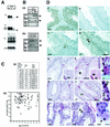
Confirmation of CytcT inactivation. (A) Northern blot analysis of the following fragments: cT, partial cDNA probe corresponding to a DNA fragment between BglII in exon III and BglII in exon IV of the Cyt cT shown in Fig. 1b; cT entire cDNA, cT probe using entire Cyt cT cDNA; cS, cS probe using entire Cyt cS cDNA; Neo, Neo gene probe; L32, ribosomal protein L32-A4, used as a standard. In the Cyt cT−/− mice, the Cyt cT probe gave no signal (arrowhead); however, the entire Cyt cT cDNA probe detected a faint shorter signal (thin arrowhead), suggesting that there was transcription until the stop codon in exon III. The expression pattern of Cyt cS in the Cyt cT−/− testis was normal and not distinguishable from Cyt cS expression in heterozygotes and wild-type mice. Samples are 10 μg of total RNA from testes. (B) Western blotting using anti-Cyt cS and Cyt cT antibodies. Sperm cells were isolated from wild-type and Cyt cT−/− mice (2 × 10⁵ sperm/lane); liver extract was from wild-type mice (100 μg/lane); testis extracts were from mice (30 μg of protein/lane). Sperm cells were purified on a Percoll gradient (Amersham Pharmacia, Uppsala, Sweden) and washed in 0.45% NaCl hypotonic solution to remove contaminating somatic cells. (a) Black arrowheads indicate the predicted 12-kDa Cyt cT or Cyt cS protein. (b) Enriched population of mitochondria extracted from testes of wild-type, heterozygous, and Cyt cT KO mice (12.5 μg of protein/lane). (C) Fertility and weight of testes. (a) Breeding record of Cyt cT KO homozygous males. Litter size is the mean number of pups in each litter. (b) Average weight of testes from each individual mouse. Wild-type (+/+), n = 50, mean = 110.8 ± 17.1 mg; heterozygote (+/−), n = 21, mean = 113.0 ± 15.7 mg; homozygote (−/−), n = 60, mean = 102.9 ± 22.2 mg. Homozygous animals and their littermate controls, either wild type or heterozygotes, were analyzed at the same time. The ages of the mice subjected to analyses were 3 to 19 months. (D) Immunohistochemistry and morphology of the Cyt cT−/− testis. Panels on the right (b, d, f, g, i, and j) show Cyt cT−/− sections, and panels on the left (a, c, e, and h) show wild-type control sections. (a to d) Immunohistochemistry of testes fixed with Bouin's and embedded in paraffin. Panels a and b were stained with anti-cT antibody; no Cyt cT protein was seen in spermatocytes and spermatids of the Cyt cT−/− testes. Panels c and d were stained with anti-cS antibody showing expression of Cyt cS in spermatogonia, Sertoli cells, and Leydig cells in both +/+ and −/− mice. The mice are 3 months old. Bar, 100 μm. (e to j) Hematoxylin-eosin staining of testis and epididymis (ep; square insets in panels e and f) fixed with formalin and embedded in OCT compound. (e to g) Three-month-old mice. Testis weight of Cyt cT−/− mice (f) was the same as control (e) weights. At this age, homozygote testes appeared to be similar to controls except for the unusual basophilic structures in the luminal region of the seminiferous tubules and epididymis (arrowheads). Panel g is an enlarged view of the basophilic structure in homozygote testis (arrowheads). (h to j) Seven-month-old mice. At this age, testis weight of Cyt cT−/− mice (i) was approximately 27% of controls (h). Seminiferous tubules showed a dramatic loss of spermatocytes, spermatids, and spermatozoa, although some seminiferous tubules in the same testis still contain mature spermatozoa (i). Spermatogonia, Sertoli cells, and Leydig cells appeared unchanged in Cyt cT−/− and wild-type mice (i and j). Bars: e, f, h, and i, 100 μm; j, 10 μm. L, Leydig cell; St, Sertoli cell; Sg, spermatogonia. (k to m) Hematoxylin-eosin staining of testes of 17- to 19-month-old wild-type mice. (k) Testis of a 17-month-old mouse, showing marked loss of germ cells. (l) Testis of a 19-month-old mouse with reduced germ cells and basophilic structures (arrowheads). (m) High magnification of the basophilic structure from the same testis as shown in panel l. Bars: k and l, 100 μm; m, 10 μm.
FIG. 3.
FIG. 3.
Apoptosis in the Cyt cT−/− testis. (a to c) TUNEL staining of testes from 4.5-month-old mice; (b) Cyt cT−/− mouse; (a) Cyt cT+/+ littermate control of animal shown in panel b (weight of the −/− testis was approximately 71% of control); (c) high magnification of image shown in panel b. Arrowheads in panel c indicate positive cells. (d) Electron microscopic view of apoptotic cells in testis of 5.5-month-old Cyt cT−/− mouse (weight of −/− testis was approximately 53% of control). (e to g) Immunostaining with anti-caspase-3 antibody on the same testis as shown in panel b. (f) Control inhibition of immunoreaction by a specific blocking peptide for the epitope; (g) high magnification of panel e. (h and i) Immunostaining with anti-caspase-9 antibody on testes from the same animals as shown in panels a and b. (j) Staining with normal rabbit immunoglobulin G (4 μg/ml) using same testis samples as shown in panels b, e, and i. Ap, apoptotic cell; P, pachytene spermatocyte; L, Leydig cell. Bars: a, b, e, and f, 100 μm; c, g, h, i, and j, 20 μm; d, 2.5 μm.
FIG. 4.
FIG. 4.
Expression of testis-specific proteins and apoptosis-related proteins. (A) Northern blot hybridization of testis RNA. (a) Analysis of testis-specific genes. The first pair of samples are from normal-sized testes of Cyt cT−/− and control (+/+) mice. The second pair of samples are from small testes of Cyt cT−/− and control (+/+) mice. Weight of the small testis was approximately 27% of its control littermate. (b) Analysis of genes related to oxidative phosphorylation or apoptosis. The first pair of samples are testes from a Cyt cT−/− mouse and his littermate control (both testes were approximately the same weight). For the second pair of samples the weight of the Cyt cT−/− testis was approximately 60% of the littermate control value. D3 is RNA from D3 ES cells and was included as a positive control for Apaf-1. (B) Western blotting of protein extracts from testes and sperm. (a and b) Testis extracts from a Cyt cT−/− mouse (weight of −/− testis was approximately 75% of the +/+ control), with antibodies against apoptosis-related proteins (AIF, Apaf-1, Nip3, and Fas) (a) or antibodies to cytochrome b, transferrin, and vimentin (b); 100 μg of protein/lane. (c) Western blotting with anti-COX1 antibody on testis extracts from aged wild-type mice (14, 17, 18, or 19 months old) and Cyt cT−/− mice (weight of −/− testis was approximately 75% of the +/+ control), and control and Cyt cT−/− mice with testes of comparable weight; 100 μg of protein/lane. (d) Sperm cells isolated from epididymides of Cyt cT−/− and control mice with anti-cT and anti-COX1 antibody. Extracts are of 3.3 × 10⁵ sperm/lane.
FIG. 5.
FIG. 5.
Reduced motility of Cyt cT−/− spermatozoa. (A) Numbers of spermatozoa collected from vas deferens (left graph) and cauda epididymis (right graph) from a single mouse. White bars indicate total spermatozoa. Control samples (cntrl) consist of 16 wild-type and four heterozygous mice (n = 20; mean = [1.1 ± 1] × 107 per mouse) while homozygote samples were from 23 mice (mean = [0.71 ± 0.40] × 107 per mouse). Bars with hatch marks show immotile spermatozoa in each sample, and percent numbers within the hatched bar show the ratio of immotile spermatozoa in the total sperm count. (B) Ratio of ATP levels in testes and isolated spermatozoa of Cyt cT−/− mice relative to values of littermate wild-type controls. The average ATP level in sperm from Cyt cT−/− mice was approximately 60% of control, while the average ATP level in testis was approximately the same as in controls (about 100%). (C) Motility assay of spermatozoa isolated from epididymis. Total numbers of spermatozoa passing through 3-μm pores in media with or without 0.33 mM oxamic acid (OX) were determined. The same assay was conducted with or without 0.05 mM iodoacetamide (IA).

Similar articles

Cited by

  • Aberrant expression of sperm‑specific glycolytic enzymes are associated with poor sperm quality.
    Liu X, Li Q, Wang W, Liu F. Liu X, et al. Mol Med Rep. 2019 Apr;19(4):2471-2478. doi: 10.3892/mmr.2019.9926. Epub 2019 Feb 1. Mol Med Rep. 2019. PMID: 30720094 Free PMC article.
  • Transcriptome research on spermatogenic molecular drive in mammals.
    Zhu ZJ, Yang S, Li Z. Zhu ZJ, et al. Asian J Androl. 2015 Nov-Dec;17(6):961-71. doi: 10.4103/1008-682X.159721. Asian J Androl. 2015. PMID: 26306849 Free PMC article. Review.
  • Diverse functions of cytochrome c in cell death and disease.
    Zhou Z, Arroum T, Luo X, Kang R, Lee YJ, Tang D, Hüttemann M, Song X. Zhou Z, et al. Cell Death Differ. 2024 Apr;31(4):387-404. doi: 10.1038/s41418-024-01284-8. Epub 2024 Mar 23. Cell Death Differ. 2024. PMID: 38521844 Free PMC article. Review.
  • Apoptotic cell death in disease-Current understanding of the NCCD 2023.
    Vitale I, Pietrocola F, Guilbaud E, Aaronson SA, Abrams JM, Adam D, Agostini M, Agostinis P, Alnemri ES, Altucci L, Amelio I, Andrews DW, Aqeilan RI, Arama E, Baehrecke EH, Balachandran S, Bano D, Barlev NA, Bartek J, Bazan NG, Becker C, Bernassola F, Bertrand MJM, Bianchi ME, Blagosklonny MV, Blander JM, Blandino G, Blomgren K, Borner C, Bortner CD, Bove P, Boya P, Brenner C, Broz P, Brunner T, Damgaard RB, Calin GA, Campanella M, Candi E, Carbone M, Carmona-Gutierrez D, Cecconi F, Chan FK, Chen GQ, Chen Q, Chen YH, Cheng EH, Chipuk JE, Cidlowski JA, Ciechanover A, Ciliberto G, Conrad M, Cubillos-Ruiz JR, Czabotar PE, D'Angiolella V, Daugaard M, Dawson TM, Dawson VL, De Maria R, De Strooper B, Debatin KM, Deberardinis RJ, Degterev A, Del Sal G, Deshmukh M, Di Virgilio F, Diederich M, Dixon SJ, Dynlacht BD, El-Deiry WS, Elrod JW, Engeland K, Fimia GM, Galassi C, Ganini C, Garcia-Saez AJ, Garg AD, Garrido C, Gavathiotis E, Gerlic M, Ghosh S, Green DR, Greene LA, Gronemeyer H, Häcker G, Hajnóczky G, Hardwick JM, Haupt Y, He S, Heery DM, Hengartner MO, Hetz C, Hildeman DA, Ichijo H, Inoue S, Jäättelä M, Janic A, Joseph B, Jost PJ, Kanneganti TD, Karin M, Kashkar H, Kaufmann T, Kelly … See abstract for full author list ➔ Vitale I, et al. Cell Death Differ. 2023 May;30(5):1097-1154. doi: 10.1038/s41418-023-01153-w. Epub 2023 Apr 26. Cell Death Differ. 2023. PMID: 37100955 Free PMC article. Review.
  • Targeted disruption of Slc2a8 (GLUT8) reduces motility and mitochondrial potential of spermatozoa.
    Gawlik V, Schmidt S, Scheepers A, Wennemuth G, Augustin R, Aumüller G, Moser M, Al-Hasani H, Kluge R, Joost HG, Schürmann A. Gawlik V, et al. Mol Membr Biol. 2008 Apr;25(3):224-35. doi: 10.1080/09687680701855405. Mol Membr Biol. 2008. PMID: 18428038 Free PMC article.

References

    1. Bourgeron, T. 2000. Mitochondrial function and male infertility. Probl. Cell Differ. 28:187-210. - PubMed
    1. Cai, L., B. F. Hales, and B. Robaire. 1997. Induction of apoptosis in the germ cells of adult male rats after exposure to cyclophosphamide. Biol. Reprod. 56:1490-1497. - PubMed
    1. Chen, G., J. Cizeau, C. Vande Velde, J. H. Park, G. Bozek, J. Bolton, L. Shi, D. Bubik, and A. Greenberg. 1999. Nix and Nip3 form a subfamily of pro-apoptotic mitochondrial proteins. J. Biol. Chem. 274:7-10. - PubMed
    1. Cummins, J. M., A. M. Jequier, and R. Kan. 1994. Molecular biology of human male infertility: links with aging, mitochondrial genetics, and oxidative stress? Mol. Reprod. Dev. 37:345-362. - PubMed
    1. Furuchi, T., K. Masuko, Y. Nishimune, M. Obinata, and Y. Matsui. 1996. Inhibition of testicular germ cell apoptosis and differentiation in mice misexpressing Bcl-2 in spermatogonia. Development 122:1703-1709. - PubMed

Publication types

MeSH terms