. 2002 Aug;22(15):5563-74.
doi: 10.1128/MCB.22.15.5563-5574.2002.

NF-kappaB1 can inhibit v-Abl-induced lymphoid transformation by functioning as a negative regulator of cyclin D1 expression

Affiliations

NF-kappaB1 can inhibit v-Abl-induced lymphoid transformation by functioning as a negative regulator of cyclin D1 expression

Yukio Nakamura et al. Mol Cell Biol. 2002 Aug.

Abstract

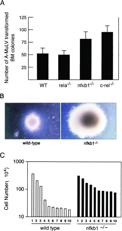
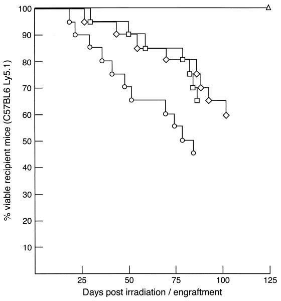
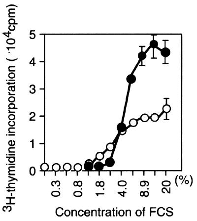
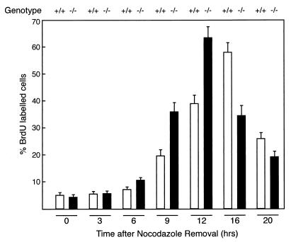
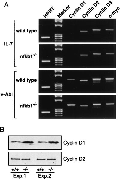
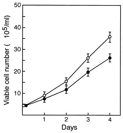
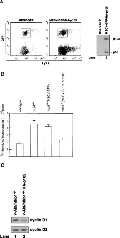
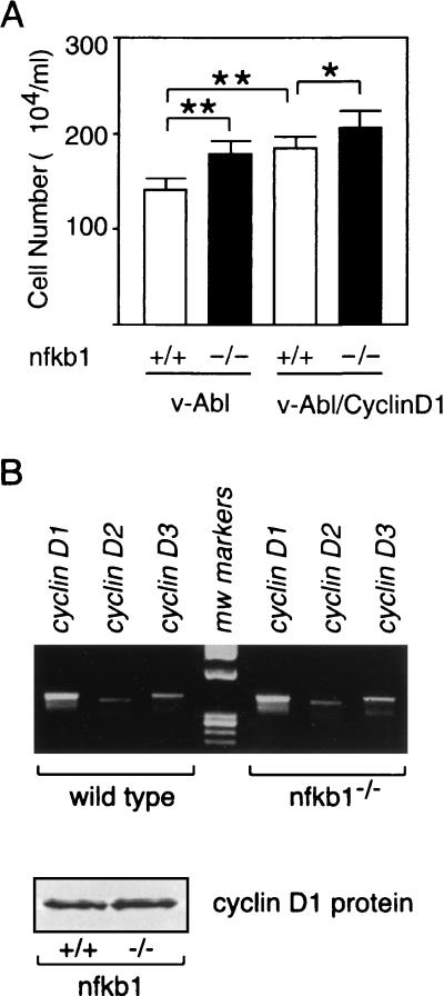
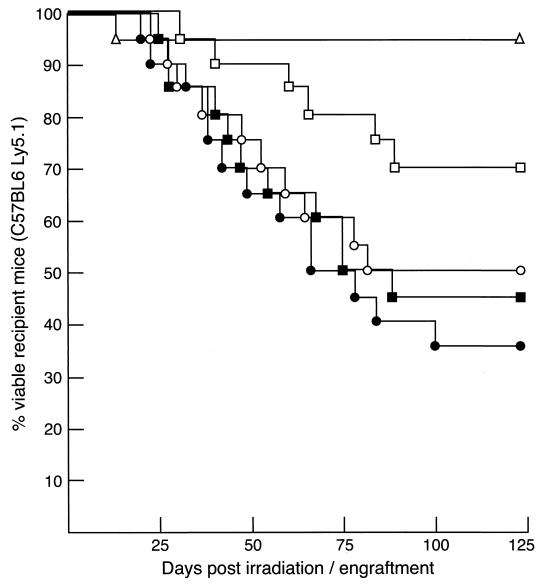
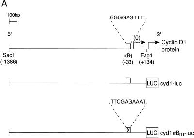
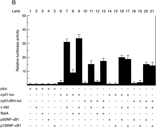
Mounting evidence implicates deregulated Rel/NF-kappaB signaling as a common feature of lymphoid malignancies. Despite the fact that they promote the survival and proliferation of normal lymphocytes, the underlying mechanisms by which various Rel/NF-kappaB proteins with different transcriptional regulatory capacities might facilitate transformation remain to be established. Here we show that the proliferation and tumorigenicity of Abelson murine leukemia virus (A-MuLV)-transformed pre-B cells are enhanced in the absence of NF-kappaB1 and that this coincides with elevated levels of cyclin D1. Support for a link between cyclin D1 expression and v-Abl transformation came from the finding that proliferation of transformed pre-B cells was reduced in the absence of cyclin D1, while enforced cyclin D1 expression increased the proliferation and tumorigenicity of wild-type transformants. A reduction in endogenous cyclin D1 levels that coincided with NF-kappaB1 transgene reversal of enhanced nfkb1(-/-) pre-B-cell transformation, coupled with NF-kappaB1 inhibition of v-Abl-induced kappaB-dependent murine cyclin D1 transcription, lends support to a model in which v-Abl-induced cyclin D1 transcription in transformed pre-B cells is controlled by Rel/NF-kappaB dimers with different activities.


Figures

FIG. 1.
FIG. 1.
A-MuLV transformation of BM cells from Rel/NF-κB mutant mice. (A) Focus assays. Equivalent numbers of wild-type (WT), c-rel−/−, rela−/−, and nfkb1−/− BM cells infected with A-MuLV were seeded in agar cultures and incubated for 12 to 14 days; then the number of compact macroscopic colonies was enumerated. No colonies were observed in mock-infected BM cultures. Data are means ± standard deviations from five experiments. (B) Colonies of A-MuLV-transformed nfkb1−/− BM cells are larger than normal. Representative examples of typical A-MuLV-transformed wild-type and nfkb1−/− BM colonies after 14 days of incubation are shown (magnification, ×20). v-Abl-transformed wild-type and nfkb1−/− colonies were typically 1.5 to 2.0 mm and 3.0 to 3.5 mm in diameter, respectively. (C) A-MuLV-transformed nfkb1−/− colonies contain more cells. Individual wild-type or nfkb1−/− colonies were picked, and cells were dispersed and enumerated. Results shown are representative of a typical experiment.
FIG. 2.
FIG. 2.
A-MuLV-infected nfkb1−/− BM cells display enhanced tumorigenicity. The survival of lethally irradiated C57BL/6-Ly5.1 mice engrafted with mock-infected (20 recipients, comprising 10 wild-type, 5 nfkb1−/−, and 5 c-rel−/− mice) (triangles) or A-MuLV-infected wild-type (20 recipients (squares), c-rel−/− (20 recipients) (diamonds), or nfkb1−/− (20 recipients) (circles) BM cells is presented as a Kaplan-Meyer plot. After engraftment, irradiated recipients were monitored daily and sacrificed for analysis either upon presenting with symptoms typical of A-MuLV-induced disease, upon detection of a palpable tumor, or before imminent death.
FIG. 3.
FIG. 3.
Enhanced proliferation of A-MuLV-transformed pre-B cells lacking NF-κB1. A-MuLV-infected wild-type (open circles) or nfkb1−/− (solid circles) B220+ BM cells (2 × 10⁴) were seeded into 96-well plates in the presence of varying concentrations of FCS and were cultured for 72 h. Cultures were then pulsed with 0.l μCi of [3H]thymidine for 5 h prior to harvesting. Data are means ± standard deviations of results from triplicate wells from three separate experiments. The absence of error bars indicates that the standard deviation is <2,500 cpm.
FIG. 4.
FIG. 4.
The duration of G1 is reduced in nfkb1−/− transformed pre-B cells. Viable v-Abl-transformed wild-type or nfkb1−/− pre-B cells previously treated with nocodazole for 24 h to induce M-phase arrest were placed in culture at a density of 5 × 10⁵/ml (a total of 106/well) and incubated for 3, 6, 9,12,16, or 20 h. Forty minutes prior to each time point, wild-type (open bars) and nfkb1−/− (solid bars) cultures were pulsed with BrdU, harvested, and fixed, and BrdU-positive cells were detected by flow cytometry using an anti-BrdU antibody. Data are means ± standard deviations of results from duplicate wells from two separate experiments.
FIG. 5.
FIG. 5.
Cyclin D1 expression induced by v-Abl is enhanced in the absence of NF-κB1. (A) Cyclin mRNA expression in primary and transformed pre-B cells. Total mRNA was isolated from equivalent numbers of primary wild-type or nfkb1−/− pre-B cells cultured in IL-7 for 5 days or BM cells 14 days after infection with A-MuLV. Expression of cyclin D1, cyclin D2, cyclin D3, c-myc, and HPRT was determined by semiquantitative RT-PCR using primers specific for each of the transcripts. PCR products were fractionated on 1.5% agarose gels. Data shown are representative of two independent experiments for IL-7-stimulated primary pre-B cells and five independent experiments for v-Abl-transformed cells. (B) Western blot analysis of cyclin D1 and cyclin D2 expression in A-MuLV-transformed pre-B cells. Total-cell extracts from equivalent numbers of wild-type and nfkb1−/− A-MuLV-transformed pre-B cells 14 days postinfection were subjected to Western blot analysis using antisera specific for cyclin D1 and cyclin D2. Results from two typical experiments are shown.
FIG. 6.
FIG. 6.
An absence of cyclin D1 reduces the proliferation of v-Abl-transformed pre-B cells. Equivalent numbers (input, 106 cells) of wild-type (open circles) or cyclin D1−/− (solid circles) B220+ BM cells infected 14 days earlier with A-MuLV were placed in culture, and viable cell numbers were determined at 24-h intervals over 96 h. Data are means ± standard deviations from two experiments performed in quadruplicate.
FIG. 7.
FIG. 7.
The proliferative response of v-Abl-transformed nfkb1−/− pre-B cells is restored to that of wild-type transformants by an NF-κB1 transgene. (A) NF-κB1 transgene expression in BM cells. BM cells isolated from lethally irradiated Ly5.l+ C57BL6 mice previously reconstituted with Ly5.2 C57BL6 nfkb1−/− BM that had been infected with MSCV-GFP or MSCV-GFP/HA-p105 virus were stained with a biotinylated Ly5.2+-specific MAb, and cells were subjected to two-color cell sorting (GFP expression detected as fluorescein isothiocyanate positive). Equivalent numbers (3 × 10⁵) of Ly5.2+ GFP+ cells infected with these retroviruses were subjected to Western blot analysis and probed with an anti-HA MAb. The precursor (p105) and mature processed (p50) forms of NF-κB1 are indicated. (B) Enforced NF-κB1 transgene expression reduces the proliferation of v-Abl-transformed nfkb1−/− pre-B cells. Equivalent numbers of wild-type, nfkb1−/−, MSCV-GFP nfkb1−/−, and MSCV-GFP HA-p105 nfkb1−/− B220+ BM cells infected 14 days earlier with A-MuLV were placed in culture for 72 h and then pulsed with 0.1 μCi of [3H]thymidine prior to harvesting. Data are means ± standard deviations of results from triplicate wells from three separate experiments. (C) Cyclin D1 expression is reduced in v-Abl-transformed nfkb1−/− pre-B cells that express an NF-κB1 transgene. Total-cell extracts from equivalent numbers of MSCV-GFP nfkb1−/− (lane 1) and MSCV-GFP HA-p105 nfkb1−/−(lane 2) A-MuLV-transformed pre-B cells 14 days postinfection were subjected to Western blot analysis using antisera specific for cyclin D1 and cyclin D2. Results are representative of those obtained from two experiments.
FIG. 8.
FIG. 8.
Enforced cyclin D1 expression increases the proliferation of v-Abl-transformed pre-B cells. (A) Proliferation of pre-B cells infected with a virus expressing v-Abl or v-Abl plus cyclin D1. Equivalent numbers (input, 106 cells) of wild-type (open bars) and nfkb1−/− (solid bars) B220+ BM cells infected 14 days earlier with a virus expressing either v-Abl or v-Abl plus cyclin D1 were placed in culture, and viable cell numbers were determined after 48 h. Data are means ± standard deviations from three experiments performed in duplicate. ∗, P < 0.01 by Student's t test; ∗∗, P < 0.001 by Student's t test. (B) Cyclin D1 expression in cells infected with the v-Abl/cyclin D1 virus. Equivalent numbers of wild-type or nfkb1−/− B220+ cells infected as described for panel A were used to determine cyclin D1 mRNA and protein levels. Total RNA was subjected to semiquantitative RT-PCR using primers specific for cyclin D1, cyclin D2, and cyclin D3, and PCR samples were fractionated on a 1.5% agarose gel. Total-protein extracts were subjected to Western blot analysis using cyclin D1-specific antisera.
FIG. 9.
FIG. 9.
Enforced cyclin D1 expression enhances the tumorigenicity of v-Abl-transformed BM cells. Lethally irradiated C57BL/6-Ly5.l+ mice were engrafted with equivalent numbers of mock-infected (20 recipients, comprising 10 wild-type, 5 nfkb1−/−, and 5 c-rel−/− mice) (triangles), v-Abl virus-infected (open symbols), or v-Abl/cyclin D1 virus-infected (closed symbols) wild-type (20 recipients) (squares) or nfkb1−/− (20 recipients) (circles) BM cells. Engrafted recipients were sacrificed for analysis upon presenting with symptoms of A-MuLV-induced disease, upon detection of a palpable tumor, or before imminent death.
FIG. 10.
FIG. 10.
NF-κB1 represses v-Abl-induced cyclin D1 transcription. (A) Schematic representation of the mouse cyclin D1 promoter and the luciferase reporter plasmids. Numbers in parentheses indicate the positions of the restriction enzyme sites in the murine cyclin D1 promoter relative to the transcription start site determined by Wood et al. (59). The open box represents the conserved NF-κB binding site κB1 (5′-GGGGAGTTTT-3′; −42 to −33), whereas the corresponding symbol with an “X” represents the mutated motif κB1m (5′TTCGAGAAAT-3′). Wavy and straight arrows represent the transcription initiation and translation start sites, respectively. The firefly luciferase gene is shown as LUC. Plasmid nomenclature is given to the right of each construct. (B) v-Abl-dependent cyclin D1 transcription is repressed by NF-κB1. 293T cells were transiently transfected with 1 μg of pluc3 (lanes 1 to 5), cyd1-luc (lanes 6 to 9 and 14 to 17), or cyd1κBm-1uc (lanes 10 to 13 and 18 to 21) together with the expression plasmid pEF-BOS containing no insert (lanes 1, 6, and 10), v-Abl (lanes 2, 7, 9, 11, 13, 16, 17, 20, and 21), RelA (lanes 3, 8, 9, 12, and 13), p50 NF-κB1 (lanes 4, 14, 16, 18, and 20), or p105 NF-κB1 (lanes 5, 15, 17, 19, and 21). The relative luciferase units for each set of transfections were normalized by cotransfection with the Renilla luciferase vector pRL-TK. Results are the mean percentages of luciferase activity ± standard deviations obtained for four separate sets of transfections.
FIG. 10.
FIG. 10.
NF-κB1 represses v-Abl-induced cyclin D1 transcription. (A) Schematic representation of the mouse cyclin D1 promoter and the luciferase reporter plasmids. Numbers in parentheses indicate the positions of the restriction enzyme sites in the murine cyclin D1 promoter relative to the transcription start site determined by Wood et al. (59). The open box represents the conserved NF-κB binding site κB1 (5′-GGGGAGTTTT-3′; −42 to −33), whereas the corresponding symbol with an “X” represents the mutated motif κB1m (5′TTCGAGAAAT-3′). Wavy and straight arrows represent the transcription initiation and translation start sites, respectively. The firefly luciferase gene is shown as LUC. Plasmid nomenclature is given to the right of each construct. (B) v-Abl-dependent cyclin D1 transcription is repressed by NF-κB1. 293T cells were transiently transfected with 1 μg of pluc3 (lanes 1 to 5), cyd1-luc (lanes 6 to 9 and 14 to 17), or cyd1κBm-1uc (lanes 10 to 13 and 18 to 21) together with the expression plasmid pEF-BOS containing no insert (lanes 1, 6, and 10), v-Abl (lanes 2, 7, 9, 11, 13, 16, 17, 20, and 21), RelA (lanes 3, 8, 9, 12, and 13), p50 NF-κB1 (lanes 4, 14, 16, 18, and 20), or p105 NF-κB1 (lanes 5, 15, 17, 19, and 21). The relative luciferase units for each set of transfections were normalized by cotransfection with the Renilla luciferase vector pRL-TK. Results are the mean percentages of luciferase activity ± standard deviations obtained for four separate sets of transfections.

Similar articles

Cited by

References

    1. Afar, D. E. H., J. McLaughlin, C. J. Sherr, O. N. Witte, and M. F. Roussel. 1995. Signaling by ABL oncogenes through cyclin D1. Proc. Natl. Acad. Sci. USA 92:9540-9544. - PMC - PubMed
    1. Baldwin, A. S., Jr. 1996. The NF-κB and IκB proteins: new discoveries and insights. Annu. Rev. Immunol. 14:649-681. - PubMed
    1. Beg, A. A., W. C. Sha, R. Bronson, S. Ghosh, and D. Baltimore. 1995. Embryonic lethality and liver degeneration in mice lacking the relA component of NF-κB. Nature 376:167-170. - PubMed
    1. Bodrug, S. E., B. J. Warner, M. L. Bath, G. J. Lindeman, A. W. Harris, and J. M. Adams. 1994. Cyclin D1 transgene impedes lymphocyte maturation and collaborates in lymphomagenesis with the myc gene. EMBO J. 13:2124-2130. - PMC - PubMed
    1. Carlesso, N., D. A. Frank, and J. D. Griffin. 1996. Tyrosyl phosphorylation and DNA binding activity of signal transducers and activators of transcription (STAT) proteins in hematopoietic cell lines transformed by Bcr/Abl. J. Exp. Med. 183:811-820. - PMC - PubMed

Publication types

MeSH terms