. 2002 Aug;22(15):5585-92.
doi: 10.1128/MCB.22.15.5585-5592.2002.

Mice lacking paternally expressed Pref-1/Dlk1 display growth retardation and accelerated adiposity

Affiliations

Mice lacking paternally expressed Pref-1/Dlk1 display growth retardation and accelerated adiposity

Yang Soo Moon et al. Mol Cell Biol. 2002 Aug.

Abstract

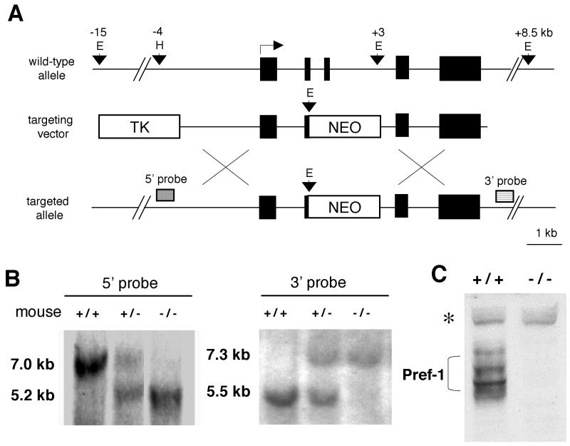
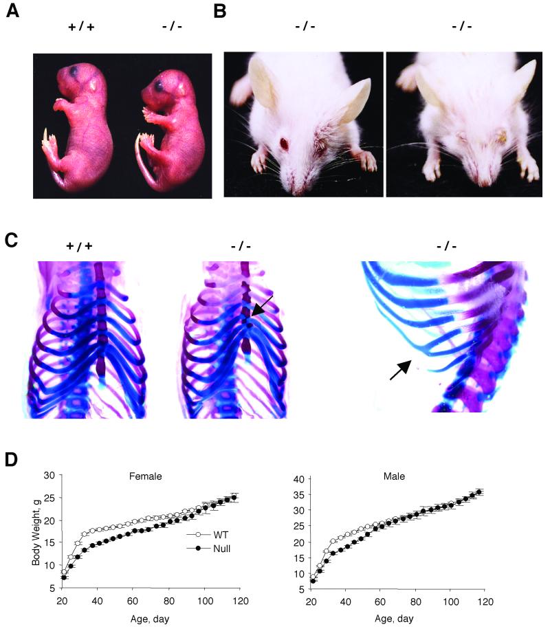
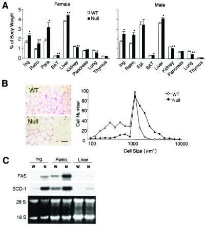
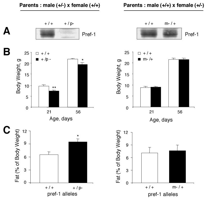
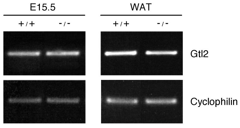
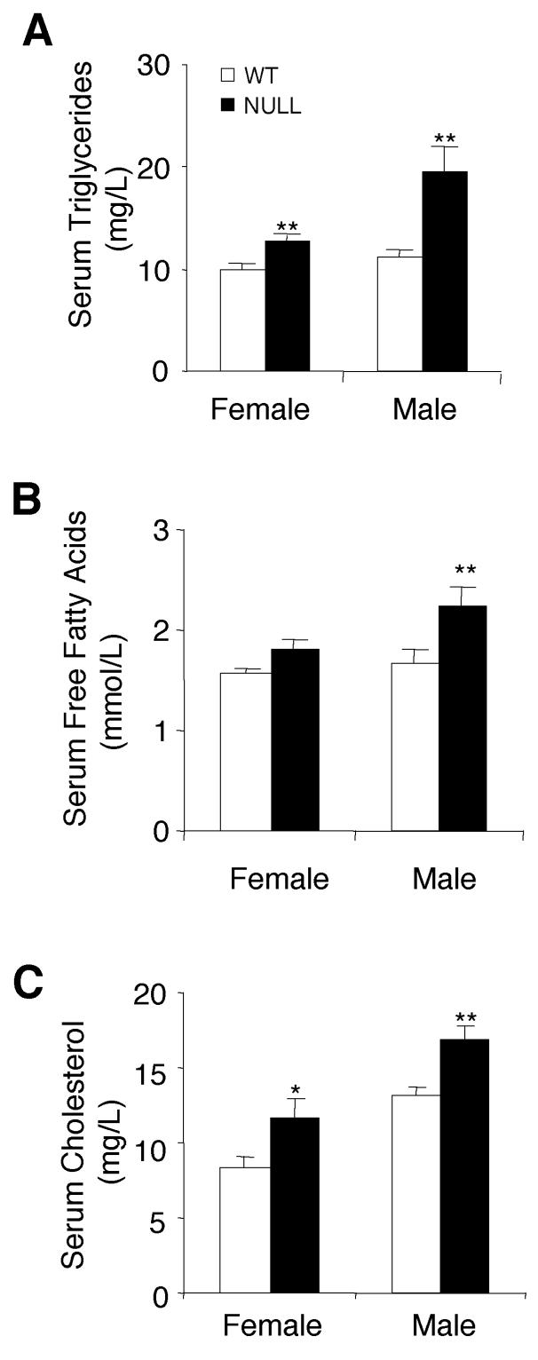
Preadipocyte factor 1 (Pref-1/Dlk1) inhibits in vitro adipocyte differentiation and has been recently reported to be a paternally expressed imprinted gene at human chromosome 14q32. Studies on human chromosome 14 deletions and maternal uniparental disomy (mUPD) 14 suggest that misexpression of a yet-to-be-identified imprinted gene or genes present on chromosome 14 causes congenital disorders. We generated Pref-1 knockout mice to assess the role of Pref-1 in growth and in vivo adipogenesis and to determine the contribution of Pref-1 in mUPD. Pref-1-null mice display growth retardation, obesity, blepharophimosis, skeletal malformation, and increased serum lipid metabolites. Furthermore, the phenotypes observed in Pref-1-null mice are present in heterozygotes that harbor a paternally inherited, but not in those with a maternally inherited pref-1-null allele. Our results demonstrate that Pref-1 is indeed paternally expressed and is important for normal development and for homeostasis of adipose tissue mass. We also suggest that Pref-1 is responsible for most of the symptoms observed in mouse mUPD12 and human mUPD14. Pref-1-null mice may be a model for obesity and other pathologies of human mUPD14.


Figures

FIG. 1.
FIG. 1.
Targeted disruption of the pref-1 gene. (A) The top diagram is a schematic representation of the wild-type pref-1 allele with five exons. Pref-1 exons are represented by black boxes. The middle diagram shows the targeting construct with pPNT-NEO and thymidine kinase (TK) as positive and negative selectable markers, respectively. The bottom diagram shows the pref-1 allele mutated by homologous recombination. The probes used for Southern blot analyses and the sizes of the restriction fragments detected are indicated. E, EcoRI; H, HindIII; X, XbaI. (B) Southern blot analysis of EcoRI-cut (3′ probe) and EcoRI-HindIII-cut (5′ probe) mouse tail DNA prepared from F2 mice generated by intermating F1 heterozygotes (pref-1+/−). The 7.0- and 5.5-kb bands represent the wild-type allele, and the 5.2- and 7.3-kb bands represent pref-1-null alleles. (C) Western blot analysis of Pref-1 in embryos (E17.5). A polyclonal antibody against mouse Pref-1 was used. The embryos were generated from crossbreeding pref-1 heterozygotes. The multiple forms of Pref-1 produced by alternative splicing and posttranslational modification (bracketed) are detected in wild-type but not in homozygous (pref-1−/−) embryos. ∗, nonspecific band indicating equal amounts of sample loading.
FIG. 2.
FIG. 2.
Pre- and postnatal growth retardation and eyelid abnormalities in mice lacking Pref-1. (A) Intrauterine growth retardation. Pref-1-null embryos are smaller than wild-type embryos at E18.5. The embryos were generated from crossbreeding Pref-1 heterozygotes (+/−). (B) Blepharophimosis. Anterior view of mice at age 27 days showing the left or both eyelid defects observed on the BALB/cJ strain background. (C) Skeletal malformation. Arrows indicate asymmetric zigzagged sternums and fusion of ribs in Pref-1-null mice. (D) Growth retardation in Pref-1-null mice of the C57BL/6J background. Female and male wild-type and Pref-1-null mice (n = 27 to 37 per group) were fed high-fat (45 kcal%) diet ad libitum from 21 days old and were weighed at 4-day intervals. Statistics were performed with a two-tailed t test. For female mice, from day 21 to day 68, P < 0.01, and from day 72 to day 76, P < 0.05. For male mice, from day 21 to day 52, P < 0.01.
FIG. 3.
FIG. 3.
Accelerated adiposity in mice lacking Pref-1. (A) Percentage of fat pad and organ weight relative to body weight. Fat depots and organs were dissected from 16-week-old wild-type (WT) and Pref-1-null mice fed a high-fat diet (n = 7 to 10 per group). Ing, inguinal fat pad; Epi, epididymal fat pad; Retro, retroperitoneal fat pad; Para., parametrial fat pad; BAT, brown adipose tissue. All values are means ± standard errors. ∗, P < 0.05; ∗∗, P < 0.01. (B) Paraffin section of inguinal fat pad from 16-week-old female mice and the distribution of fat cell size. The size of at least 300 cells per sample (mean of four mice per group) was determined with NIH Image software. The distribution of cell volume is shown. The scale bar represents 50 μm. (C) Expression of adipocyte marker genes in adipose tissues and liver. Total RNA was extracted from adipose tissue of 16-week-old wild-type (W) and Pref-1-null (N) mice fed a high-fat diet.
FIG. 4.
FIG. 4.
Mice with maternal or paternal inheritance of the pref-1-knockout allele (m−/+ or +/p−) have phenotypes similar to those of wild-type or null mice, respectively. (A) Pref-1 protein expression in E17.5 embryos generated from matings of male heterozygotes (+/−) and female wild-type mice or vice versa. Pref-1 protein is expressed in m−/+ heterozygotes, but not in +/p− heterozygotes. (B) Body weights of F1 littermates were measured at 21 and 56 days of age. Values are means ± standard errors with n = 5 to 13 per group. ∗, P < 0.05; ∗∗, P < 0.01. (C) Percentage of fat pad mass (total white fat, including inguinal, retroperitoneal, and reproductive fat) relative to body weight (16 weeks old; n = 7 to 9 per group). All values are means ± standard errors. ∗, P < 0.05. Statistics were performed with a two-tailed t test for panels B and C.
FIG. 5.
FIG. 5.
Expression of the gtl2 gene in wild-type and Pref-1-null mice. The expression of gtl2 was measured from whole embryo (E15.5) or white adipose tissue (WAT) (16 weeks) by RT-PCR. As reference for quantitative gene expression, cyclophilin was amplified simultaneously under conditions identical to those used for gtl2.
FIG. 6.
FIG. 6.
Altered serum lipid metabolite levels in mice lacking Pref-1. (A, B, and C) Levels of triglycerides, free fatty acids, and cholesterol in serum were measured from Pref-1-null and wild-type (WT) mice at 16 weeks of age after feeding on a high-fat diet from 3 to 16 weeks. Values are means ± standard errors with n = 5 to 8 per group. Statistics were performed with a two-tailed t test. ∗, P < 0.05; ∗∗, P < 0.01.

Similar articles

Cited by

References

    1. Berends, M. J., R. Hordijk, H. Scheffer, J. C. Oosterwijk, D. J. Halley, and N. Sorgedrager. 1999. Two cases of maternal uniparental disomy 14 with a phenotype overlapping with the Prader-Willi phenotype. Am. J. Med. Genet. 84:76-79. - PubMed
    1. Carlsson, C., D. Tornehave, K. P. Lindberg, N. Galante, B. Billestrup, L. Michelsen, I. Larsson, and J. H. Nielsen. 1997. Growth hormone and prolactin stimulate the expression of rat preadipocyte factor-1/delta-like protein in pancreatic islets: molecular cloning and expression pattern during development and growth of the endocrine pancreas. Endocrinology 138:3940-3948. - PubMed
    1. Charlier, C., K. Segers, D. Wagenaar, L. Karim, S. Berghmans, O. Jaillon, T. Shay, J. Weissenbach, N. Cockett, G. Gyapay, and M. Georges. 2001. Human-ovine comparative sequencing of a 250-kb imprinted domain encompassing the callipyge (clpg) locus and identification of six imprinted transcripts: DLK1, DAT, GTL2, PEG11, antiPEG11, and MEG8. Genome Res. 11:850-862. - PMC - PubMed
    1. Charlier, C., K. Segers, L. Karim, T. Shay, G. Gyapay, N. Cockett, and M. Georges. 2001. The callipyge mutation enhances the expression of coregulated imprinted genes in cis without affecting their imprinting status. Nat. Genet. 27:367-369. - PubMed
    1. Fokstuen, S., C. Ginsburg, M. Zachmann, and A. Schinzel. 1999. Maternal uniparental disomy 14 as a cause of intrauterine growth retardation and early onset of puberty. J. Pediatr. 134:689-695. - PubMed

Publication types

MeSH terms

LinkOut - more resources