. 2002 Aug;22(15):5616-25.
doi: 10.1128/MCB.22.15.5616-5625.2002.

Major histocompatibility complex class II transcriptional platform: assembly of nuclear factor Y and regulatory factor X (RFX) on DNA requires RFX5 dimers

Affiliations

Major histocompatibility complex class II transcriptional platform: assembly of nuclear factor Y and regulatory factor X (RFX) on DNA requires RFX5 dimers

Nabila Jabrane-Ferrat et al. Mol Cell Biol. 2002 Aug.

Abstract

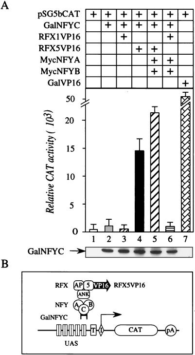
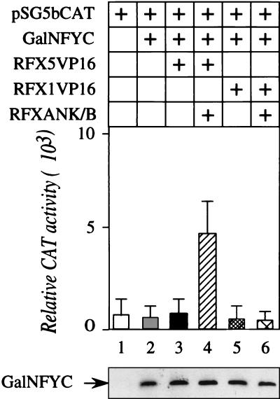
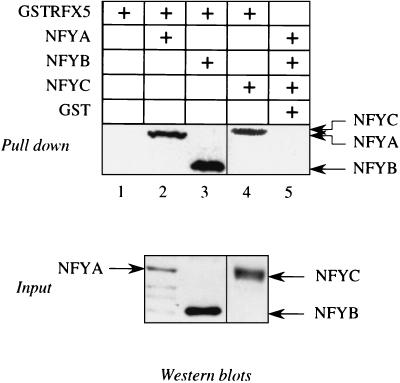
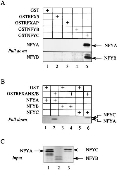
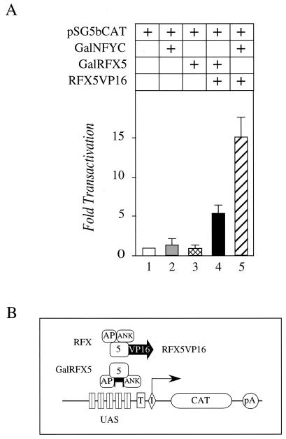
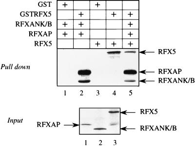
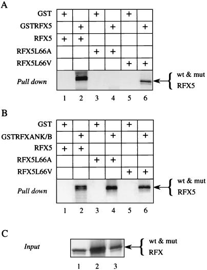
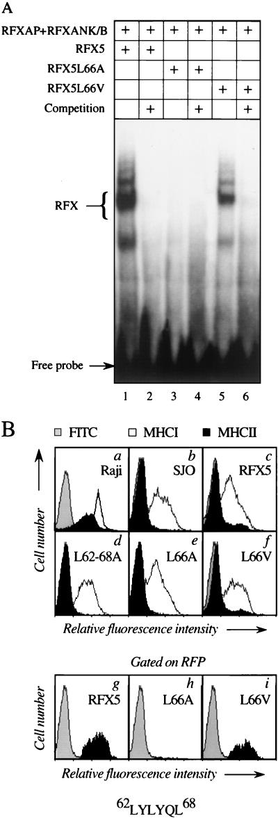
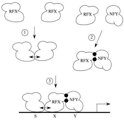
Major histocompatibility complex class II (MHC-II) genes are regulated in a B-cell-specific and gamma interferon-inducible manner. Conserved upstream sequences (CUS) in their compact promoters bind nuclear factor Y (NFY) and regulatory factor X (RFX) complexes. These DNA-bound proteins form a platform that attracts the class II transactivator, which initiates and elongates MHC-II transcription. In this report, we analyzed the complex assembly of these DNA-bound proteins. First, we found that NFY can interact with RFX in cells. In particular, NFYA and NFYC bound RFXANK/B in vitro. Next, RFX5 formed dimers in vivo and in vitro. Within a leucine-rich stretch N-terminal to the DNA-binding domain in RFX5, the leucine at position 66 was found to be critical for this self-association. Mutant RFX5 proteins that could not form dimers also did not support the formation of higher-order DNA-protein complexes on CUS in vitro or MHC-II transcription in vivo. We conclude that the MHC-II transcriptional platform begins to assemble off CUS and then binds DNA via multiple, spatially constrained interactions. These findings offer one explanation of why in the Bare Lymphocyte Syndrome, which is a congenital severe combined immunodeficiency, MHC-II promoters are bare when any subunit of RFX is mutated or missing.


Figures

FIG. 1.
FIG. 1.
NFY and RFX interact in cells. (A) The interaction between NFY and RFX activates transcription from a synthetic target promoter. As indicated by + signs on the top, COS cells were cotransfected with the plasmid reporter pSG5bCAT (0.5 μg) (lanes 1 to 7) with or without 1 μg of the plasmid effector coding for the hybrid GalNFYC protein as the bait (lanes 2 to 6). Additional combinations of the following prey proteins were used: the hybrid RFX1VP16 (lane 3) and RFX5VP16 (lanes 4 and 5) proteins or NFYA (lanes 5 and 6) and NFYB (lanes 5 and 6). The GalVP16 chimera (lane 7) was used as the positive control. The total amount of DNA was held constant at 5 μg. CAT assays were performed 48 h after the transfection. Relative CAT values are represented in the bar graph. They represent the average of two experiments performed in duplicate, and the standard error of the mean is represented by error bars. The bottom panel shows the expression levels of the GalNFYC chimera as monitored by Western blotting using the anti-Myc monoclonal antibody 9E10. (B) Schematic representation of our mammalian two-hybrid system. pG5bCAT plasmid reporter contained five Gal4 DNA-binding sites (UAS) upstream of a minimal promoter containing a TATA box (T) and an initiator (I) sequence and directed the expression of the CAT reporter gene (CAT), which terminated with a poly(A) signal (pA). The GalNFYC chimera was used as the bait. The hybrid RFX5VP16 protein was used as the prey. Exogenous expression of NFYA and NFYB was under the control of a cytomegalovirus promoter. All subunits of NFY carried the Myc epitope tag.
FIG. 2.
FIG. 2.
Complementation of Bequit cell line with RFXANK/B restored transcription from pG5bCAT. As indicated by + signs on the top, Bequit cells were cotransfected by electroporation with 4 μg of pSG5bCAT and 10 μg of each plasmid effector. The total amount of DNA was constant (40 μg). CAT activity was determined as described in the legend for Fig. 1. The bottom panel shows expression levels of the GalNFYC chimera as monitored by Western blotting using the 9E10 monoclonal antibody.
FIG. 3.
FIG. 3.
RFX5 interacts with NFY subunits in COS cells. As indicated by + signs on the top, the GSTRFX5 fusion protein was expressed alone (lane 1) or in combination with the Myc epitope-tagged NFYA (lane 2), NFYB (lane 3), or NFYC (lane 4) proteins. GST was also used as a control with the cocktail of NFYA, NFYB, and NFYC (lane 5). At 48 h after the transfection, total cell lysates were used for a GST pull-down assay using glutathione-Sepharose beads. Western blotting with the anti-Myc monoclonal antibody 9E10 revealed bound proteins after SDS-PAGE. Additionally, expression levels of NFYA, NFYB, and NFYC were determined from 10% of cell lysates by Western blotting with 9E10 monoclonal antibody (Input). Arrows indicate the position of different NFY proteins.
FIG. 4.
FIG. 4.
The interaction between RFX5 and NFY subunits is mediated through other subunits of the RFX complex. (A) Neither RFX5 nor RFXAP binds subunits of NFY in a cell-free system. GST, GSTRFX5, and GSTRFXAP fusion proteins were produced in E. coli. 35S-labeled NFYA (top panel) or NFYB (bottom panel), which were produced from RRL in vitro, were combined with GST alone or with the GST fusion proteins (1 μg) and selected on glutathione-Sepharose beads. Bound proteins were resolved with SDS-PAGE and revealed by autoradiography. The + signs in the grids indicate the presence of different proteins in the assay. (B) RFXANK/B interacts with subunits of NFY in vitro. Labeled subunits of NFY produced from RRL were incubated with GST alone (lanes 1, 3, and 5) or the GSTRFXANK/B chimera (lanes 2, 4, and 6), and bound proteins were analyzed as described for panel A. The presence of NFYA (lanes 1 and 2), NFYB (lanes 3 and 4), and NFYC (lanes 5 and 6) is indicated by the + signs. (C) Ten percent of input NFYA, NFYB, and NFYC proteins were separated on SDS-PAGE and revealed by autoradiography. Arrows point to positions of different proteins.
FIG. 5.
FIG. 5.
RFX5 associates with itself in cells. (A) As indicated by + signs on the top, COS cells were transfected with 0.5 μg of pSG5bCAT and 1 μg of each plasmid effector that expressed GalNFYC (lanes 2 and 5), GalRFX5 (lanes 3 and 4), and RFX5VP16 (lanes 4 and 5) chimeras. (B) Schematic representation of the heterologous tethering system used to assay the self-association of RFX5 in vivo. pSG5bCAT was coexpressed with RFX5 fused to the Gal4 DBD (GalRFX5) as the bait and the hybrid RFX5VP16 protein as the prey.
FIG. 6.
FIG. 6.
Dimerization of RFX5 does not interfere with its association with other RFX subunits. 35S-labeled RFXAP (lanes 1, 2, and 5), RFXANK/B (lanes 1, 2, and 5), and RFX5 (lanes 3, 4, and 5) proteins were combined with GST alone (lanes 1 and 3) or with the hybrid GSTRFX5 protein (lanes 2, 4, and 5) in a GST pull-down assay. The + signs mark the expression of appropriate proteins. Ten percent of the input labeled proteins was included. Arrows point to the position of expressed proteins as determined by SDS-PAGE and autoradiography.
FIG. 7.
FIG. 7.
Point mutation in the leucine-rich stretch inhibits the formation of RFX5 dimers. (A) GST (lanes 1, 3, and 5) or the hybrid GSTRFX5 protein (lanes 2, 4, and 6) were combined with 35S-labeled RFX5 (lanes 1 and 2), mutant RFX5L66A (lanes 3 and 4), or mutant RFX5L66V (lanes 5 and 6) proteins in a GST pull-down assay. Bound proteins were then separated by SDS-PAGE and revealed by autoradiography. (B) Alanine mutations do not prevent the interaction between RFX5 and other subunits of RFX. As indicated by + signs above the gel, GST (lanes 1, 3, and 5) or the hybrid GSTRFXANK/B protein (lanes 2, 4, and 6) was combined with the 35S-labeled wild-type or mutant RFX5 proteins. Bound proteins were revealed as described in the legend for panel A. (C) Ten percent of the input of RFX5 (lane 1), mutant RFX5L66A (lane 2), and mutant RFX5L66V (lane 3) proteins were analyzed by SDS-PAGE.
FIG. 8.
FIG. 8.
RFX5 dimers are critical for the assembly of the RFX complex on the DRA promoter and activation of MHC-II transcription. (A) Wild-type subunits of RFX and mutant RFX5 proteins were produced in vitro. As indicated by the + sign on the top, EMSA was carried out by combining a DNA probe that contained S and X boxes with the wild-type RFXAP and RFXANK/B proteins and wild-type (lanes 1 and 2) or mutant RFX5 (lanes 3, 4, 5, and 6) proteins. The positions of the complex between DNA and RFX and the free probe are indicated. Equivalent amounts of the subunits of RFX were used for each assay. The sequence of the leucine-rich stretch is presented below the FACS profiles. (B) The mutant RFX5L66V protein rescues the expression of MHC-II determinants in SJO cells. The expression of DR (black histogram) and MHC-I proteins (included as a positivecontrol; white histogram) was analyzed by FACS in Raji cells (a), untransfected SJO cells (b), and SJO cells which were transfected with plasmids that directed the expression of RFX5 (c), mutant RFX5 (L62-68A) (d), mutant RFX5 (L66A) (e), and mutant RFX5 (L66V) (f) proteins. Several profiles were also gated on RFP that was coexpressed with RFX5 (g), mutant RFX5 (L66A) (h), and mutant RFX5 (L66V) (i) proteins. The gray histogram represents background staining obtained with the FITC-coupled secondary antibody.
FIG. 9.
FIG. 9.
A model for the assembly of the transcriptional platform on MHC-II promoters. Subunits of both NFY and RFX form trimers. Next, RFX dimers form (1), and NFY and RFX begin to assemble in solution (2). Finally, the complex binds many points on the same face of the double helix (3). DNA provides another surface that strengthens these protein-protein interactions. Although the binding of the RFX dimer on S and X boxes is presented, this dimer could also bind to each S and X box, thus forming a tetramer or an even higher-order complex on CUS.

Similar articles

Cited by

References

    1. Alberti, S., S. Oehler, B. von Wilcken-Bergmann, H. Kramer, and B. Muller-Hill. 1991. Dimer-to-tetramer assembly of Lac repressor involves a leucine heptad repeat. New Biol. 3:57-62. - PubMed
    1. Bellorini, M., J. C. Dantonel, J. B. Yoon, R. G. Roeder, L. Tora, and R. Mantovani. 1996. The major histocompatibility complex class II Ea promoter requires TFIID binding to an initiator sequence. Mol. Cell. Biol. 16:503-512. - PMC - PubMed
    1. Boss, J. M. 1999. A common set of factors control the expression of the MHC class II, invariant chain, and HLA-DM genes. Microbes Infect. 1:847-853. - PubMed
    1. Boss, J. M., and J. L. Strominger. 1986. Regulation of a transfected human class II major histocompatibility complex gene in human fibroblasts. Proc. Natl. Acad. Sci. USA 83:9139-9143. - PMC - PubMed
    1. Bottazzo, G. F., R. Pujol-Borrell, T. Hanafusa, and M. Feldmann. 1983. Role of aberrant HLA-DR expression and antigen presentation in induction of endocrine autoimmunity. Lancet ii:1115-1119. - PubMed

Publication types

MeSH terms