

Differences in mandibular distraction osteogenesis after corticotomy and osteotomy
- PMID: 12102418
- PMCID: PMC4950923
- DOI: 10.1054/ijom.2001.0193
Differences in mandibular distraction osteogenesis after corticotomy and osteotomy
Abstract
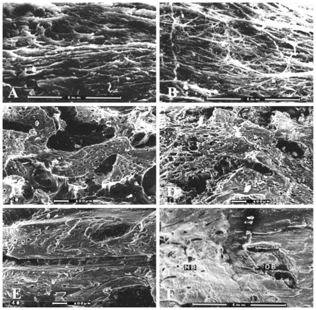
Corticotomy or osteotomy was performed on opposing sides of the mandibles in 18 goats. A custom-made distractor was used to lengthen the mandible at a rate of 1 mm/day for 10 days (total 10 mm elongation). Six goats were sacrificed respectively at 2, 4 and 8 weeks after completion of distraction. The distracted calluses were harvested and processed for radiographic, histologic, and scanning electron microscopic evaluation as well as Ca/P ratio analysis. The regenerate bone in the corticotomy side showed more bone formation and earlier mineralization than in the osteotomy side. The results of this study suggest that preservation of intramedullary vessels is beneficial to bone regeneration following mandibular osteodistraction, and that performing corticotomy may be a simple but effective way to promote the maturity of the distracted callus and shorten the time for fixation.
Figures


Similar articles
-
Transplantation of osteoblast-like cells to the distracted callus in the rabbit mandible.Plast Reconstr Surg. 2007 Feb;119(2):500-7. doi: 10.1097/01.prs.0000246374.53516.78. Plast Reconstr Surg. 2007. PMID: 17230082
-
Acceleration of callus maturation using rhOP-1 in mandibular distraction osteogenesis in a rat model.Int J Oral Maxillofac Surg. 2003 Oct;32(5):528-33. Int J Oral Maxillofac Surg. 2003. PMID: 14759113
-
[Ultrastructural and Ca/P changes of the regenerated bone after mandibular distraction in goats].Hua Xi Kou Qiang Yi Xue Za Zhi. 2001 Feb;19(1):9-10, 31. Hua Xi Kou Qiang Yi Xue Za Zhi. 2001. PMID: 12539625 Chinese.
-
[Changes in the inferior alveolar vessels and angiogenesis following mandibular lengthening with different rates of distraction].Hua Xi Kou Qiang Yi Xue Za Zhi. 2002 Jun;20(3):203-5, 224. Hua Xi Kou Qiang Yi Xue Za Zhi. 2002. PMID: 12600067 Chinese.
-
Periosteal Distraction Osteogenesis: An Effective Method for Bone Regeneration.Biomed Res Int. 2016;2016:2075317. doi: 10.1155/2016/2075317. Epub 2016 Dec 18. Biomed Res Int. 2016. PMID: 28078283 Free PMC article. Review.
Cited by
-
Are endogenous BMPs necessary for bone healing during distraction osteogenesis?Clin Orthop Relat Res. 2009 Dec;467(12):3190-8. doi: 10.1007/s11999-009-1065-6. Clin Orthop Relat Res. 2009. PMID: 19760469 Free PMC article.
-
Treatment of Class II open bite complicated by an ankylosed maxillary central incisor.Angle Orthod. 2011 Jul;81(4):726-35. doi: 10.2319/102010-578.1. Epub 2011 Feb 21. Angle Orthod. 2011. PMID: 21341998 Free PMC article.
-
The effect of heparan sulfate application on bone formation during distraction osteogenesis.PLoS One. 2013;8(2):e56790. doi: 10.1371/journal.pone.0056790. Epub 2013 Feb 15. PLoS One. 2013. PMID: 23457615 Free PMC article.
References
-
- Alberius P, Isaksson S, Klinge B, Sjogren S, Jonsson J. Regeneration of cranial suture and bone plate lesions in rabbits. J Craniomaxillofac Surg. 1990;18:271–279. - PubMed
-
- Cancedda R, Descalzi CF, Castagnola P. Chondrocyte differentiation. Int Rev Cytol. 1995;159:265–358. - PubMed
-
- Costantino PD, Shybut G, Friedman CD, Pelzer HJ, Masini M, Shindo ML, Sisson GA. Segmental mandibular regeneration by distraction osteogenesis. Arch Otolaryngol Head Neck Surg. 1990;119:535–545. - PubMed
-
- Ganey TM, Klotch DW, Slater-Haase AS, Sasse J. Evaluation of distraction osteogenesis by scanning electron microscopy. Otolaryngol Head and Neck Surg. 1994;111:265–272. - PubMed
-
- Hagiwara T, Bell WH. Effect of electrical stimulation on mandibular distraction osteogenesis. J Craniomaxillo Surg. 2000;28:12–19. - PubMed
Publication types
MeSH terms
Substances
Grants and funding
LinkOut - more resources
Full Text Sources
Miscellaneous
