. 2002 Jun 28:2:16.
doi: 10.1186/1471-2180-2-16.

Interaction of the Yersinia pestis type III regulatory proteins LcrG and LcrV occurs at a hydrophobic interface

Affiliations

Interaction of the Yersinia pestis type III regulatory proteins LcrG and LcrV occurs at a hydrophobic interface

Jyl S Matson et al. BMC Microbiol. .

Erratum in

  • BMC Microbiol. 2002 Sep 5;2(1):25.

Abstract

Background: Secretion of anti-host proteins by Yersinia pestis via a type III mechanism is not constitutive. The process is tightly regulated and secretion occurs only after an appropriate signal is received. The interaction of LcrG and LcrV has been demonstrated to play a pivotal role in secretion control. Previous work has shown that when LcrG is incapable of interacting with LcrV, secretion of anti-host proteins is prevented. Therefore, an understanding of how LcrG interacts with LcrV is required to evaluate how this interaction regulates the type III secretion system of Y. pestis. Additionally, information about structure-function relationships within LcrG is necessary to fully understand the role of this key regulatory protein.

Results: In this study we demonstrate that the N-terminus of LcrG is required for interaction with LcrV. The interaction likely occurs within a predicted amphipathic coiled-coil domain within LcrG. Our results demonstrate that the hydrophobic face of the putative helix is required for LcrV interaction. Additionally, we demonstrate that the LcrG homolog, PcrG, is incapable of blocking type III secretion in Y. pestis. A genetic selection was utilized to obtain a PcrG variant capable of blocking secretion. This PcrG variant allowed us to locate a region of LcrG involved in secretion blocking.

Conclusion: Our results demonstrate that LcrG interacts with LcrV via hydrophobic interactions located in the N-terminus of LcrG within a predicted coiled-coil motif. We also obtained preliminary evidence that the secretion blocking activity of LcrG is located between amino acids 39 and 53.


Figures

Figure 1
Figure 1
Schematic representation of LcrG truncations fused to the GAL4 activation domain of plasmid pACT2. All constructs were cotransformed with full-length LcrV cloned into cyh2-deleted pAS2-1 to produce a GAL4-DNA binding domain-LcrV chimera in S. cerevisiae strain Y187. The ability of each truncation to bind LcrV was determined qualitatively by X-gal hydrolysis from filter-lift assays and quantitatively by the level of β-galactosidase activity expressed as Miller units in yeast liquid cultures. Final β-galactosidase activities are calculated from the average of at least four independent measurements.
Figure 2
Figure 2
Pairwise BLAST alignment of LcrG and PcrG. Amino acid sequence alignment of LcrG from Y. pestis (NCBI RefSeq, NP_395166; ) and PcrG from P. aeruginosa (NCBI RefSeq, NP_250396; ). Overall the two proteins are 43% identical and 56% similar. The sequences were aligned using the BLAST algorithm as implemented for pairwise alignments [37]. The region highlighted in yellow (amino acids 7–40) is the smallest region identified that interacts with LcrV. The region in blue (amino acids 39–53) corresponds to a previously described deletion that eliminated secretion blocking activity [5]. The amino acids identified with red asterisks are residues identified as participating in the LcrG-LcrV interaction (A16, S23, and L30). The residue marked with the blue asterisk (F48) is required for secretion blocking activity of LcrG.
Figure 3
Figure 3
Helical wheel projection of residues 14–31 of LcrG. Residues 14 to 31 of LcrG are shown in a helical wheel projection created with the WheelApp applet; . Basic residues are shown in pink, acidic residues in green, neutral amino acids in blue, and hydrophobic amino acids in yellow. Residues in LcrG that are critical for LcrV interaction are designated with an asterisk.
Figure 4
Figure 4
Copurification of LcrG variants with His6-tagged LcrV. Cells of Y. pestis KIM8-3002.8 (ΔlcrGV2) containing plasmids pAra-HT-V and pAraG18K (lanes 1 to 3), pJM131 (LcrG S23R) (lanes 4 to 6), pJM99 (LcrG L30R) (lanes 7 to 9), or pJM89 (LcrG A16R) (lanes 10 to12) were grown in TMH with calcium and induced with 0.2% (wt/vol) arabinose prior to temperature shift to 37°C. Cultures were harvested after 4 h of growth at 37°C, and cellular extracts were disintegrated using a French press (20,000 lb/in2). Unbroken cells and large debris were removed by centrifugation (14,000 × g) for 10 min and the cleared extracts (lanes 1, 4, 7, and 10) were applied to a Talon column. Proteins that did not bind were collected as the flowthrough fraction (lanes 2, 5, 8, and 11). Proteins were eluted from the column with 50 mM imidazole and collected (lanes 3, 6, 9, and 12). All protein samples were resolved by SDS-PAGE in a 12.5% polyacrylamide gel after dilution in 2X SDS sample buffer and analyzed by immunoblotting with α-LcrG and α-LcrV. Proteins were visualized by probing with alkaline phosphatase-conjugated secondary antibodies and developing with NBT-BCIP.
Figure 5
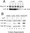
Figure 5
PcrG interacts with LcrV, but is incapable of blocking Yops secretion. (A) Cells of Y. pestis KIM8-3002.8 (Δ lcrGV2) containing plasmids pAra-HT-V and pAraG18K (lanes 1 to 3) or pJM132 (lanes 4–6) were grown in TMH and harvested as described for Fig. 4. The cleared cellular extracts (lanes 1 and 4) were applied to a Talon column and proteins that did not bind were collected as the flowthrough fraction (lanes 2 and 5). Proteins were eluted from the column with 50 mM imidazole and collected (lanes 3 and 6). Protein samples were resolved by SDS-PAGE in a 12.5% polyacrylamide gel and analyzed by immunoblotting with α-LcrG and α-LcrV. (B) Cells of Y. pestis KIM8-3002.8 (ΔlcrGV2) containing plasmids pBAD18-Kan (vector; lanes 1 and 2), pAraG18-K (+LcrG; lanes 3 and 4), pJM132 (+PcrG; lanes 5 and 6), and pJM133 (+PcrG F42L) were grown in TMH with or without calcium. Arabinose was added to 0.2% (wt/vol) to each of the cultures immediately prior to temperature shift to 37°C to induce the expression of LcrG, PcrG, or PcrG F42L from the plasmids. Cultures were harvested after 4 h of growth at 37°C, and samples were fractionated into whole-cell and cell-free culture supernatants. Culture supernatant samples were separated by SDS-PAGE in a 12.5% polyacrylamide gel and analyzed by immunoblotting with α-YopM, α-LcrV, and α-YopE. Proteins were visualized by probing with alkaline phosphatase-conjugated secondary antibodies and developing with NBT-BCIP.
Figure 6
Figure 6
Identification of an LcrG residue required for secretion blocking activity. Cells of Y. pestis KIM8-3002.8 (ΔlcrGV2) containing plasmids pBAD18-Kan (vector; lanes 1 and 2), pAraG18-K (+LcrG; lanes 3 and 4), pJM143 (+L39R; lanes 5 and 6), and pJM144 (+F48R; lanes 7 and 8) were grown in TMH with or without calcium. Arabinose was added at 0.2% (wt/vol) to each of the cultures immediately prior to temperature shift to 37°C to induce the expression of the LcrG variants from the plasmids. Cultures were harvested after 4 h of growth at 37°C, and samples were fractionated into whole-cell (A) and cell-free culture supernatants (B). Samples were separated by SDS-PAGE in a 12.5% polyacrylamide gel and analyzed by immunoblotting with α-YopM, α-LcrV, α-YopN, α-YopD, α-YopE, and α-LcrG. Proteins were visualized by probing with alkaline phosphatase-conjugated secondary antibodies and developing with NBT-BCIP.

Similar articles

Cited by

References

    1. Perry RD, Straley SC, Fetherston JD, Rose DJ, Gregor J, Blattner FR. DNA sequencing and analysis of the low-Ca2+-response plasmid pCD1 of Yersinia pestis KIM5. Infect Immun. 1998;66:4611–4623. - PMC - PubMed
    1. Cornelis GR, Boland A, Boyd AP, Geuijen C, Iriarte M, Neyt C, Sory M-P, Stainier I. The Virulence plasmid of Yersinia, an antihost genome. Microbiol Mol Biol Rev. 1998;62:1315–1352. - PMC - PubMed
    1. Straley SC, Perry RD. Environmental modulation of gene expression and pathogenesis in Yersinia. Trends Microbiol. 1995;3:310–317. doi: 10.1016/S0966-842X(00)88960-X. - DOI - PubMed
    1. Nilles ML, Williams AW, Skrzypek E, Straley SC. Yersinia pestis LcrV forms a stable complex with LcrG and may have a secretion-related regulatory role in the low-Ca2+ response. J Bacteriol. 1997;179:1307–1316. - PMC - PubMed
    1. Skrzypek E, Straley SC. LcrG, a secreted protein involved in negative regulation of the low-calcium response in Yersinia pestis. J Bacteriol. 1993;175:3520–3528. - PMC - PubMed

Publication types

MeSH terms