. 2002 Jul 8:3:18.
doi: 10.1186/1471-2164-3-18. Epub 2002 Jul 8.

Expression and genomic analysis of midasin, a novel and highly conserved AAA protein distantly related to dynein

Affiliations

Expression and genomic analysis of midasin, a novel and highly conserved AAA protein distantly related to dynein

Joan E Garbarino et al. BMC Genomics. .

Abstract

Background: The largest open reading frame in the Saccharomyces genome encodes midasin (MDN1p, YLR106p), an AAA ATPase of 560 kDa that is essential for cell viability. Orthologs of midasin have been identified in the genome projects for Drosophila, Arabidopsis, and Schizosaccharomyces pombe.

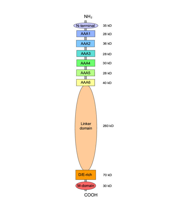
Results: Midasin is present as a single-copy gene encoding a well-conserved protein of approximately 600 kDa in all eukaryotes for which data are available. In humans, the gene maps to 6q15 and encodes a predicted protein of 5596 residues (632 kDa). Sequence alignments of midasin from humans, yeast, Giardia and Encephalitozoon indicate that its domain structure comprises an N-terminal domain (35 kDa), followed by an AAA domain containing six tandem AAA protomers (approximately 30 kDa each), a linker domain (260 kDa), an acidic domain (approximately 70 kDa) containing 35-40% aspartate and glutamate, and a carboxy-terminal M-domain (30 kDa) that possesses MIDAS sequence motifs and is homologous to the I-domain of integrins. Expression of hemagglutamin-tagged midasin in yeast demonstrates a polypeptide of the anticipated size that is localized principally in the nucleus.

Conclusions: The highly conserved structure of midasin in eukaryotes, taken in conjunction with its nuclear localization in yeast, suggests that midasin may function as a nuclear chaperone and be involved in the assembly/disassembly of macromolecular complexes in the nucleus. The AAA domain of midasin is evolutionarily related to that of dynein, but it appears to lack a microtubule-binding site.


Figures

Figure 1
Figure 1
Diagram of domain structure in midasin The values of molecular mass indicated are for midasin in humans. In other organisms, the principal difference is the length of the linker domain. In Giardia and Encephalitozoon, the N-terminal domain is truncated to ~25 residues. The mass of short regions of polypeptide joining the six protomers in the AAA domain (~10 kDa each) is not included in the values shown.
Figure 2
Figure 2
Dot-matrix plots showing level of sequence conservation as a function of residue position in midasin The amino acid sequence of midasin in yeast, Saccharomyces cerevisiae (vertical) is compared with those in fission yeast, Schizosaccharomyces pombe (horizontal, left) and in humans (horizontal, right). Closely spaced dots forming a diagonal line indicate well-conserved regions of sequence. Horizontal dashed lines have been added to indicate the abrupt changes in conservation level that correspond to putative domain boundaries. The off-diagonal lines in the AAA domain correspond to sequence similarity between the six AAA protomers comprising this domain. The rectangular area containing numerous off-diagonal dots in the D/E-rich domain derives from the conserved high percentage of aspartate and glutamate residues in this domain, although the actual sequences are only moderately conserved. Note the short region of relatively well-conserved sequence near the middle of the otherwise less conserved linker domain. The matrices were generated with a stringency of 26% in a sliding window of 40 residues. Abbreviations: Sacce, Saccharomyces cerevisiae; pombe, Schizosaccharomyces pombe.
Figure 3
Figure 3
Sequence alignment of the six AAA protomers in midasin from humans, yeast and Giardia The alignment shows two aspects of sequence conservation: (1) the level of conservation for each single AAA protomer compared to the same protomer in other organisms; (2) the level of conservation between the six AAA protomers (AAA1, AAA2, AAA3, AAA4, AAA5, AAA6) compared one against another. The Motif line above the alignment indicates the sequence motifs that characterize members of the AAA ATPase family [5]. All AAA motifs occur in regions of relatively well-conserved sequence, with the exception of Box II (not shown) which is poorly conserved in midasin. Critical residues interacting with bound nucleotide in other AAA proteins are indicated by: ▲, residues in the Walker A and B motifs required for binding and hydrolysis of ATP [17]; ●, Asn in sensor 1 and Arg in sensor 2 believed to sense the γ-phosphate status [5]; ■, Asp and Arg in Boxes VI and VII that are believed to sense the γ-phosphate status in adjacent protomers of the hexameric structure [6,15,18]. The Structure line below the alignment indicates the secondary structural elements corresponding to the sequence motifs in other AAA ATPases whose structures have been resolved at the atomic level [5]. Colors indicate level of consensus for each residue position. In this context, consensus is defined as sequence conservation of a single AAA protomer in the three organisms analyzed; it does not refer to comparisons between different AAA protomers. Color key: red/yellow, invariant; blue/blue, majority consensus of identical residues; green/green, majority consensus of highly conserved residues; green/white, weakly conserved residues; gray/white, non-conserved residues. Organisms : HUMAN, Homo sapiens; SACCE: Sacchararomyces cerevisiae; GIARD, Giardia intestinalis.
Figure 4
Figure 4
Tree of sequence relatedness for the six AAA protomers in midasin from humans, yeast and Giardia Asterisks indicate nodes supported with a bootstrap probability of 95% or better, plus signs indicate nodes with probability of 75–95%. The tree was calculated from the multiple alignment in Fig. 3.
Figure 5
Figure 5
Distribution of aspartate and glutamate residues as a function of residue position in midasin The distribution is shown as the mole fraction of (aspartate + glutamate) in a sliding window of 31 residues.
Figure 6
Figure 6
Multiple sequence alignment of midasin M-domains with selected magnesium chelatases and integrin I-domains The conserved residues in the MIDAS sequence motifs (hhhhDxSxS, [S/T] and hhhh[S/T]DG) are indicated by ▲ under the alignment. In all midasins and chelatases, the MIDAS-containing domain is located at the COOH-terminus of the protein (asterisk). The Structure line below the alignment indicates the position of secondary structural elements in the I-domain of integrin α2 (pdb: 1AOX_A). The coloring indicates the level of consensus for each residue position in the sets of three midasins, three chelatases, and three integrins, with each set being considered separately. Color key: red/yellow, invariant; blue/blue, majority of identical residues; green/green, majority of highly conserved residues; green/white, weakly conserved residues; gray/white, non-conserved residues. Abbreviations: MIDA, midasin; YEAST, Saccharomyces cerevisiae; GIARD, Giardia intestinalis; BCHD_RHOCA, D-subunit of Mg chelatase from Rhodobacter capsulatus (accession P26157); BCHD_CHLFL, D-subunit of Mg chelatase from Chloroflexus aurantiacus (accession AAG15217); CHLD_ARATH, D-subunit of Mg chelatase from Arabidopsis thaliana (accession Q9SJE1). ITAL, I-domain of integrin αL (accession P20701). ITAM, I-domain of integrin αM (accession P11215); ITA2, I-domain of integrin α2 (accession P17301). All residue numbers indicate position in full-length subunits.
Figure 7
Figure 7
Sequence alignment of the AAA protomers in midasin with those in other branches of AAA-ATPase family The alignment is limited to residues contained in the P-loop, Walker B, and sensor 1 motifs of AAA2 and AAA3 protomers of midasin from humans and yeast, together with the corresponding residues in the AAA1 protomer of cytoplasmic dynein [6], and in a variety of other AAA proteins. The positions and lengths of the P-loop, Walker B, and sensor 1 motifs are taken from the review of Neuwald et al [5]. The color used for each residue in the alignment indicates its similarity to that present at the corresponding position in yeast cytoplasmic dynein. Color key: Yellow/red, all AAA proteins shown have identical residue; blue/blue, identical to AAA1 of dynein; green/green, closely similar to AAA1 of dynein; green/white, moderately similar; gray/white, not similar.
Figure 8
Figure 8
Tree of sequence relatedness of AAA protomers in midasin to those in other branches of AAA family The numbers beside the nodes in the tree represent the statistical significance of the node, assayed by the number of times the identical node appeared in 1000 bootstrap trials [57]. Nodes lacking a number are less than 90% significant. The tree is calculated for the alignment in Fig. 7.
Figure 9
Figure 9
Electrophoresis gel showing expression of HA-tagged midasin in haploid yeast Preparations made from wild type yeast (wt) and from yeast with a HA epitope tag on the MDN1 gene (HA). Gel A: S0, supernatant fraction after homogenizing yeast with glass beads and centrifuging at 2000 × g for 2 min; S1, P1, supernatant and pellet fractions after centrifugation of S0 fraction at 18,000 × g for 20 min. Gel B: S2, P2, supernatant and pellet fractions obtained by resuspending P1 fraction in 8 M urea and centrifuging at 150,000 × g for 20 min. The midasin polypeptide (arrow) migrates somewhat more slowly than the dynein heavy chain (dyn) used as a molecular mass standard of ~525 kDa. The 45 kDa band in the 8 M urea supernatant is a cross-reacting protein present in wild type yeast. The 3–8% polyacrylamide gels were electrophoresed in the presence of 0.1% Na dodecyl SO4 and blotted onto Immobilon membrane. The blot was stained with monoclonal antibody against the HA epitope. The molecular mass standards were run in a noncontiguous lane of gel A.
Figure 10
Figure 10
Light microscopy of immunostained yeast cellsA: cells in interphase and during division; B, C, selected cells in which the region staining for midasin is larger (arrows) than the region staining for DNA. "Comet tails" are conspicuous in several of these cells, with short tails remaining post-division into early interphase. All fields show haploid yeast containing the MDN1(HA) gene double-stained with HA-antibody and DAPI. D, differential interference images; M, localization of HA-tagged midasin; P, localization of DNA, as stained by DAPI. All antibody-stained and DAPI fields are matched levels taken from through-focal series with steps of 0.3 μm.

Similar articles

Cited by

References

    1. Winzeler EA. et al.Functional characterization of the S. cerevisiae genome by gene deletion and parallel analysis. Science. 1999;3:901–906. doi: 10.1126/science.285.5429.901. - DOI - PubMed
    1. Bassler J. et al.Identification of a 60S preribosomal particle that is closely linked to nuclear export. Mol Cell. 2001;3:517–529. - PubMed
    1. Gavin AC. et al.Functional organization of the yeast proteome by systematic analysis of protein complexes. Nature. 2002;3:141–147. doi: 10.1038/415141a. - DOI - PubMed
    1. Ho Y. et al.Systematic identification of protein complexes in Saccharomyces cerevisiae by mass spectrometry. Nature. 2002;3:180–183. doi: 10.1038/415180a. - DOI - PubMed
    1. Neuwald AF, Aravind L, Spouge JL, Koonin EV. AAA+: A class of chaperone-like ATPases associated with the assembly, operation, and disassembly of protein complexes. Genome Res. 1999;3:27–43. - PubMed