. 2002 Jun 27:3:11.
doi: 10.1186/1471-2156-3-11.

The Tnfrh1 (Tnfrsf23) gene is weakly imprinted in several organs and expressed at the trophoblast-decidua interface

Affiliations

The Tnfrh1 (Tnfrsf23) gene is weakly imprinted in several organs and expressed at the trophoblast-decidua interface

Lorraine Clark et al. BMC Genet. .

Abstract

Background: The Tnfrh1 gene (gene symbol Tnfrsf23) is located near one end of a megabase-scale imprinted region on mouse distal chromosome 7, about 350 kb distant from the nearest known imprinting control element. Within 20 kb of Tnfrh1 is a related gene called Tnfrh2 (Tnfrsf22) These duplicated genes encode putative decoy receptors in the tumor necrosis factor (TNF) receptor family. Although other genes in this chromosomal region show conserved synteny with genes on human Chr11p15.5, there are no obvious human orthologues of Tnfrh1 or Tnfrh2.

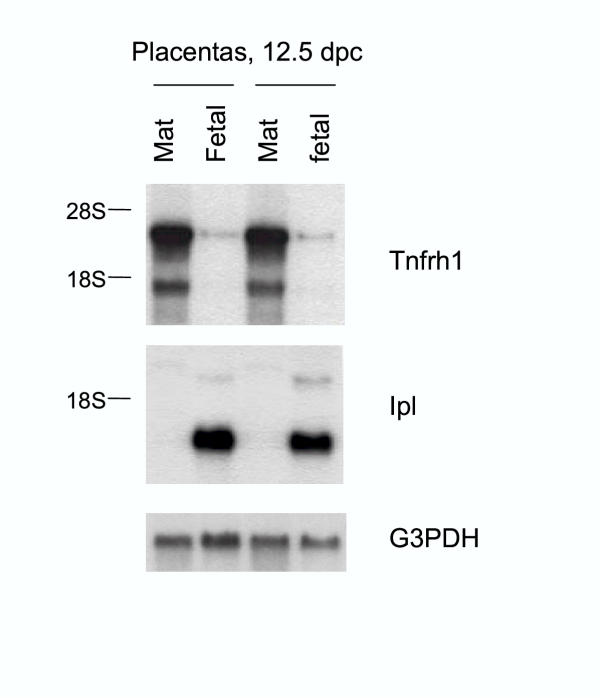
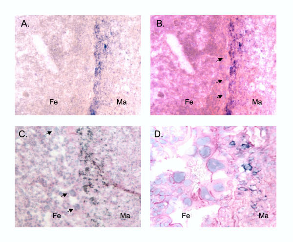
Results: We analyzed Tnfrh1 for evidence of parental imprinting, and characterized its tissue-specific expression. Tnfrh1 mRNA is detectable in multiple adult and fetal tissues, with highest expression in placenta, where in situ hybridization reveals a distinctive population of Tnfrh1-positive cells in maternal decidua, directly beneath the trophoblast giant cells. In offspring of interspecific mouse crosses, Tnfrh1 shows a consistent parent-of-origin-dependent allelic expression bias, with relative repression, but not silencing, of the paternal allele in several organs including fetal liver and adult spleen.

Conclusions: Genes preferentially expressed in the placenta are predicted to evolve rapidly, and Tnfrh1 appears to be an example of this phenomenon. In view of its strong expression in cells at the fetal-maternal boundary, Tnfrh1 warrants further study as a gene that might modulate immune or trophic interactions between the invasive placental trophoblast and the maternal decidua. The preferential expression of Tnfrh1 from the maternal allele indicates weak functional imprinting of this locus in some tissues.


Figures

Figure 1
Figure 1
Genes and PCR strategies.A, Map showing the positions of genes in the telomeric half of the distal mouse Chr7 imprinted domain. Direction of transcription is shown by the arrows, and imprinting is color-coded, with red indicating preferential expression of the maternal allele and blue indicating expression of the paternal allele. The Obph1 gene is coded as orange, since it shows an allelic expression bias only in the placenta, which may indicate either imprinting or expression from maternal cells. Genes that do not show an allelic expression bias, or for which complete information is not available, are in black. The KvDMR1 differentially methylated sequence, that acts as an imprinting control element and gives rise to the Kcnq1ot1 non-translated RNA, is indicated by the green bar. B, Structure of the Tnfrh1 and Tnfrh2 genes. PCR primers (arrows) are shown above the exons (rectangles). The yellow and orange shading highlights the non-conserved first exons. The last exons are highly conserved, and the remaining exons show intermediate conservation. Genetic polymorphisms in Tnfrh1 are indicated in violet (polymorphisms that distinguish CAST from BL/6) or maroon (polymorphisms that distinguish MOLD from BL/6). For analysis of allele-specific mRNA expression, we used primer pairs (1,2) or, alternatively (1,3). Estimation of relative expression from Tnfrh1 vs. Tnfrh2 by RT-PCR was done with primers 1, 4 and 5. Primers 1 and 6 were used to generate a Tnfrh1-specific cDNA probe.
Figure 2
Figure 2
Amino acid sequence similarity between mouse Tnfrh1 and Tnfrh2 proteins and the extracellular domains of the prototypical TNF receptor family members TNFR and Fas/CD95. Amino acids with complete conservation are shaded, and the conserved cysteine residues are in bold. The sequence of TNFR is from F. catus and the sequence of Fas is from S. scrofa. Proteins from these species were chosen for this illustration because they yielded the most significant BLAST scores in alignments with Tnfrh1 and Tnfrh2.
Figure 3
Figure 3
Expression of Tnfrh1 in adult and fetal mouse tissues.Tnfrh1 mRNA is detected as two alternative transcripts, both of which are present in most tissues. Both transcripts are most abundant in the placenta. The Tnfrh1 partial cDNA used to probe these blots has no detectable sequence identity with Tnfrh2 over the 5'-most 65 nt, but has 91% identity with the Tnfrh2 over the remaining 450 nt. Stripping and rehybridization at high stringency, with a smaller 5' probe with 72% overall sequence identity over 297 nt gave an identical pattern of bands. Ethidium bromide (EtBr) staining of ribosomal RNA is a loading control. The lanes contain 20 micrograms of total RNA; blots were exposed overnight.
Figure 4
Figure 4
Tissue-specific ratios of Tnfrh1 to Tnfrh2 expression. cDNAs were amplified using upstream primers specific for the divergent first exons of either Tnfrh1 or Tnfrh2 (Fig.1, primers 1 and 5), with a shared downstream primer (Fig.1, primer 4). The predicted sizes for the PCR products derived from each gene are indicated. Several organs and tissues, including whole placentas at 12.5 and 14.5 dpc (asterisks), show preferential expression of Tnfrh1.
Figure 5
Figure 5
Tnfrh1 mRNA is restricted to the deep layers of the placenta. Two placentas from 12.5 dpc conceptuses were frozen and divided into superficial (fetal) and deep (maternal; mat) halves prior to RNA extraction. The Ipl cDNA probe is a specific marker for the labyrinthine trophoblast and therefore hybridizes to the lanes from the superficial halves of the placentas.
Figure 6
Figure 6
Expression of Tnfrh1 is restricted to a discrete sub-population of cells near the fetal-maternal boundary.A, low-magnification image of 10.5 dpc placenta subjected to ISH with the Tnfrh1-antisense probe, showing a band of Tnfrh1-positive cells near the fetal-maternal junction. Signal in other areas is not higher than that seen with the sense control probe (not shown). Fe=fetal side of placenta, Ma=maternal side. B, the same section stained with hematoxylin-eosin (HE) to show cellular morphology. The arrows indicate trophoblast giant cells at the fetal-maternal boundary. The in situ image has been superimposed on the HE image, revealing Tnfrh1-positive cells restricted to the zone immediately under the trophoblast giant cells. C, superimposed ISH and HE images obtained from a placenta at 12.5 dpc, showing a distribution of Tnfrh1-positive cells similar to that at the earlier stage. Trophoblast giant cells are indicated by arrows. D, high-magnification field of another section at 12.5 dpc, which was first subjected to ISH and then lightly counterstained with periodic acid Schiff (PAS). The ISH reaction product persists under these conditions, revealing Tnfrh1-positive cells in close proximity to trophoblast giant cells.
Figure 7
Figure 7
Preferential expression of the maternal allele of Tnfrh1 in fetal liver from CAST × BL/6 reciprocal crosses.A, Analysis of the MboII RFLP, in which the BL/6 allele is undigested and the CAST allele yields two fragments. There is a strong maternal allele bias in placenta, and a weaker but still obvious parent-of-origin dependent biases in fetal liver. This imprinting effect is superimposed on a non-imprinted bias for higher expression of the BL/6 allele. The allelic bias in placenta reflects expression from maternal cells, but the bias in fetal liver reflects parental imprinting. Similar results were obtained with multiple aliquots of cDNA (not shown). B=BL/6, C=CAST. (Note that only the internal comparisons of alleles in single PCR reactions are valid; the stronger band obtained from the CAST maternal kidney should not be compared to that from the BL/6 maternal kidney for intensity of expression, since these were separate RT-PCR reactions). B, Direct sequencing of RT-PCR products. This shows an allelic expression bias that depends on the direction of the cross, consistent with the RFLP results.
Figure 8
Figure 8
Preferential expression of the maternal allele of Tnfrh1 in fetal and adult tissues from MOLD × BL/6 reciprocal crosses.A, SSCP analysis of RT-PCR products from the BL/6 and MOLD alleles. The mixing experiment validates this method. There is a bias towards greater expression of the maternal allele in several organs, indicated by a relative increase in the MOLD allele intensity in the MxB lanes compared to the BxM lanes. This is superimposed on a non-imprinted baseline of hyper-expression of the BL/6 allele. The allelic bias in placenta reflects expression from maternal cells, but the parent-of-origin dependent bias in other tissues reflects imprinting. Brackets indicate duplicate or triplicate assays using independent aliquots of cDNA. B=BL/6, M=MOLD. B, Direct sequencing of RT-PCR products. Consistent with the SSCP analysis, this shows an expression bias favoring the maternal allele, which is superimposed on a baseline of hyper-expression of the BL/6 allele.
Figure 9
Figure 9
Lack of allelic expression bias for Obph1 in fetal and adult tissues from MOLD × BL/6 reciprocal crosses.A, Separation of BL/6 and MOLD alleles of Obph1 by SSCP analysis of RT-PCR products. This shows equal biallelic expression in all organs, except for the placenta. B, Confirmation of the SSCP data by direct sequencing of Obph1 cDNAs. An allelic bias is only present in the placenta.

Similar articles

Cited by

References

    1. Maher ER, Reik W. Beckwith-Wiedemann syndrome: imprinting in clusters revisited. J Clin Invest. 2000;105:247–252. - PMC - PubMed
    1. Ferguson-Smith AC, Surani MA. Imprinting and the epigenetic asymmetry between parental genomes. Science. 2001;293:1086–1089. doi: 10.1126/science.1064020. - DOI - PubMed
    1. Yuan L, Qian N, Tycko B. An extended region of biallelic gene expression and rodent-human synteny downstream of the imprinted H19 gene on chromosome 11p15.5. Hum Mol Genet. 1996;5:1931–1937. doi: 10.1093/hmg/5.12.1931. - DOI - PubMed
    1. Engemann S, Strodicke M, Paulsen M, Franck O, Reinhardt R, Lane N, Reik W, Walter J. Sequence and functional comparison in the Beckwith-Wiedemann region: implications for a novel imprinting centre and extended imprinting. Hum Mol Genet. 2000;9:2691–2706. doi: 10.1093/hmg/9.18.2691. - DOI - PubMed
    1. Hu RJ, Lee MP, Johnson LA, Feinberg AP. A novel human homologue of yeast nucleosome assembly protein, 65 kb centromeric to the p57KIP2 gene, is biallelically expressed in fetal and adult tissues. Hum Mol Genet. 1996;5:1743–1748. doi: 10.1093/hmg/5.11.1743. - DOI - PubMed

Publication types

MeSH terms

Associated data